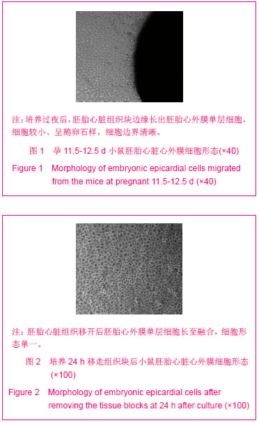
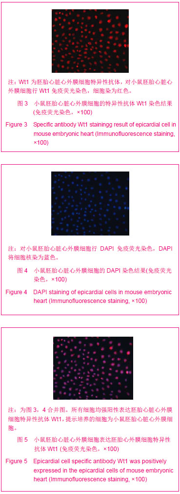
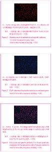
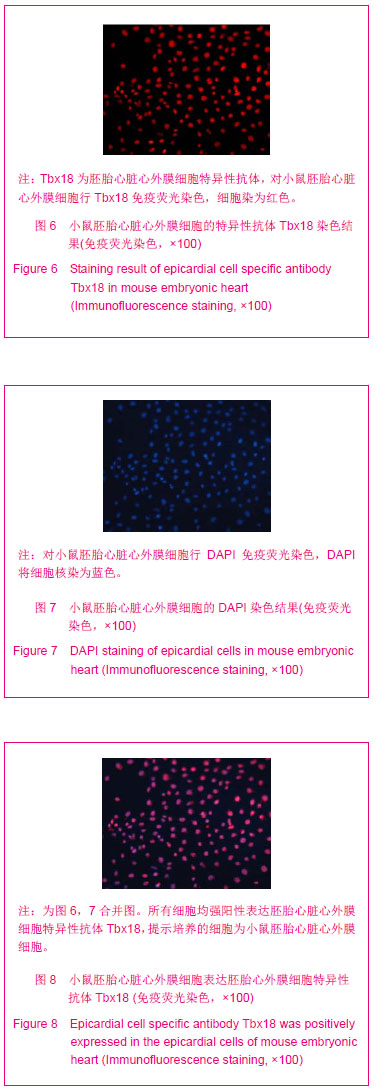
| [1]Lui KO, Bu L, Li RA, et al. Pluripotent stem cell-based heart regeneration: from the developmental and immunological perspectives. Birth Defects Res C Embryo Today. 2012; 96(1): 98-108.
[2]Ludwig M, Steinhoff G, Li J. The regenerative potential of angiotensin AT2 receptor in cardiac repair. Can J Physiol Pharmacol. 2012;90(3):287-293.
[3]Abdelwahid E, Siminiak T, Guarita-Souza LC, et al. Stem cell therapy in heart diseases: a review of selected new perspectives, practical considerations and clinical applications. Curr Cardiol Rev. 2011;7(3):201-212.
[4]Sánchez A, Schimmang T, García-Sancho J. Cell and tissue therapy in regenerative medicine. Adv Exp Med Biol. 2012; 741: 89-102.
[5]Gnecchi M, Danieli P, Cervio E. Mesenchymal stem cell therapy for heart disease. Vascul Pharmacol. 2012; 57(1): 48-55.
[6]Rangappa S, Makkar R, Forrester J. Review article: current status of myocardial regeneration: new cell sources and new strategies. J Cardiovasc Pharmacol Ther. 2010;15(4): 338-343.
[7]Menasché P. Embryonic stem cells in the treatment of severe cardiac insufficiency. Biol Aujourdhui. 2012;206(1):31-44.
[8]Pal SN, Kofidis T. New cell therapies in cardiology. Expert Rev Cardiovasc Ther. 2012;10(8):1023-1037.
[9]Gittenberger-de Groot AC, Winter EM, Bartelings MM, et al. The arterial and cardiac epicardium in development, disease and repair. Differentiation. 2012;84(1):41-53.
[10]Barile L, Messina E, Giacomello A, et al. Endogenous cardiac stem cells. Prog Cardiovasc Dis. 2007;50(1):31-48.
[11]Collins JM, Russell B. Stem Cell Therapy for Cardiac Repair. J Cardiovasc Nurs. 2009;24(2):93-97.
[12]Cai CL, Martin JC, Sun Y, et al. A myocardial lineage derives from Tbx18 epicardial cells. Nature. 2008;454 (7200): 104-108.
[13]Wu X, Ferrara C, Shapiro E, et al. Bmp7 expression and null phenotype in the urogenital system suggest a role in re-organization of the urethral epithelium. Gene Expr Patterns. 2009;9(4):224-230.
[14]Hassane S, Claij N, Jodar M, et al. Pkd1-inactivation in vascular smooth muscle cells and adaptation to hypertension. Lab Invest. 2011;91(1):24-32.
[15]Kim J, Rubin N, Huang Y, et al. In vitro culture of epicardial cells from adult zebrafish heart on a fibrin matrix. Nat Protoc. 2012; 7(2):247-255.
[16]Zhou B, Pu WT. Isolation and characterization of embryonic and adult epicardium and epicardium-derived cell. Methods Mol Biol. 2012;843:155-168.
[17]余德立,余资江. 大鼠视网膜干细胞的增殖和多向分化[J].中国组织工程研究,2012,16(6):1015-1018.
[18]于滋超,郭开今.不同年龄段大鼠脊髓源性神经干细胞的体外培养及鉴定[J].中国组织工程研究,2012,16(45):8506-8509.
[19]Mazo M, Araña M, Pelacho B,et al. Mesenchymal stem cells and cardiovascular disease: a bench to bedside roadmap. Stem Cells Int. 2012;2012:175979.
[20]Choi SH, Junq SY, Kwon SM, et al. Perspectives on stem cell therapy for cardiac regeneration. Cir J. 2012;76(6):1307- 1312.
[21]Didié M, Christalla P, Rubart M,et al. Parthenogenetic stem cells for tissue-engineered heart repair. J Clin Invest. 2013; 123(3):1285-1298.
[22]Hindley C, Philpott A. The cell cycle and pluripotency. Biochem J. 2013;15(12):135-143.
[23]Tsubouchi T, Fisher AG. Reprogramming and the pluripotent stem cell cycle. Curr Top Dev Biol. 2013;104:223-241.
[24]Medvedev SP, Pokushalov EA, Zakian SM. Epigenetics of pluripotent cells. Acta Naturae. 2012;4(4):28-46.
[25]杨新春,易方方,蔡军,等.由胚胎干细胞分化的心肌细胞移植对心肌梗死大鼠左心室重构及心功能的影响[J].中华器官移植杂志,2007,28(9):530-533.
[26]Ensenat-Waser R, Pellicer A, Simon C. Reprogrammed induced pluripotent stem cells: how suitable could they be in reproductive medicine? Fertil Steril. 2009;91(4):971-974.
[27]Ferreira LM, Mostajo-Radji MA. How induced pluripotent stem cells are redefining personalized medicine. Gene. 2013; 520 (1): 1-6.
[28]Simara P, Motl JA, Kaufman DS. Pluripotent stem cells and gene therapy. Transl Res. 2013;161(4):284-292.
[29]Dawn B, Stein AB,Urbanek K, et al.Cardiac stem cells delivered intravascularly traverse the vessel barrier, regenerate infarcted myocardium, and improve cardiac function. Proc Natl Acad Sci USA. 2005;102(10):3766-3771.
[30]Oh H, Brasfute SB, Gallardo TD, et al. Cardiac progenitor cells from adult myocardium: homing, differentiation, and fusion after infarction. Proc Natl Acad Sci USA. 2003; 100(21):12313-12318.
[31]Cai CL, Liang X, Shi Y, et al. Isl1 identifies a cardiac progenitor population that proliferates prior to differentiation and contributes a majority of cells to the heart. Dev Cell. 2003;5(6):877-889.
[32]Klaus A, Müller M, Schulz H, et al. Wnt/β-catenin and Bmp signals control distinct sets of transcription factors in cardiac progenitor cells. Proc Natl Acad Sci U S A. 2012;109(27): 10921-10926.
[33]Pandur P, Sirbu IO, Kühl SJ, et al. Islet1-expressing cardiac progenitor cells: a comparison across species. Dev Genes Evol. 2013;223(1-2):117-129.
[34]Messina E, De Angelis L, Frati G, et al. Isolation and expansion of adult cardiac stem cells from human and murine heart.Circ Res. 2004;95(9):911-921.
[35]Pérez-Pomares JM, de la Pompa JL. Signaling during epicardium and coronary vessel development. Circ Res. 2011;109(12):1429-1442.
[36]Hinkel R, Trenkwalder T, Kupatt C. Molecular and cellular mechanisms of thymosin β4-mediated cardioprotection. Ann N Y Acad Sci. 2012;1269:102-109.
[37]Nakano H, Williams E, Hoshijima M, et al. Cardiac origin of smooth muscle cells in the inflow tract.J Mol Cell Cardiol. 2011;50(2):337-345.
[38]Lepilina A, Coon AN, Kikuchi K, et al. A dynamic epicardial injury response supports progenitor cell activity during zebrafish heart regeneration. Cell.2006,127(3):607-619.
[39]Limana F, Capogrossi MC, Germani A. The epicardium in cardiac repair: from the stem cell view. Pharmacol Ther. 2011;129(1):82-96.
[40]Kim J, Wu Q, Zhang Y, et al. PDGF signaling is required for epicardial function and blood vessel formation in regenerating zebrafish hearts. Proc Natl Acad Sci U S A. 2010;107(40): 17206-17210.
[41]González-Rosa JM, Martín V, Peralta M, et al. Extensive scar formation and regression during heart regeneration after cryoinjury in zebrafish. Development. 2011;138(9):1663-1674. |