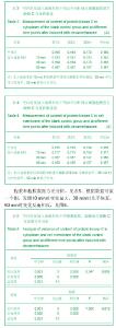
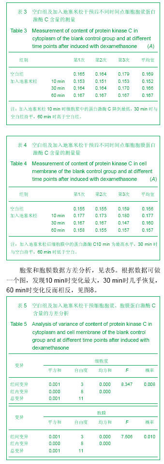
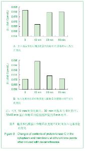
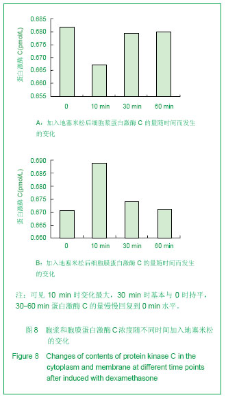
| [1] Pennington JM, Millar J, L Jones CP, et al. Simultaneous real-time amperometric measurement of catecholamines and serotonin at carbon fibre 'dident' microelectrodes. J Neurosci Methods. 2004;140(1-2):5-13.
[2] Marciniak D, Furey C, Shaffer JW. Osteonecrosis of the femoral head. A study of 101 hips treated with vascularized fibular grafting. J Bone Joint Surg Am. 2005;87(4):742-747.
[3] Hong D, Chen HX, Yu HQ, et al. Quantitative proteomic analysis of dexamethasone-induced effects on osteoblast differentiation, proliferation, and apoptosis in MC3T3-E1 cells using SILAC. Osteoporos Int. 2011;22(7):2175-2186.
[4] Kósa JP, Kis A, Bácsi K, et al. The protective role of bone morphogenetic protein-8 in the glucocorticoid-induced apoptosis on bone cells. Bone. 2011;48(5):1052-1057.
[5] De Vries F, Bracke M, Leufkens HG,et al.. Fracture risk with intermittent high-dose oral glucocorticoid therapy. Arthritis & Rheumatism. 2007;56(1):208-214.
[6] Ross EJ, Linch DC. Cushing's syndrome-killing disease: discriminatory value of signs and symptoms aiding early diagnosis. Lancet.1982;2(8299):646–9. PMID:125785
[7] Adinoff AD, Hollister JR. Steroid-induced fractures and bone loss in patients with asthma. N Engl J Med. 1983;309(5): 265-268.
[8] LoCascio V, Bonucci E, Imbimbo B, et al. Bone loss in response to long-term glucocorticoid therapy. Bone Miner. 1990;8(1):39-51.
[9] Weinstein RS, Jilka RL, Parfitt AM, et al. Inhibition of osteoblastogenesis and promotion of apoptosis of osteoblasts and osteocytes by glucocorticoids. Potential mechanisms of their deleterious effects on bone. J Clin Invest. 1998;102(2):274-282.
[10] Zauli G, Rimondi E, Celeghini C, et al. Dexamethasone counteracts the anti-osteoclastic, but not the anti-leukemic, activity of TNF-related apoptosis inducing ligand (TRAIL). J Cell Physiol. 2010;222(2):357-364.
[11] Canalis E, Bilezikian JP, Angeli A, et al. Perspectives on glucocorticoid-induced osteoporosis. Bone. 2004;34(4): 593-598.
[12] Rubin MR, Bilezikian JP. cal review 151: The role of parathyroid hormone in the pathogenesis of glucocorticoid-I nduced osteoporosis: a re-examination of the evidence. J Clin Endocrinol Metab. 2002;87(9):4033-4041.
[13] Roth J, Palm C, Scheunemann I, et al. Musculoskeletal abnormalities of the forearm in patients with juvenile idiopathic arthritis relate mainly to bone geometry. Arthritis Rheum. 2004;50(4):1277-1285.
[14] O'Brien CA, Jia D, Plotkin LI, et al. Glucocorticoids act directly on osteoblasts and osteocytes to induce their apoptosis and reduce bone formation and strength. Endocrinology. 2004; 145(4):1835-1841.
[15] Wang FS, Ko JY, Weng LH, et al. Inhibition of glycogen synthase kinase-3beta attenuates glucocorticoid-induced bone loss. Life Sci. 2009;85(19-20):685-692.
[16] Hill PA, Tumber A. Ceramide-induced cell death/survival in murine osteoblasts. J Endocrinol. 2010;206(2):225-233.
[17] Weinstein RS, Manolagas SC. Apoptosis and osteoporosis. Am J Med. 2000;108(2):153-164.
[18] Weinstein RS, Jilka RL, Parfitt AM, et al. Inhibition of osteoblastogenesis and promotion of apoptosis of osteoblasts and osteocytes by glucocorticoids. Potential mechanisms of their deleterious effects on bone. J Clin Invest. 1998;102(2): 274-282.
[19] Canalis E. Clinical review 83: Mechanisms of glucocorticoid action in bone: implications to glucocorticoid-induced osteoporosis. J Clin Endocrinol Metab. 1996;81(10): 3441-3447.
[20] Weinstein RS, Jilka RL, Parfitt AM, et al. Inhibition of osteoblastogenesis and promotion of apoptosis of osteoblasts and osteocytes by glucocorticoids. Potential mechanisms of their deleterious effects on bone. J Clin Invest. 1998;102(2): 274-282.
[21] Smith E, Coetzee GA, Frenkel B. Glucocorticoids inhibit cell cycle progression indifferentiating osteoblasts via glycogen synthase kinase-3beta. J Biol Chem. 2002;277(20): 18191-18197.
[22] Weinstein RS, Nicholas RW, Manolagas SC. Apoptosis of osteocytes in glucocorticoid-induced osteonecrosis of the hip. J Clin Endocrinol Metab. 2000;85(8):2907-2912.
[23] Calder JD, Pearse MF, Revell PA. The extent of osteocyte death in the proximal femur of patients with osteonecrosis of the femoral head. J Bone Joint Surg Br. 2001;83(3):419-422.
[24] Eberhardt AW, Yeager-Jones A, Blair HC. Regional trabecular bone matrix degeneration and osteocyte death in femora of glucocorticoid- treated rabbits. Endocrinology. 2001;142(3): 1333-1340.
[25] Jih-Yang Ko , Feng-Sheng Wang, Increased Dickkopf-1 expression accelerates bone cell apoptosis in femoral head osteonecrosis, Bone 46 (2010) 584–591.
[26] Hampson G, Fogelman I. Clinical role of bisphosphonate therapy. Int J Womens Health. 2012;4:455-469.
[27] Leclerc N, Noh T, Cogan J, et al. Opposing effects of glucocorticoids and Wnt signaling on Krox20 and mineral deposition in osteoblast cultures. J Cell Biochem. 2008;103(6): 1938-1951.
[28] Camps M, Nichols A, Arkinstall S. Dual specificity phosphatases: a gene family for control of MAP kinase function. FASEB J. 2000;14(1):6-16.
[29] Xu N, Liu H, Qu F, et al. Hypoxia inhibits the differentiation of mesenchymal stem cells into osteoblasts by activation of Notch signaling. Exp Mol Pathol. 2013;94(1):33-39.
[30] Canalis E. Notch signaling in osteoblasts. Sci Signal. 2008; 1(17):pe17.
[31] Kitase Y, Barragan L, Qing H, et al. Mechanical induction of PGE2 in osteocytes blocks glucocorticoid-induced apoptosis through both the β-catenin and PKA pathways. J Bone Miner Res. 2010;25(12):2657-2668.
[32] Lombardi G, Di Somma C, Rubino M, et al. The roles of parathyroid hormone in bone remodeling: prospects for novel therapeutics. J Endocrinol Invest. 2011;34(7 Suppl):18-22.
[33] Davis RJ. Signal transduction by the JNK group of MAP kinases. Cell. 2000;103(2):239-252.
[34] [Heath JK, Atkinson SJ, Meikle MC, et al. Mouse osteoblasts synthesize collagenase in response to bone resorbing agents. Biochim Biophys Acta. 1984;802(1):151-154.
[35] Lin GL, Hankenson KD. Integration of BMP, Wnt, and notch signaling pathways in osteoblast differentiation. J Cell Biochem. 2011;112(12):3491-3501.
[36] Lombardi G, Di Somma C, Rubino M, et al. The roles of parathyroid hormone in bone remodeling: prospects for novel therapeutics. J Endocrinol Invest. 2011;34(7 Suppl):18-22. |