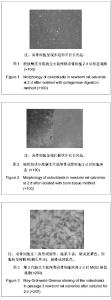
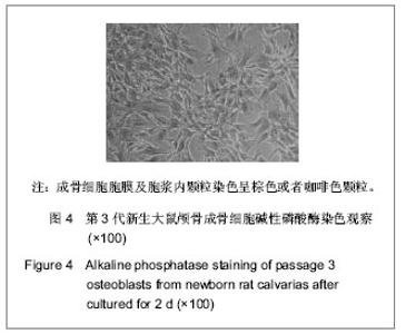
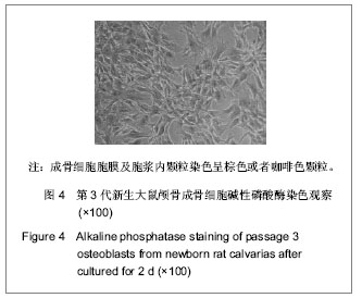
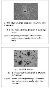
| [1] 李晓峰,赵劲民,苏伟,等. 大鼠成骨细胞的原代培养和鉴定[J]. 中国组织工程研究与临床康复,2011,15(6):990-994.
[2] Xing ZC, Han SJ, Shin YS, et al. Enhanced osteoblast responses to poly(methyl methacrylate)/hydroxyapatite electrospun nanocomposites for bone tissue engineering. J Biomater Sci Polym Ed. 2013;24(1):61-76.
[3] Czekanska EM, Stoddart MJ, Richards RG, et al. In search of an osteoblast cell model for in vitro research. Eur Cell Mater. 2012;24:1-17.
[4] 姚振强,曾才铭,唐景清. 骨膜成骨细胞修复骨缺损的实验研究[J]. 中国修复重建外科杂志,1996,10(3):173-175.
[5] 茹嘉,李强. 最新进展的转基因技术在骨与软骨缺损修复中的应用[J]. 中国组织工程研究,2013,17(2):353-357.
[6] Orriss IR, Taylor SE, Arnett T R. Rat osteoblast cultures. Methods Mol Biol.2012;816:31-41.
[7] Ciarelli TE, Tjhia C, Rao DS, et al. Trabecular packet-level lamellar density patterns differ by fracture status and bone formation rate in white females. Bone. 2009;45(5):903-908.
[8] Thiele F, Cohrs CM, Przemeck GK, et al. In vitro analysis of bone phenotypes in Col1a1 and Jagged1 mutant mice using a standardized osteoblast cell culture system. J Bone Miner Metab. 2013;31(3):293-303.
[9] Gallet M, Saidi S, Hay E, et al. Repression of osteoblast maturation by ERRalpha accounts for bone loss induced by estrogen deficiency. PLoS One. 2013;8(1):e54837.
[10] Jayakumar P, Di Silvio L. Osteoblasts in bone tissue engineering. Proc Inst Mech Eng H. 2010;224(12):1415-1440.
[11] Costa R, Ribeiro C, Lopes AC, et al. Osteoblast, fibroblast and in vivo biological response to poly(vinylidene fluoride) based composite materials. J Mater Sci Mater Med. 2013; 24(2):395-403.
[12] Peck WA, Birge SJ Jr, Fedak SA. Bone cells: biochemical and biological studies after enzymatic isoliation. Science. 1964; 146(3650): 1476-1477.
[13] Wong GL, Cohn DV. Target cells in bone for parathormone and calcitonin are different: enrichment for each cell type by sequential digestion of mouse calvaria and selective adhesion to polymeric surfaces. Proc Natl Acad Sci USA. 1975;72(8): 3167-3171.
[14] Robey PG, Termine JD. Human bone cells in vitro. Calcif Tissue Int.1985;37(5):453-460.
[15] Masquelier D, Herbert B, Hauser N, et al. Morphologic characterization of osteoblast-like cell cultures isolated from newborn rat calvaria. Calcif Tissue Int.1990;47(2):92-104.
[16] Morriss-Kay GM, Wilkie AO. Growth of the normal skull vault and its alteration in craniosynostosis: insights from human genetics and experimental studies. J Anat. 2005;207(5): 637-653.
[17] 张涌泉,郭征,白建萍. 利用漂浮组织块获得高纯度兔成骨细胞的方法[J]. 科学技术与工程,2006,6(17):2640-2643.
[18] 鄂玲玲,刘洪臣,王东胜. 贴壁组织块反复消化法培养新生大鼠下颌骨成骨细胞及鉴定 [J]. 华西口腔医学杂志,2009,27(2): 130-134.
[19] 张巍,史册,倪世磊,等. 新生大鼠下颌骨成骨细胞的分离、培养与鉴定[J]. 北京口腔医学,2011,19(4):318-321.
[20] 陈洁,兰由玉,何成松,等. 骨组织块法和酶消化法联合培养人成骨细胞[J].中国组织工程研究与临床康复,2012,16(7): 1145-1149.
[21] Clarke MS, Sundaresan A, Vanderburg CR, et al. A three-dimensional tissue culture model of bone formation utilizing rotational co-culture of human adult osteoblasts and osteoclasts. Acta Biomater. 2013;9(8):7908-7916.
[22] Linsley C, Wu B, Tawil B. The effect of fibrinogen, collagen type I, and fibronectin on mesenchymal stem cell growth and differentiation into osteoblasts. Tissue Eng Part A. 2013; 19(11-12):1416-1423.
[23] Dygai AM, Zyuz'kov GN, Zhdanov VV, et al. Specific activity of electron-beam synthesis immobilized hyaluronidase on G-CSF induced mobilization of bone marrow progenitor cells. Stem Cell Rev. 2013;9(2):140-147.
[24] 吴曼,侯建明,杨海燕,等. 乳铁蛋白对体外培养原代大鼠成骨细胞增生与分化的影响[J]. 中华骨质疏松和骨矿盐疾病杂志, 2013, 5(1):44-49.
[25] 黄芳,那思家,王健平,等. 聚乙酸-聚乙醇酸共聚物复合同种异体细胞修复软骨缺损[J].中国组织工程研究,2012,16(51): 9523-9528.
[26] 刘印,田京. 骨合成促进剂治疗骨质疏松症的优势[J]. 中国组织工程研究,2013,17(15):2803-2810.
[27] Nakamura M, Nagai A, Hentunen T, et al. Surface electric fields increase osteoblast adhesion through improved wettability on hydroxyapatite electret. ACS Appl Mater Interfaces. 2009;1(10):2181-2189.
[28] Fang Z, Sun T, Yadav SK. Research progress of bone morphogenetic protein and liability of ossification of posterior longitudinal ligament. Zhongguo Xiu Fu Chong Jian Wai Ke Za Zhi.2012;26(10):1255-1258.
[29] Peng Y, Shi K, Wang L, et al. Characterization of Osterix protein stability and physiological role in osteoblast differentiation. PLoS One. 2013;8(2):e56451.
[30] Lo KW, Kan HM, Ashe KM, et al. The small molecule PKA-specific cyclic AMP analogue as an inducer of osteoblast-like cells differentiation and mineralization. J Tissue Eng Regen Med. 2012;6(1):40-48.
[31] Uchihashi K, Aoki S, Matsunobu A, et al. Osteoblast migration into type I collagen gel and differentiation to osteocyte-like cells within a self-produced mineralized matrix: a novel system for analyzing differentiation from osteoblast to osteocyte. Bone. 2013;52(1):102-110.
[32] 虞冀哲,杨勇,刘朝旭,等. 共培养条件下电磁场干预对大鼠成骨细胞及骨髓间充质干细胞成骨分化的影响[J]. 中华物理医学与康复杂志,2013,35(4):250-255. |