Chinese Journal of Tissue Engineering Research
Previous Articles Next Articles
Anatomical measurements of proximal tibia of anterior and posterior cruciate ligament-retaining knee prosthesis
He Pei-heng, Xu Dong-liang, Zuo Jian-wei, Li Shuai-hua, Wa Qing-de
- Department of Joint Surgery, the First Affiliated Hospital of Sun Yat-sen University, Guangzhou 510080, Guangdong Province, China
-
Received:2013-03-05Revised:2013-04-23Online:2013-06-25Published:2013-06-25 -
Contact:Xu Dong-liang, Doctoral supervisor, Professor, Chief physician, Department of Joint Surgery, the First Affiliated Hospital of Sun Yat-sen University, Guangzhou 510080, Guangdong Province, China xdl1234hph@sina.com -
About author:He Pei-heng☆, Studying for doctorate, Department of Joint Surgery, the First Affiliated Hospital of Sun Yat-sen University, Guangzhou 510080, Guangdong Province, China hepeiheng1234@sina.com -
Supported by:“5010” Clinical Research Fund of Sun Yat-sen University, No.2010005
CLC Number:
Cite this article
He Pei-heng, Xu Dong-liang, Zuo Jian-wei, Li Shuai-hua, Wa Qing-de. Anatomical measurements of proximal tibia of anterior and posterior cruciate ligament-retaining knee prosthesis[J]. Chinese Journal of Tissue Engineering Research, doi: 10.3969/j.issn.2095-4344.2013.26.004.
share this article
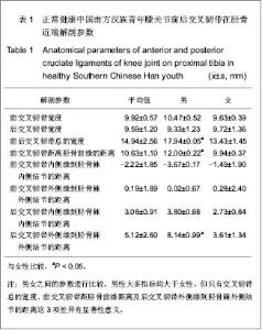
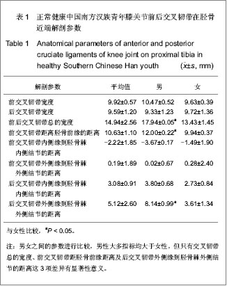
2.1 前后交叉韧带在胫骨近端解剖参数均值及标准差 男女之间的参数进行比较,结果显示交叉韧带总的宽度、前交叉韧带距胫骨前缘距离及后交叉韧带外侧缘到胫骨棘外侧结节的距离差异有显著性意义(P < 0.05),男性均值明显大于女性,其他指标测量结果男女之间比较,差异无显著性意义,见表1。"
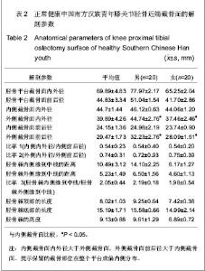
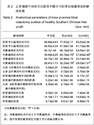
2.2 胫骨近端截骨面的解剖参数均值及标准差 见表2。"
| [1] Seo JG, Moon YW, Park SH, et al. A case-control study of spontaneous patellar fractures following primary total knee replacement. J Bone Joint Surg Br. 2012;94(7):908-913. [2] Weinstein AM, Rome BN, Reichmann WM, et al. Estimating the burden of total knee replacement in the United States. J Bone Joint Surg Am. 2013;95(5):385-392. [3] Weber O, Goost H, Mueller M, et al. Mid-term results after post-traumatic knee joint replacement in elderly patients. Z Orthop Unfall. 2011r;149(2):166-172.[4] Judge A, Arden NK, Cooper C, et al. Predictors of outcomes of total knee replacement surgery. Rheumatology (Oxford). 2012;51(10):1804-1813. [5] Tungtrongjit Y, Weingkum P, Saunkool P. The effect of preoperative quadriceps exercise on functional outcome after total knee arthroplasty. J Med Assoc Thai. 2012t;95 Suppl 10: S58-66. [6] Judd DL, Eckhoff DG, Stevens-Lapsley JE. Muscle strength loss in the lower limb after total knee arthroplasty. Am J Phys Med Rehabil. 2012;91(3):220-226. [7] Dennis DA, Komistek RD, Mahfouz MR, et al. Multicenter determination of in vivo kinematics after total knee arthroplasty. Clin Orthop Relat Res. 2003;(416):37-57. [8] Kitagawa A, Tsumura N, Chin T, et al. In vivo comparison of knee kinematics before and after high-flexion posterior cruciate-retaining total knee arthroplasty. J Arthroplasty. 2010; 25(6):964-969.[9] Hosseini A, Van de Velde S, Gill TJ, et al. Tibiofemoral cartilage contact biomechanics in patients after reconstruction of a ruptured anterior cruciate ligament. J Orthop Res. 2012; 30(11):1781-1788. [10] Chen CH, Li JS, Hosseini A,et al. Anteroposterior stability of the knee during the stance phase of gait after anterior cruciate ligament deficiency. Gait Posture. 2012;35(3):467-471. [11] Moro-oka TA, Muenchinger M, Canciani JP, et al. Comparing in vivo kinematics of anterior cruciate-retaining and posterior cruciate-retaining total knee arthroplasty. Knee Surg Sports Traumatol Arthrosc. 2007;15(1):93-99. [12] Mikashima Y, Tomatsu T, Horikoshi M, et al. In vivo deep-flexion kinematics in patients with posterior-cruciate retaining and anterior-cruciate substituting total knee arthroplasty. Clin Biomech (Bristol, Avon). 2010;25(1):83-87.[13] Koyonos L, Stulberg SD, Moen TC, et al. Sources of error in total knee arthroplasty. Orthopedics. 2009;32(5):317. [14] Perillo-Marcone A, Taylor M. Effect of varus/valgus malalignment on bone strains in the proximal tibia after TKR: an explicit finite element study. J Biomech Eng. 2007;129(1): 1-11.[15] Jenny JY, Jenny G. Preservation of anterior cruciate ligament in total knee arthroplasty. Arch Orthop Trauma Surg. 1998; 118(3):145-148.[16] Lozano-Calderón SA, Shen J, Doumato DF, et al. Cruciate-retaining vs posterior-substituting inserts in total knee arthroplasty: functional outcome comparison. J Arthroplasty. 2013;28(2):234-242. [17] Luo CF. Reference axes for reconstruction of the knee. Knee. 2004;11(4):251-257. [18] Nowakowski AM, Müller-Gerbl M, Valderrabano V. Assessment of knee implant alignment using coordinate measurement on three-dimensional computed tomography reconstructions. Surg Innov. 2012;19(4):375-384.[19] Nowakowski AM, Müller-Gerbl M, Valderrabano V. Surgical approach for a new knee prosthesis concept (TSTP) retaining both cruciate ligaments. Clin Anat. 2010v;23(8):985-991.[20] Nowakowski AM, Stangel M, Grupp TM, et al. Investigating the primary stability of the transversal support tibial plateau concept to retain both cruciate ligaments during total knee arthroplasty. J Appl Biomater Funct Mater. 2012 ;10(2): e127-135. [21] Siebold R, Ellert T, Metz S, et al. Tibial insertions of the anteromedial and posterolateral bundles of the anterior cruciate ligament: morphometry, arthroscopic landmarks, and orientation model for bone tunnel placement. Arthroscopy. 2008;24(2):154-161. [22] Voos JE, Mauro CS, Wente T, et al. Posterior cruciate ligament: anatomy, biomechanics, and outcomes. Am J Sports Med. 2012;40(1):222-231. [23] Han GB, Zhang S, Chen JQ, et al. Zhongguo Zuzhi Gongcheng Yanjiu. 2012;16(35):6489-6464.韩贵宾,张寿,陈建强,等.国人胫骨截面矢量化测量与解剖型假体形态的设计[J].中国组织工程研究,2012,16(35):6489- 6464.[24] Zheng YF, Qu TB, Guo XY, et al. Zhongguo Jiaoxing Waike Zazhi. 2011;19(13):1118-1121.郑寅峰,曲铁兵,郭璇瑛,等.国人胫骨平台截骨面与国人膝关节假体的涵盖率分析[J].中国矫形外科杂志,2011,19(13):1118- 1121.[25] Purnell ML, Larson AI, Clancy W. Anterior cruciate ligament insertions on the tibia and femur and their relationships to critical bony landmarks using high-resolution volume-rendering computed tomography. Am J Sports Med. 2008;36(11):2083-2090.[26] Hwang MD, Piefer JW, Lubowitz JH. Anterior cruciate ligament tibial footprint anatomy: systematic review of the 21st century literature. Arthroscopy. 2012;28(5):728-734. [27] Cheng CK, Lung CY, Lee YM, et al. A new approach of designing the tibial baseplate of total knee prostheses. Clin Biomech (Bristol, Avon). 1999;14(2):112-117. [28] Zhang YD, Liu YR, Zhao GZ, et al. Zhongguo Zuzhi Gongcheng Yanjiu. 2012;16(35):6466-6470.张艳东,刘奕蓉,赵国志,等.成人距骨数字化计算机三维模型解剖学测量及对个性化治疗的意义[J].中国组织工程研究,2012, 16(35):6466-6470.[29] Xu P, Li YL, Chen WD, et al. Zhongguo Zuzhi Gongcheng Yanjiu yu Linchuang Kangfu. 2011;15(43):8006-8009.许鹏,李彦林,陈文栋,等. MRI影像下股骨髁间窝三维数字化解剖学数据与实体解剖测量值的差异[J].中国组织工程研究与临床康复,2011,15(43):8006-8009.[30] Hitt K, Shurman JR 2nd, Greene K, et al. Anthropometric measurements of the human knee: correlation to the sizing of current knee arthroplasty systems. J Bone Joint Surg Am. 2003;85-A Suppl 4:115-122. [31] Kwak DS, Surendran S, Pengatteeri YH, et al. Morphometry of the proximal tibia to design the tibial component of total knee arthroplasty for the Korean population. Knee. 2007; 14(4):295-300. [32] Chang TW, Huang CH, McClean CJ,et al. Morphometrical measurement of resected surface of medial and lateral proximal tibia for Chinese population. Knee Surg Sports Traumatol Arthrosc. 2012p;20(9):1730-1735.[33] Yang B, Yu JK, Zheng ZZ, et al. Computed Tomography Morphometric Study of Gender Differences in Osteoarthritis Proximal Tibias. J Arthroplasty. 2012. pii: S0883- 5403(12) 00564-5. [34] Liu Z, Yuan G, Zhang W, et al. Anthropometry of the Proximal Tibia of Patients With Knee Arthritis in Shanghai. J Arthroplasty. 2013. pii: S0883-5403(13)00112-5.[35] Skovgaard C, Holm B, Troelsen A,et al. No effect of fibrin sealant on drain output or functional recovery following simultaneous bilateral total knee arthroplasty. Acta Orthop. 2013;84(2):153-158. [36] Chong DY, Hansen UN, van der Venne R, et al. The influence of tibial component fixation techniques on resorption of supporting bone stock after total knee replacement. J Biomech. 2011;44(5):948-954. [37] Roh YW, Jang J, Choi WC, et al. Preservation of the posterior cruciate ligament is not helpful in highly conforming mobile-bearing total knee arthroplasty: a randomized controlled study. |
| [1] | Chen Junming, Yue Chen, He Peilin, Zhang Juntao, Sun Moyuan, Liu Youwen. Hip arthroplasty versus proximal femoral nail antirotation for intertrochanteric fractures in older adults: a meta-analysis [J]. Chinese Journal of Tissue Engineering Research, 2021, 25(9): 1452-1457. |
| [2] | Wu Zijian, Hu Zhaoduan, Xie Youqiong, Wang Feng, Li Jia, Li Bocun, Cai Guowei, Peng Rui. Three-dimensional printing technology and bone tissue engineering research: literature metrology and visual analysis of research hotspots [J]. Chinese Journal of Tissue Engineering Research, 2021, 25(4): 564-569. |
| [3] | Li Xiaozhuang, Duan Hao, Wang Weizhou, Tang Zhihong, Wang Yanghao, He Fei. Application of bone tissue engineering materials in the treatment of bone defect diseases in vivo [J]. Chinese Journal of Tissue Engineering Research, 2021, 25(4): 626-631. |
| [4] | Chen Song, He Yuanli, Xie Wenjia, Zhong Linna, Wang Jian. Advantages of calcium phosphate nanoparticles for drug delivery in bone tissue engineering research and application [J]. Chinese Journal of Tissue Engineering Research, 2021, 25(22): 3565-3570. |
| [5] | Zhang Zhenhua, Liu Zichen, Yu Baoqing. Status and problems of polycaprolactone and its composite materials in bone tissue engineering [J]. Chinese Journal of Tissue Engineering Research, 2021, 25(22): 3571-3577. |
| [6] | Xu Hui, Kang Bingxin, Zhong Sheng, Gao Chenxin, Zhao Chi, Qiu Guowei, Sun Songtao, Xie Jun, Xiao Lianbo, Shi Qi. Pressing local acupoints plus adjustion of the knee joint in a sitting position for treating knee osteoarthritis: a randomized controlled trial [J]. Chinese Journal of Tissue Engineering Research, 2021, 25(2): 216-221. |
| [7] | Wang Liu, Song Dongzhe, Huang Dingming. Bone morphogenetic protein 9 regulates stem cell differentiation and bone regeneration [J]. Chinese Journal of Tissue Engineering Research, 2021, 25(19): 3064-3070. |
| [8] | Li Yanle, Yue Xiaohua, Nie Zhen, Zhang Junwei, Li Zhaohui, Nie Weizhi, Jiang Hongjiang. Characteristics and application of bioabsorbable materials in orthopedics [J]. Chinese Journal of Tissue Engineering Research, 2021, 25(16): 2612-2617. |
| [9] | Liu Zige, Liu Xinrui, Li Yan, Song Guorui, Zhang Chen, Chen Desheng. In vitro experiment of tetrandrine on the model of osteolysis induced by wear particles around the prosthesis [J]. Chinese Journal of Tissue Engineering Research, 2021, 25(15): 2358-2363. |
| [10] | Han Ningning, Zuo Jinfu, Sun Miao, Tang Shengjian, Liu Fangjun. Application and progress of umbilical cord mesenchymal stem cells in bone tissue engineering [J]. Chinese Journal of Tissue Engineering Research, 2021, 25(13): 2079-2086. |
| [11] | Meng Maohua, Li Ying, Chen Xin, Cheng Lu, Dong Qiang. Effects and mechanisms of enamel matrix derivatives on osteogenic differentiation of bone marrow mesenchymal stem cells [J]. Chinese Journal of Tissue Engineering Research, 2021, 25(13): 2108-2113. |
| [12] | Wang Xiankang, Zhang Yuejing, Yang You, Liu Jun. Simulation analysis of wear performance for tibial insert of unicompartmental knee arthroplasty prosthesis under gait load [J]. Chinese Journal of Tissue Engineering Research, 2021, 25(12): 1831-1835. |
| [13] | Li Dongdong, Liao Hongbing . MicroRNA-214 is involved in the regulation of bone metabolism [J]. Chinese Journal of Tissue Engineering Research, 2021, 25(11): 1779-1784. |
| [14] | Zhou Qi, Gao Yi, Wei Kang, Li Jun, Xu Jianda, Jiang Yang, Qu Yuxing. Total knee arthroplasty for rheumatoid arthritis: knee function and biochemical index changes [J]. Chinese Journal of Tissue Engineering Research, 2020, 24(9): 1337-1341. |
| [15] | Zhang Shengmin, Liu Chao. Research progress in osteogenic differentiation of adipose-derived stem cells induced by bioscaffold materials [J]. Chinese Journal of Tissue Engineering Research, 2020, 24(7): 1107-1116. |
| Viewed | ||||||
|
Full text |
|
|||||
|
Abstract |
|
|||||