Chinese Journal of Tissue Engineering Research ›› 2013, Vol. 17 ›› Issue (22): 3991-3997.doi: 10.3969/j.issn.2095-4344.2013.22.001
Bone mineral density and pathologic study of osteoporotic vertebral compression fracture
Cui Jiang-chao, Sun Jian-min, Cui Xin-gang, Jiang Zhen-song
- Department of Spine Surgery, Provincial Hospital of Shandong University, Jinan 250012, Shandong Province, China
-
Online:2013-05-28Published:2013-05-28 -
Contact:Sun Jian-min, M.D., Professor, Master’s supervisor, Department of Spine Surgery, Provincial Hospital of Shandong University, Jinan 250012, Shandong Province, China spine163@163.com -
About author:Cui Jiang-chao★, Studying for master’s degree, Department of Spine Surgery, Provincial Hospital of Shandong University, Jinan 250012, Shandong Province, China hykdcyfm@sina.com
CLC Number:
Cite this article
Cui Jiang-chao, Sun Jian-min, Cui Xin-gang, Jiang Zhen-song. Bone mineral density and pathologic study of osteoporotic vertebral compression fracture[J]. Chinese Journal of Tissue Engineering Research, 2013, 17(22): 3991-3997.
share this article
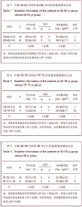
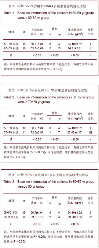
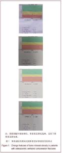
2.1 参与者数量分析 参与临床试验的患者共45例,按年龄分为4组,资料均完整,病史详细,数据准确可靠,均进行结果分析,未出现数据资料的丢失等。 2.2 各组基线资料比较 见表1-3。各组间患者年龄构成比差异有显著性意义(P < 0.05),在60-69岁组与其他组可见明显性别差异,而体质量指数在各组之间差异无显著性意义(P > 0.05)。"
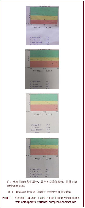
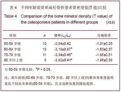
2.3 不同年龄组间所测定的骨质疏松骨折患者骨密度值比较 各年龄组间腰椎平均骨密度值下降差异存在显著性意义(P < 0.05),且腰椎骨密度降低比股骨近端较为明显。高年龄组(60-69岁组、70-79岁组、80岁以上组)的椎体骨密度值明显低于较低年龄组(50-59岁组),且呈逐渐加重的降低趋势;各组间的椎体密度值差异有显著性意义(P < 0.05),见图1和表4。"
"
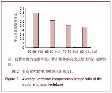
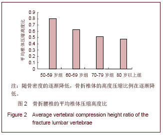
椎体骨密度的大小直接影响椎体的抗变形能力[5,9-10] ,结果发现椎体骨质密度与压缩比值程度存在明显的正相关关系,见图2,与肖越勇等[11]的实验研究相一致,即椎体骨密度下降明显者所引起的椎体压缩变形程度更为严重,而椎体破坏直接表现为病理形态学的变化。"
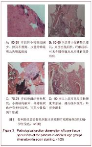
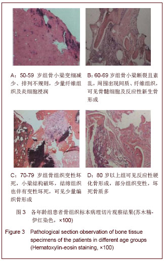
2.4 病理切片观察结果 45例患者共取骨组织标本47个,未发现有肿瘤、结核等病例。 50-59岁组共10例,主要表现为骨小梁变细减少、小梁之间连接断续,间隔增宽、排列不规则,有少量纤维增生及血块组织,见图3A。 60-69岁组共18例,病理主要表现为可见少量坏死的骨组织,骨小梁断裂、排列紊乱,周围间质、纤维组织增生,可见骨髓细胞,有反应性新生骨形成,见图3B;1例出现周围软组织肌纤维母样细胞反应性增生。 70-79岁组共11例,其病理示有较多骨组织变性坏死,骨小梁结构破坏紊乱不规则,周围结缔组织也伴有变性坏死,有少量编织骨形成,见图3C。 80岁以上组6例,病理见反应性硬化骨形成,部分组织变性伴坏死,可有慢性炎细胞,坏死骨质较多,见图3D。 2.5 骨密度与病理特征的相关性 通过病史、影像及骨密度检查,所有45例椎体压缩骨折均为单纯性骨质疏松性骨折。4个年龄组骨密度测量显示随着时间的延长,椎体内的密度逐渐降低,且下降的趋势加重;病理组织学可见早期有较明显的血块组织及急慢性炎细胞浸润,骨小梁随骨折的不断加重出现变细减少、稀疏、断裂等,其排列出现移位、不规则、甚至紊乱等,同时周围可见有纤维软骨组织、结缔组织等增生,也有少量的反应性新生骨。 Konermann等[12]以及Ebbesen等[13]的研究证实椎体的骨质疏松早先发生在椎体中部区域,而到后期才波及到椎体周边骨质;在实验所观察的骨显微结构切片中得到相同的结论,椎体深部所取标本可见骨组织的结构与序列破坏较为严重,而浅层标本尚可见存在一定结构的骨小梁及纤维结缔组织。 "
| [1]An Z,Yang DZ,Zhang ZJ,et al. Zhongguo Guzhi Shusong Zazhi. 2002;8(1):82-83.安珍,杨定焯,张祖君,等.骨质疏松性脊椎压缩性骨折流行病学调查分析[J]. 中国骨质疏松杂志, 2002,8(1):82-83.[2]Li NH,Qu PZ,Zhu HM,et al. Zhonghua Yixue Zazhi: Yingwenban. 2002;115(5):773-775.李宁华,区品中,朱汉民,等.中国部分地区中老年人群骨质疏松症患病率研究[J].中华医学杂志:英文版,2002,115(5):773- 775.[3]Ettinger MP. Aging bone and osteoporosis: strategies for preventing fractures in the elderly. Arch Intern Med. 2003; 163(18):2237-2246.[4]Fechtenbaum J, Cropet C, Kolta S,et al.The severity of vertebral fractures and health-related quality of life in osteoporotic postmenopausal women. Osteoporos Int. 2005;16(12):2175-2179.[5]Xu WH,Ma Y,Li Y,et al. Jiangxi Yixueyuan Xuebao. 2005; 45(5):21-23,27.徐文华,马勇,李云,等.椎体骨密度与椎体生物力学的关系[J]. 江西医学院学报, 2005, 45(5):21-23,27.[6]Xu SW,Gu GH,Zhao GF,et al. Zhonghua Jizhen Yixue Zazhi. 2002;11(5):324-326.徐少文,顾耕华,赵光锋,等.实验性骨质疏松性骨折愈合中的骨密度及组织学观察[J].中华急诊医学杂志,2002,11(5):324-326.[7]Wu L,Wu YN,Shen Y,et al. Yunnan Yiyao. 2007;28(1):11-14.吴玲,吴亚楠,沈芸,等.骨质疏松性骨折与老年保健[J].云南医药, 2007,28(1):11-14.[8]Hao YQ,Dai KR. Zhongguo Guzhi Shusong Zazhi. 2005;11(3): 273-277.郝永强,戴尅戎.骨质疏松性骨折愈合与骨量、骨结构及力学性能相关性的实验研究[J].中国骨质疏松杂志, 2005, 11(3):273-277.[9]Hulme PA, Boyd SK, Ferguson SJ. Regional variation in vertebral bone morphology and its contribution to vertebral fracture strength.Bone. 2007;41(6):946-957.[10]Vega E, Ghiringhelli G, Mautalen C,et al. Bone mineral density and bone size in men with primary osteoporosis and vertebral fractures.Calcif Tissue Int. 1998;62(5):465-469.[11]Xiao YY,Zhang JS,Hua BX,et al. Zhongguo Yixue Yingxiang Jishu. 2002; 18(7):625-627.肖越勇,张金山,华伯勋,等. 定量CT骨密度测量预测椎体压缩骨折的实验研究[J].中国医学影像技术, 2002, 18(7):625-627.[12]Konermann W, Stubbe F, Link T,et al. Axial compressive strength of thoraco-lumbar vertebrae--an experimental biomechanical study. Z Orthop Ihre Grenzgeb. 1999;137(3): 223-231.[13]Ebbesen EN, Thomsen JS, Beck-Nielsen H,et al. Age- and gender-related differences in vertebral bone mass, density, and strength.J Bone Miner Res. 1999;14(8):1394-1403.[14]Xu L.Zhongguo Guzhi Shusong Zazhi. 2000;6(2):44-47.徐苓. 骨质疏松症的流行病学调查[J].中国骨质疏松杂志, 2000, 6(2):44-47.[15]Bai B,Jazrawi LM,Kummer FJ,et al. The use of an injectable, biodegradable calcium phosphate bone substitute for the prophylactic augmentation of osteoporotic vertebrae and the management of vertebral compression fractures.Spine (Phila Pa 1976). 1999;24(15):1521-1526.[16]Tohmeh AG, Mathis JM, Fenton DC, et al. Biomechanical efficacy of unipedicular versus bipedicular vertebroplasty for the management of osteoporotic compression fractures.Spine (Phila Pa 1976). 1999;24(17):1772-1776.[17]Feltrin GP, Macchi V, Saccavini C,et al. Fractal analysis of lumbar vertebral cancellous bone architecture.Clin Anat. 2001;14(6):414-417.[18]Heaney RP. Pathophysiology of osteoporosis. Endocrinol Metab Clin North Am. 1998;27(2):255-265.[19]Briggs AM, Greig AM, Wark JD.The vertebral fracture cascade in osteoporosis: a review of aetiopathogenesis. Osteoporos Int. 2007;18(5):575-584.[20]Kumasaka S,Asa K, Kawamata R, et al. Relationship between bone mineral density and bone stiffness in bone fracture. Oral Radiol. 2005; 21:38-40.[21]McDonnell P,McHugh PE, O'Mahoney D.Vertebral osteoporosis and trabecular bone quality.Ann Biomed Eng. 2007;35(2):170-189.[22]Jager PL,Jonkman S, Koolhaas W, et al. Combined vertebral fracture assessment and bone mineral density measurement: a new standard in the diagnosis of osteoporosis in academic populations.Osteoporos Int. 2011;22(4):1059-1068.[23]Shen NJ, Kuhn V, Eckstein F, et al.Zhongguo Guzhi Shusong Zazhi. 2002; 8(3):222-225.沈宁江, Kuhn V, Eckstein F, et al. 老年及不同老龄段脊柱椎体骨密度测定研究[J]. 中国骨质疏松杂志, 2002, 8(3):222-225.[24]Antonacci MD, Mody DR, Rutz K,et al. A histologic study of fractured human vertebral bodies. J Spinal Disord Tech. 2002; 15(2):118-126.[25]Diamond TH, Clark WA, Kumar SV. Histomorphometric analysis of fracture healing cascade in acute osteoporotic vertebral body fractures. Bone. 2007;40(3):775-780.[26]Chen H, Shoumura S, Emura S,et al. Regional variations of vertebral trabecular bone microstructure with age and gender.Osteoporos Int. 2008;19(10):1473-1483.[27]Legrand E, Chappard D, Pascaretti C,et al.Trabecular bone microarchitecture, bone mineral density, and vertebral fractures in male osteoporosis. J Bone Miner Res. 2000;15(1): 13-19.[28]Ravn P, Rix M, Andreassen H,et al. High bone turnover is associated with low bone mass and spinal fracture in postmenopausal women. Calcif Tissue Int. 1997;60(3): 255-260.[29]Zhang SX,Guo XQ,Qiu YJ,et al. Shiyong Fangshexue Zazhi. 2008; 24(10):1414-1417.张淑娴,郭新全,邱玉金,等.兔椎体骨折愈合过程的X线表现与病理学变化对照研究[J].实用放射学杂志, 2008, 24(10):1414- 1417.[30]Li SP. Zhongguo Zuzhi Gongcheng Yanjiu yu Linchuang Kangfu. 2011;15(20):3767-3770.李素萍.骨质疏松动物模型的研究现状[J].中国组织工程研究与临床康复, 2011,15(20):3767-3770. |
| [1] | Tang Hui, Yao Zhihao, Luo Daowen, Peng Shuanglin, Yang Shuanglin, Wang Lang, Xiao Jingang. High fat and high sugar diet combined with streptozotocin to establish a rat model of type 2 diabetic osteoporosis [J]. Chinese Journal of Tissue Engineering Research, 2021, 25(8): 1207-1211. |
| [2] | Li Zhongfeng, Chen Minghai, Fan Yinuo, Wei Qiushi, He Wei, Chen Zhenqiu. Mechanism of Yougui Yin for steroid-induced femoral head necrosis based on network pharmacology [J]. Chinese Journal of Tissue Engineering Research, 2021, 25(8): 1256-1263. |
| [3] | Hou Guangyuan, Zhang Jixue, Zhang Zhijun, Meng Xianghui, Duan Wen, Gao Weilu. Bone cement pedicle screw fixation and fusion in the treatment of degenerative spinal disease with osteoporosis: one-year follow-up [J]. Chinese Journal of Tissue Engineering Research, 2021, 25(6): 878-883. |
| [4] | Li Shibin, Lai Yu, Zhou Yi, Liao Jianzhao, Zhang Xiaoyun, Zhang Xuan. Pathogenesis of hormonal osteonecrosis of the femoral head and the target effect of related signaling pathways [J]. Chinese Journal of Tissue Engineering Research, 2021, 25(6): 935-941. |
| [5] | Xiao Fangjun, Chen Shudong, Luan Jiyao, Hou Yu, He Kun, Lin Dingkun. An insight into the mechanism of Salvia miltiorrhiza intervention on osteoporosis based on network pharmacology [J]. Chinese Journal of Tissue Engineering Research, 2021, 25(5): 772-778. |
| [6] | Liu Bo, Chen Xianghe, Yang Kang, Yu Huilin, Lu Pengcheng. Mechanism of DNA methylation in exercise intervention for osteoporosis [J]. Chinese Journal of Tissue Engineering Research, 2021, 25(5): 791-797. |
| [7] | Zhong Yuanming, Wan Tong, Zhong Xifeng, Wu Zhuotan, He Bingkun, Wu Sixian. Meta-analysis of the efficacy and safety of percutaneous curved vertebroplasty and unilateral pedicle approach percutaneous vertebroplasty in the treatment of osteoporotic vertebral compression fracture [J]. Chinese Journal of Tissue Engineering Research, 2021, 25(3): 456-462. |
| [8] | Nie Shaobo, Li Jiantao, Sun Jien, Zhao Zhe, Zhao Yanpeng, Zhang Licheng, Tang Peifu. Mechanical stability of medial support nail in treatment of severe osteoporotic intertrochanteric fracture [J]. Chinese Journal of Tissue Engineering Research, 2021, 25(3): 329-333. |
| [9] | Feng Guancheng, Fang Jianming, Lü Haoran, Zhang Dongsheng, Wei Jiadong, Yu Bingbing. How does bone cement dispersion affect the early outcome of percutaneous vertebroplasty [J]. Chinese Journal of Tissue Engineering Research, 2021, 25(22): 3450-3457. |
| [10] | Liu Chang, Li Datong, Liu Yuan, Kong Lingbo, Guo Rui, Yang Lixue, Hao Dingjun, He Baorong. Poor efficacy after vertebral augmentation surgery of acute symptomatic thoracolumbar osteoporotic compression fracture: relationship with bone cement, bone mineral density, and adjacent fractures [J]. Chinese Journal of Tissue Engineering Research, 2021, 25(22): 3510-3516. |
| [11] | Cai Qunbin, Yang Lijuan, Li Qiumin, Chen Xinmin, Zheng Liqin, Huang Peizhen, Lin Ziling, Jiang Ziwei . Feasibility of internal fixation removal of intertrochanteric fractures in elderly patients based on fracture mechanics [J]. Chinese Journal of Tissue Engineering Research, 2021, 25(21): 3313-3318. |
| [12] | Liu Yulin, Li Guotai. Combined effects of hyperbaric oxygen, vibration training and astaxanthin on bone mineral density, glucose metabolism and oxidative stress in diabetic osteoporosis rats [J]. Chinese Journal of Tissue Engineering Research, 2021, 25(20): 3117-3124. |
| [13] | Lin Haishan, Mieralimu Muertizha, Li Peng, Ma Chao, Wang Li. Correlation between skeletal muscle fiber characteristics and bone mineral density in postmenopausal women with hip fractures [J]. Chinese Journal of Tissue Engineering Research, 2021, 25(20): 3144-3149. |
| [14] | Hu Guang, Guan Zhiyu, Zhang Kaiwei . Mechanism underlying the interventional effect of Panax Notoginsenosides on ovariectomized osteoporotic fracture rats [J]. Chinese Journal of Tissue Engineering Research, 2021, 25(2): 172-177. |
| [15] | Geng Bin, Xia Yayi. Involvement of ERK5 signaling pathway in osteoporosis development in mice [J]. Chinese Journal of Tissue Engineering Research, 2021, 25(2): 178-185. |
| Viewed | ||||||
|
Full text |
|
|||||
|
Abstract |
|
|||||