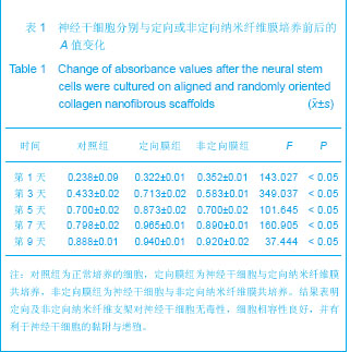
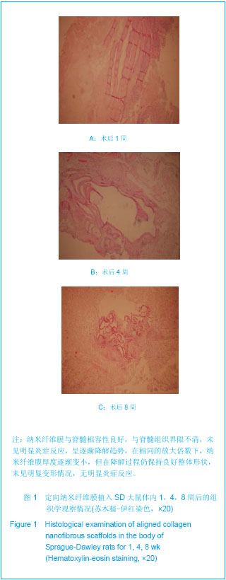
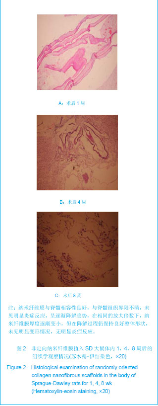
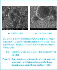
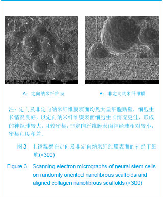
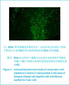
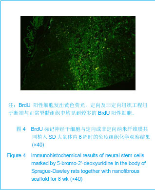
| [1] Cao H,Liu T,Chew SY. The application of nanofibrous scaffolds in neural tissue engineering.Adv Drug Deliv Rev. 2009;61(12):1055-1064.[2] Jiang ZG,Li YH,Bi XB,et al.Zhongshan Daxue Yanjiusheng Xuekan. 2007;28(2): 20-25.江志羔,黎银焕,毕小彬,等.生物材料应用于脊髓损伤修复的研究进展[J].中山大学研究生学刊,2007,28(2): 20-25.[3] Saracino GA,Cigognini D,Silva D,et al.Nanomaterials design and tests for neural tissue engineering. Chem Soc Rev. 2012. In press.[4] Wang Y,Yao M,Zhou C,et al.Erythropoietin promotes spinal cord-derived neural progenitor cell proliferation by regulating cell cycle. Neuroscience.2010;167(3):750e7.[5] Wang M,Zhai P,Chen X,et al.Bioengineered scaffolds for spinal cord repair. Tissue Eng Part B Rev. 2011;17(3): 177-194.[6] Madigan NN,McMahon S,O'Brien T,et al.Current tissue engineering and novel therapeutic approaches to axonal regeneration following spinal cord injury using polymer scaffolds.Respir Physiol Neurobiol.2009;169(2):183-199.[7] Liu Y, Zhang BA, Song Y, et al. Bone marrow mesenchymal stem cell transplantation for treatment of spinal cord injury: an in vivo magnetic resonance imaging tracking study. Neural Regen Res. 2011;6(13):978-982.[8] Li L, Lü G, Wang YF, et al. Glial cell-derived neurotrophic factor mRNA expression in a rat model of spinal cord injury following bone marrow stromal cell transplantation. Neural Regen Res 2008;3(10):1056-1059.[9] Kumar PR,Khan N,Vivekanandhan S,et al.Nanofibers: effective generation by electrospinning and their applications.J Nanosci Nanotechnol.2012;12(1):1-25.[10] Cho Y,Borgens RB.Polymer and nano-technology applications for repair and reconstruction of the central nervous system.Exp Neurol. 2012;233(1):126-144.[11] Prabhakaran MP,Ghasemi-Mobarakeh L,Ramakrishna S.Electrospun composite nanofibers for tissue regeneration.J Nanosci Nanotechnol.2011;11(4):3039-3057.[12] Xie J,MacEwan MR,Schwartz AG,et al.Electrospun nanofibersforneural tissue engineering. Nanoscale. 2010; 2(1):35-44.[13] Christens on EM,Anseth KS,van den Beucken JJ,et al.Nanobiomaterial applications in orthopedics.J Orthop Res.2007;1:11-22.[14] Subramanian A,Krishnan UM,Sethuraman S.Development of biomaterial scaffold for nerve tissue engineering: Biomaterial mediated neural regeneration. J Biomed Sci. 2009;16:108.[15] Sedaghati T,Yang SY,Mosahebi A,et al. Nerve regeneration with aid of nanotechnology and cellular engineering. Biotechnol Appl Biochem. 2011;58(5):288-300.[16] Oray BN,Kelly S,Konobeck T,et al.Novel propylene oxide-treated bovine pericardium as soft tissue repair material and potential scaffold for tissue engineering. Surg Technol Int. 2009;18:47-54.[17] Landgren H, Curtis MA. Locating and labeling neural stem cells in the brain. J Cell Physiol. 2011;226(1):1-7.[18] Yun MJ,Wang LZ,Wang YC, et al.In vitro growth, differentiation and biological characteristics of neural stem cells.Neural Regen Res 2006;1(4):364-367.[19] Lehner B,Sandner B, Marschallinger J, et al. The dark side of BrdU in neural stem cell biology: detrimental effects on cell cycle, differentiation and survival. Cell Tissue Res. 2011; 345(3): 313-328. |