Chinese Journal of Tissue Engineering Research ›› 2013, Vol. 17 ›› Issue (20): 3643-3649.doi: 10.3969/j.issn.2095-4344.2013.20.005
Previous Articles Next Articles
Transforming growth factor beta 1 and beta 3 expression during skin wound healing of rats
Zhao Jia-jia, Yang Pi-bo, Han Chuan-huo, Liu Jia-rong
- Department of Stomatology, Wuhan Union Hospital, Tongji Medical College, Huazhong University of Science and Technology, Wuhan 430022, Hubei Province, China
-
Received:2012-11-19Revised:2012-12-19Online:2013-05-14Published:2013-05-14 -
Contact:Liu Jia-rong, M.D., Associate professor, Department of Stomatology, Wuhan Union Hospital, Tongji Medical College, Huazhong University of Science and Technology, Wuhan 430022, Hubei Province, China kqyyljr@aliyun.com -
About author:Zhao Jia-jia☆, Studying for doctorate, Department of Stomatology, Wuhan Union Hospital, Tongji Medical College, Huazhong University of Science and Technology, Wuhan 430022, Hubei Province, China zhaojiajia0130@163.com -
Supported by:the National Natural Science Foundation of China, No. 81000424; a grant from the Department of Health, Hubei Province, No. QJX2010-9; the Fund for Distinguished Young Scholars of Hubei Province, No. 2011CDA082
CLC Number:
Cite this article
Zhao Jia-jia, Yang Pi-bo, Han Chuan-huo, Liu Jia-rong. Transforming growth factor beta 1 and beta 3 expression during skin wound healing of rats[J]. Chinese Journal of Tissue Engineering Research, 2013, 17(20): 3643-3649.
share this article
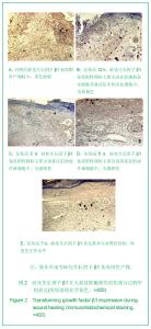
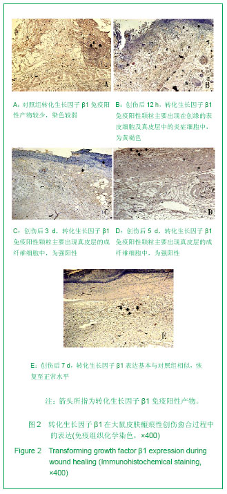
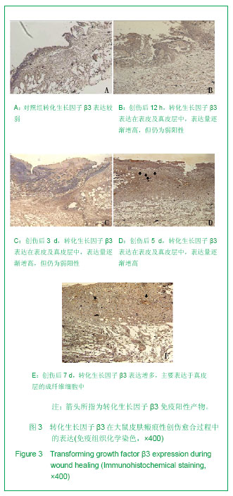
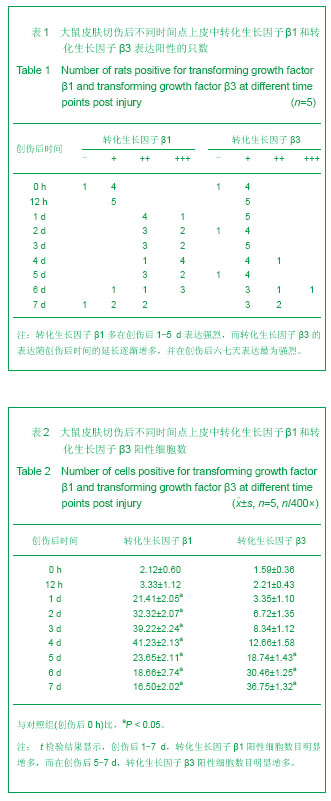
2.1 实验动物数量分析 实验共纳入45只SD大鼠,均进入结果分析。 2.2 大鼠皮肤瘢痕性创伤愈合过程中转化生长因子β1和转化生长因子β3的表达情况 免疫组织化学染色显示,在正常皮肤(对照组)中,转化生长因子β1和转化生长因子β3表达量均呈弱阳性,在创伤后12 h时,转化生长因子β1免疫阳性颗粒主要出现在创缘的表皮细胞及真皮层的炎症细胞中,为黄褐色,并在随后的1-5 d中持续高表达,为强阳性,在创伤后第4天达到高峰,该阶段主要表达在成纤维细胞中,随后转化生长因子β1的表达逐渐减弱。与转化生长因子β1相比,转化生长因子β3在伤后5 d内一直维持在较低水平,免疫组织化学染色为弱阳性,在伤后第6天时开始,其表达量开始增高,免疫阳性颗粒主要出现在真皮层的成纤维细胞及细胞外基质中,见图2,3。"

"
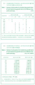
2.3 大鼠皮肤瘢痕性创伤愈合过程中转化生长因子β1和转化生长因子β3表达量的变化 在正常皮肤(对照组)中,转化生长因子β1和转化生长因子β3表达量均呈弱阳性,且在创伤后12 h,两者的阳性细胞数与对照组比较差异无显著性意义(P > 0.05)。创伤后1 d,转化生长因子β1表达明显增加,显示为阳性,且在创伤后24-96 h一直持续在阳性至强阳性水平,转化生长因子β1阳性细胞数明显多于对照组(P < 0.05)。创伤修复后期(96- 168 h),转化生长因子β1表达量逐渐降低,但仍维持在阳性至弱阳性水平,且转化生长因子β1阳性细胞数仍高于对照组(P < 0.05),见表1,2。 而在创伤愈合早期及中期(24-120 h),转化生长因子β3的表达量不如转化生长因子β1明显,一直呈现弱阳性,但是,在创伤修复后期(120-168 h),转化生长因子β3表达量逐渐增高,染色强度上升至阳性,甚至是强阳性水平,转化生长因子β3阳性细胞数也明显高于对照组(P < 0.05),见表1,2。"
| [1] Taipale J, Saharinen J, Keski-Oja J. Extracellular matrix-associated transforming growth factor-beta: role in cancer cell growth and invasion. Adv Cancer Res. 1998;75: 87-134.[2] Faler BJ, Macsata RA, Plummer D, et al. Transforming growth factor-beta and wound healing. Perspect Vasc Surg Endovasc Ther. 2006;18(1):55-62.[3] Chin D, Boyle GM, Parsons PG, et al. What is transforming growth factor-beta (TGF-beta)? Br J Plast Surg. 2004;57(3): 215-221.[4] Massagué J. The transforming growth factor-beta family. Annu Rev Cell Biol. 1990;6:597-641.[5] Shah M, Foreman DM, Ferguson MW. Neutralisation of TGF-beta 1 and TGF-beta 2 or exogenous addition of TGF-beta 3 to cutaneous rat wounds reduces scarring. J Cell Sci. 1995;108(Pt 3):985-1002.[6] Frank S, Madlener M, Werner S. Transforming growth factors beta1, beta2, and beta3 and their receptors are differentially regulated during normal and impaired wound healing. J Biol Chem. 1996;271(17):10188-10193.[7] Desmouliere A, Geinoz A, Gabbiani F, et al. Transforming growth factor-beta 1 induces alpha-smooth muscle actin expression in granulation tissue myofibroblasts and in quiescent and growing cultured fibroblasts. J Cell Biol. 1993; 122(1):103-111.[8] Deng L, Nong XL. Zhongguo Shaoshang Chuangshang Zazhi. 2009;21(3):223-226.邓凌,农晓琳.病理性瘢痕与细胞因子相关性研究进展[J].中国烧伤创伤杂志, 2009,21(3):223-226.[9] Shi HJ, Huang DY. Sichuan Jiepouxue Zazhi. 2011;19(1): 45-47.石慧娟,黄大元.成纤维细胞及其相关因素在无瘢痕愈合中的研究进展[J].四川解剖学杂志,2011,19(1):45-47.[10] Liu H, Wei W. Zhongguo Yaolixue Tongbao. 2007;23(5): 561-565.刘浩,魏伟.TGFβ信号转导通路及以其为靶点的肝纤维化治疗[J].中国药理学通报,2007,23(5):561-565.[11] Lévesque M, Villiard E, Roy S. Skin wound healing in axolotls: a scarless process. J Exp Zool B Mol Dev Evol. 2010;314(8): 684-697.[12] Honardoust D, Varkey M, Marcoux Y, et al. Reduced decorin, fibromodulin, and transforming growth factor-β3 in deep dermis leads to hypertrophic scarring. J Burn Care Res. 2012; 33(2):218-227.[13] Brunner G, Blakytny R. Extracellular regulation of TGF-beta activity in wound repair: growth factor latency as a sensor mechanism for injury. Thromb Haemost. 2004;92(2):253-261.[14] Lin SJ, Lerch TF, Cook RW, et al. The structural basis of TGF-beta, bone morphogenetic protein, and activin ligand binding. Reproduction. 2006;132(2):179-190.[15] Jagadeesan J, Bayat A. Transforming growth factor beta (TGFbeta) and keloid disease. Int J Surg. 2007;5(4):278-285.[16] Kobayashi T, Liu X, Wen FQ, et al. Smad3 mediates TGF-beta1-induced collagen gel contraction by human lung fibroblasts. Biochem Biophys Res Commun. 2006;339(1): 290-295.[17] Liu Q, Hu ZY, Guo PN. Baotou Yixueyuan Xuebao. 2011;27(5): 135-138.刘琦,胡志英,郭鹏年.瘢痕疙瘩的发病机制及治疗进展[J].包头医学院学报,2011,27(5):135-138.[18] Martin P. Wound healing-aiming for perfect skin regeneration. Science. 1997;276(5309):75-81.[19] Singer AJ, Clark RA. Cutaneous wound healing. N Engl J Med. 1999;341(10):738-746.[20] He YJ, Liu Y, Wang JH, et al. Zhongguo Meirong Yixue. 2010; 19(1):53-55.何永静,刘垠,王继华,等.TGF-β1对UVA照射人皮肤成纤维细胞MMP-1和MMP-3 mRNA表达的影响[J].中国美容医学,2010, 19(1):53-55.[21] Xia P, Tong WX, Yu YM, et al. Chongqing Yike Daxue Xuebao. 2010;35(8):1167-1171.夏鹏,童文祥,喻永敏,等.大鼠皮肤切创愈合过程中VEGF, TGF-β1蛋白的表达[J].重庆医科大学学报,2010,35(8): 1167-1171.[22] Bellemare J, Roberge CJ, Bergeron D, et al. Epidermis promotes dermal fibrosis: role in the pathogenesis of hypertrophic scars. J Pathol. 2005;206(1):1-8.[23] Ghosh A K. TGF-β signaling and its inhibition: implication in fibrosis. Current Enzyme Inhibition. 2010;6(2):54-56.[24] Jinnin M. Mechanisms of skin fibrosis in systemic sclerosis. J Dermatol. 2010;37(1):11-25.[25] Yang L, Guo SZ. Zhongguo Linchuang Kangfu. 2002;6(4): 470-471.杨力,郭树忠.成纤维细胞与创伤修复的生物学过程[J].中国临床康复,2002,6(4):470-471.[26] Zhao MQ, Zhang G. Zhongguo Meirong Yixue. 2008;17(7): 1101-1103.赵明权,张刚.无瘢痕愈合的细胞外基质研究进展[J].中国美容医学, 2008,17(7):1101-1103.[27] Yan GC, Pei YH. Zhongguo Meirong Yixue. 2010;19(8): 1251-1255.闫贵春,裴银辉.瘢痕发生机制研究进展[J].中国美容医学, 2010, 19(8):1251-1255.[28] Wang JH, Liu Y, He YJ, et al. Zhongguo Meirong Yixue. 2009; 18(3):338-341.王继华,刘垠,何永静,等.TGF-β1对UVA照射皮肤成纤维细胞I型,III型胶原合成和表达的影响[J].中国美容医学,2009,18(3): 338-341.[29] Feng YQ, Wang YB. Zhongguo Meirong Yixue. 2008;17(7): 1095-1097.冯永强,王一兵.胚胎成纤维细胞的生物学特点[J].中国美容医学,2008,17(7):1095-1097.[30] Qiu L, Jin XQ, Kingston PA, et al. Zhongguo Zuzhi Gongcheng Yanjiu yu Linchuang Kangfu. 2008;12(21): 4012-4016.邱林,金先庆, Kingston PA,等. TGF-β3c2s2基因转染骨髓间充质干细胞对创面愈合中转化生长因子β表达的影响及意义[J].中国组织工程研究与临床康复,2008,12(21):4012-4016.[31] Campbell BH, Agarwal C, Wang JH. TGF-beta1, TGF-beta3, and PGE(2) regulate contraction of human patellar tendon fibroblasts. Biomech Model Mechanobiol. 2004;2(4):239-245.[32] Hsu M, Peled ZM, Chin GS, et al. Ontogeny of expression of transforming growth factor-beta 1 (TGF-beta 1), TGF-beta 3, and TGF-beta receptors I and II in fetal rat fibroblasts and skin. Plast Reconstr Surg. 2001;107(7):1787-1794.[33] Rolfe KJ, Irvine LM, Grobbelaar AO, et al. Differential gene expression in response to transforming growth factor-beta1 by fetal and postnatal dermal fibroblasts. Wound Repair Regen. 2007;15(6):897-906.[34] Zhang G, Tan J, Li GF. Xiandai Yixue Jinzhan. 2008;8(6): 1175-1117.张刚,谭军,李高峰.细胞因子抗瘢痕的研究进展[J].现代医学进展, 2008,8(6):1175-1117. |
| [1] | Pu Rui, Chen Ziyang, Yuan Lingyan. Characteristics and effects of exosomes from different cell sources in cardioprotection [J]. Chinese Journal of Tissue Engineering Research, 2021, 25(在线): 1-. |
| [2] | Li Jing, Xie Jianshan, Cui Huilin, Cao Ximei, Yang Yanping, Li Hairong. Expression and localization of diacylglycerol kinase zeta and protein kinase C beta II in mouse back skin with different coat colors [J]. Chinese Journal of Tissue Engineering Research, 2021, 25(8): 1196-1200. |
| [3] | Fan Quanbao, Luo Huina, Wang Bingyun, Chen Shengfeng, Cui Lianxu, Jiang Wenkang, Zhao Mingming, Wang Jingjing, Luo Dongzhang, Chen Zhisheng, Bai Yinshan, Liu Canying, Zhang Hui. Biological characteristics of canine adipose-derived mesenchymal stem cells cultured in hypoxia [J]. Chinese Journal of Tissue Engineering Research, 2021, 25(7): 1002-1007. |
| [4] | Zhang Mi, Wu Saixuan, Dong Ming, Lu Ying, Niu Weidong. Expression of interleukin-24 in a mouse model of periapical periodontitis [J]. Chinese Journal of Tissue Engineering Research, 2021, 25(5): 679-684. |
| [5] | Yang Yang, Yao Yu, Shen Xiaotian, Liu Jiajia, Xue Jianhua. Expression and significance of interleukin-21 in intervertebral disc degeneration [J]. Chinese Journal of Tissue Engineering Research, 2021, 25(5): 690-694. |
| [6] | Ma Binxiang, He Wanqing, Zhou Guangchao, Guan Yonglin. Triptolide improves motor dysfunction in rats following spinal cord injury [J]. Chinese Journal of Tissue Engineering Research, 2021, 25(5): 701-706. |
| [7] | Nie Huijuan, Huang Zhichun. The role of Hedgehog signaling pathway in transforming growth factor beta1-induced myofibroblast transdifferentiation [J]. Chinese Journal of Tissue Engineering Research, 2021, 25(5): 754-760. |
| [8] | Ye Haimin, Ding Linghua, Kong Weihao, Huang Zutai, Xiong Long. Role and mechanism of hierarchical microchanneled bone scaffolds in promoting osteogenesis and angiogenesis [J]. Chinese Journal of Tissue Engineering Research, 2021, 25(4): 621-625. |
| [9] | Zhang Zhenkun, Li Zhe, Li Ya, Wang Yingying, Wang Yaping, Zhou Xinkui, Ma Shanshan, Guan Fangxia. Application of alginate based hydrogels/dressings in wound healing: sustained, dynamic and sequential release [J]. Chinese Journal of Tissue Engineering Research, 2021, 25(4): 638-643. |
| [10] | Liu Jinwei, Chen Yunzhen, Wan Chunyou. Changes of osteogenic growth factors in the broken end of bone nonunion under stress [J]. Chinese Journal of Tissue Engineering Research, 2021, 25(23): 3619-3624. |
| [11] | Tian Guangzhao, Yang Zhen, Zha Kangkang, Sun Zhiqiang, Li Xu, Sui Xiang, Huang Jingxiang, Guo Quanyi, Liu Shuyun. Regulatory effect of decellularized cartilage matrix on macrophage polarization [J]. Chinese Journal of Tissue Engineering Research, 2021, 25(22): 3545-3550. |
| [12] | Gan Lili, Xiong Na, Liu Yanfei. Hydrogel as drug scaffold in skin wound repair: challenges of clinical application possibilities [J]. Chinese Journal of Tissue Engineering Research, 2021, 25(22): 3578-3583. |
| [13] | Liu Fang, Shan Zhengming, Tang Yulei, Wu Xiaomin, Tian Weiqun. Effects of hemostasis and promoting wound healing of ozone sustained-release hydrogel [J]. Chinese Journal of Tissue Engineering Research, 2021, 25(22): 3445-3449. |
| [14] | Wang Hao, Chen Mingxue, Li Junkang, Luo Xujiang, Peng Liqing, Li Huo, Huang Bo, Tian Guangzhao, Liu Shuyun, Sui Xiang, Huang Jingxiang, Guo Quanyi, Lu Xiaobo. Decellularized porcine skin matrix for tissue-engineered meniscus scaffold [J]. Chinese Journal of Tissue Engineering Research, 2021, 25(22): 3473-3478. |
| [15] | Yang Li, Li Xueli, Song Jinghui, Yu Huiqian, Wang Weixia. Effect of cryptotanshinone on hypertrophic scar of rabbit ear and its related mechanism [J]. Chinese Journal of Tissue Engineering Research, 2021, 25(20): 3150-3155. |
| Viewed | ||||||
|
Full text |
|
|||||
|
Abstract |
|
|||||