Chinese Journal of Tissue Engineering Research ›› 2013, Vol. 17 ›› Issue (11): 1909-1916.doi: 10.3969/j.issn.2095-4344.2013.11.002
Previous Articles Next Articles
Co-culture of subchondral osteoblasts and chondrocytes from human knee joint
Zhang Li-zhi1, Jiang Yao2, Mao Yan-jie2, Hang Cheng-long2
- 1 Department of Orthopedic Surgery, Shanghai Yangpu District Central Hospital, Yangpu Hospital Affiliated to Tongji University, Shanghai 200090, China
2 Shanghai Sixth People’s Hospital, Shanghai Jiao Tong University, Shanghai 200233, China
-
Received:2012-10-28Revised:2013-01-08Online:2013-03-12Published:2013-03-12 -
About author:Zhang Li-zhi☆, Doctor, Attending physician, Lecturer, Department of Orthopedic Surgery, Shanghai Yangpu District Central Hospital, Yangpu Hospital Affiliated to Tongji University, Shanghai 200090, China zhanglizhi001@sohu.com
CLC Number:
Cite this article
Zhang Li-zhi, Jiang Yao, Mao Yan-jie, Hang Cheng-long. Co-culture of subchondral osteoblasts and chondrocytes from human knee joint[J]. Chinese Journal of Tissue Engineering Research, 2013, 17(11): 1909-1916.
share this article
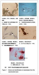
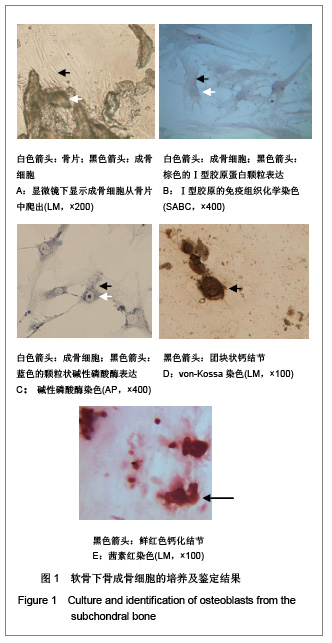
2.1 软骨下骨成骨细胞的培养及鉴定 Ⅰ型胶原酶序列消化后贴壁培养方法培养软骨下骨成骨细胞,大约3周后,细胞爬出骨片,贴附于培养瓶底,细胞可呈多种形态,如三角形、梭形、多角形、立方形等,见图1A;4-6周时,细胞可融合成片。Ⅰ型胶原蛋白的免疫组化染色显示,原代培养的成骨细胞的细胞质内可见颗粒状棕色阳性染色,见图1B;碱性磷酸酶染色显示阳性,细胞的胞浆中可见蓝色的细小颗粒,见图1C;Von-Kossa染色显示细胞外基质出现黑色团块状钙结节,见图1D;茜素红染色显示呈阳性,镜下观察有结节形成,见图1E。"
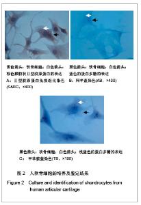
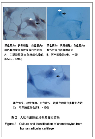
2.2 软骨细胞的培养及鉴定 采用Ⅱ型胶原酶和胰蛋白酶联合消化软骨组织后贴壁培养方法培养软骨细胞,可见消化后细胞多为圆形、半透明,细胞核清晰,逐渐贴附于培养瓶底,培养2周后,可见大部分软骨细胞贴壁生长良好,4周后,细胞可生长至融合状态。原代软骨细胞呈圆形、多角形,立方形或三角形等形状,核为圆形或椭圆形。Ⅱ型胶原蛋白免疫组化检测原代培养的细胞胞浆中可见棕色颗粒状阳性染色,见图2A;阿辛蓝染色和甲苯胺蓝染色可见软骨细胞外基质的蛋白多糖呈浅蓝色和蓝色,见图2B,C。"

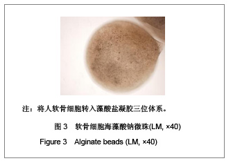
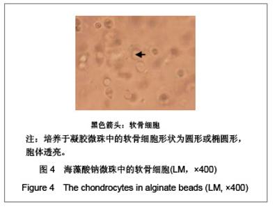
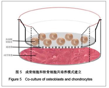
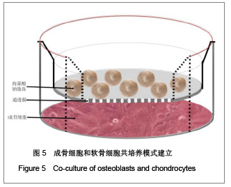
2.3 成骨细胞和软骨细胞共培养模式建立 成功构建了软骨细胞海藻酸钠微珠,软骨细胞转入藻酸盐凝胶三维培养体系,见图3。 培养于凝胶微珠中的软骨细胞形状为圆形或椭圆形,胞体透亮,见图4。 成骨细胞培养成膜状结构,应用多孔插入器对成骨细胞和软骨细胞的共培养,培养液中的可溶性分子可经过多孔插入器进行信息交流,见图5。"
"

"
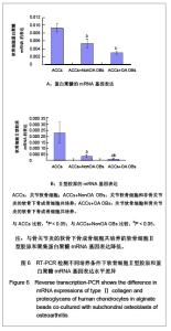
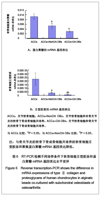
2.4 RT-PCR检测不同成骨细胞共培养的软骨细胞Ⅱ型胶原和蛋白聚糖mRNA水平基因表达差异 骨关节炎软骨下骨成骨细胞共培养的软骨细胞蛋白聚糖表达量(3.04±0.33)×10-3,Ⅱ型胶原表达量(1.22±0.54)×10-5;非骨关节炎软骨下骨成骨细胞共培养的软骨细胞蛋白聚糖表达量为(5.36±1.14)×10-3 ,Ⅱ型胶原表达量(3.75±0.8)×10-5;非共培养的软骨细胞蛋白聚糖的表达量为(9.38±1.13)×10-3 ,Ⅱ型胶原的表达量为(23.1±8.5)×10-5。在成骨细胞共培养的软骨细胞中蛋白聚糖的表达量和Ⅱ型胶原的表达量均低于非共培养的软骨细胞中的表达;在骨关节炎软骨下骨成骨细胞共培养的软骨细胞中的表达明显低于非骨关节炎成骨细胞共培养的软骨细胞中的表达(P < 0.05),见图6。"
| [1] Madry H, Grün UW, Knutsen G. Cartilage repair and joint preservation: medical and surgical treatment options. Dtsch Arztebl Int. 2011;108(40):669-677.[2] Jiang YZ, Zhang SF, Qi YY, et al. Cell transplantation for articular cartilage defects: principles of past, present, and future practice. Cell Transplant. 2011;20(5):593-607.[3] Trewenack AJ, Please CP, Landman KA. A continuum model for the development of tissue-engineered cartilage around a chondrocyte. Math Med Biol. 2009;26(3):241-262.[4] Neogi T.Clinical significance of bone changes in osteoarthritis.Ther Adv Musculoskelet Dis. 2012;4(4): 259-267.[5] Mansfield JC, Winlove CP. A multi-modal multiphoton investigation of microstructure in the deep zone and calcified cartilage. J Anat. 2012;220(4):405-416.[6] Henrotin Y, Pesesse L, Sanchez C.Subchondral bone and osteoarthritis: biological and cellular aspects. Osteoporos Int. 2012;23 Suppl 8:847-851.[7] Zhang LZ, Zheng HA, Jiang Y,et al. Mechanical and biologic link between cartilage and subchondral bone in osteoarthritis. Arthritis Care Res (Hoboken). 2012;64(7):960-967.[8] Quasnichka HL, Anderson-MacKenzie JM, Bailey AJ. Subchondral bone and ligament changes precede cartilage degradation in guinea pig osteoarthritis. Biorheology. 2006; 43(3-4):389-397.[9] Anderson-MacKenzie JM, Quasnichka HL, Starr RL,et al. Fundamental subchondral bone changes in spontaneous knee osteoarthritis. Int J Biochem Cell Biol. 2005;37(1): 224-236.[10] Mahjoub M, Berenbaum F, Houard X.Why subchondral bone in osteoarthritis? The importance of the cartilage bone interface in osteoarthritis.Osteoporos Int. 2012;23 Suppl 8: 841-846.[11] Kwan Tat S, Lajeunesse D, Pelletier JP,et al.Targeting subchondral bone for treating osteoarthritis: what is the evidence. Best Pract Res Clin Rheumatol. 2010;24(1):51-70.[12] Castañeda S, Roman-Blas JA, Largo R,et al. Subchondral bone as a key target for osteoarthritis treatment.Biochem Pharmacol. 2012;83(3):315-323.[13] Zhang LZ, Mei J, Qian ZK,et al.The role of VE-cadherin in osteosarcoma cells. Pathol Oncol Res. 2010;16(1):111-117.[14] Lin J, Fransen M, Kang X,et al. Marked disability and high use of nonsteroidal antiinflammatory drugs associated with knee osteoarthritis in rural China: a cross-sectional population-based survey. Arthritis Res Ther. 2010;12(6): R225.[15] Goldring MB. Chondrogenesis, chondrocyte differentiation, and articular cartilage metabolism in health and osteoarthritis.Ther Adv Musculoskelet Dis. 2012;4(4): 269-285.[16] Henrotin Y, Pesesse L, Sanchez C.Subchondral bone in osteoarthritis physiopathology: state-of-the art and perspectives. Biomed Mater Eng. 2009;19(4-5):311-316. [17] Goldring SR. Alterations in periarticular bone and cross talk between subchondral bone and articular cartilage in osteoarthritis.Ther Adv Musculoskelet Dis. 2012;4(4): 249-258.[18] Cox LG, van Donkelaar CC, van Rietbergen B,et al. Decreased bone tissue mineralization can partly explain subchondral sclerosis observed in osteoarthritis. Bone. 2012; 50(5):1152-1161.[19] Lories RJ, Luyten FP.The bone-cartilage unit in osteoarthritis. Nat Rev Rheumatol. 2011;7(1):43-49.[20] Goldring MB, Goldring SR. Articular cartilage and subchondral bone in the pathogenesis of osteoarthritis. Ann N Y Acad Sci. 2010;1192:230-237.[21] Cox LG, van Rietbergen B, van Donkelaar CC,et al. Bone structural changes in osteoarthritis as a result of mechanoregulated bone adaptation: a modeling approach.Osteoarthritis Cartilage. 2011;19(6):676-682.[22] Mastbergen SC, Lafeber FP. Changes in subchondral bone early in the development of osteoarthritis. Arthritis Rheum. 2011;63(9):2561-2563.[23] Funck-Brentano T, Lin H, Hay E,et al. Targeting bone alleviates osteoarthritis in osteopenic mice and modulates cartilage catabolism. PLoS One. 2012;7(3):e33543.[24] Sanchez C, Deberg MA, Bellahcène A,et al. Phenotypic characterization of osteoblasts from the sclerotic zones of osteoarthritic subchondral bone. Arthritis Rheum. 2008; 58(2): 442-455.[25] Heiligenstein S, Cucchiarini M, Laschke MW,et al. In vitro and in vivo characterization of non-biomedical and biomedical grade alginates for articular chondrocyte transplantation. Tissue Eng Part C Methods. 2011. [Epub ahead of print]. [26] Cohen J, Zaleski KL, Nourissat G,et al. Survival of porcine mesenchymal stem cells over the alginate recovered cellular method. J Biomed Mater Res A. 2011;96(1):93-99.[27] Heiligenstein S, Cucchiarini M, Laschke MW,et al. In vitro and in vivo characterization of nonbiomedical- and biomedical-grade alginates for articular chondrocyte transplantation.Tissue Eng Part C Methods. 2011;17(8): 829-842.[28] Domm C, Schünke M, Steinhagen J,et al. Influence of various alginate brands on the redifferentiation of dedifferentiated bovine articular chondrocytes in alginate bead culture under high and low oxygen tension.Tissue Eng. 2004;10(11-12): 1796-1805.[29] Baghaban Eslaminejad M, Taghiyar L, Falahi F.Q uantitative analysis of the proliferation and differentiation of rat articular chondrocytes in alginate 3D culture. Iran Biomed J. 2009; 13(3):153-160.[30] Hoemann CD, Lafantaisie-Favreau CH, Lascau-Coman V,et al. The cartilage-bone interface.J Knee Surg. 2012;25(2): 85-97.[31] Suri S, Walsh DA. Osteochondral alterations in osteoarthritis. Bone. 2012;51(2):204-211.[32] Burr DB, Radin EL. Microfractures and microcracks in subchondral bone: are they relevant to osteoarthrosis. Rheum Dis Clin North Am. 2003;29(4):675-685.[33] Pesesse L, Sanchez C, Henrotin Y. Osteochondral plate angiogenesis: a new treatment target in osteoarthritis.Joint Bone Spine. 2011;78(2):144-149.[34] Massicotte F, Fernandes JC, Martel-Pelletier J,et al. Modulation of insulin-like growth factor 1 levels in human osteoarthritic subchondral bone osteoblasts. Bone. 2006; 38(3):333-341.[35] Hulejová H, Baresová V, Klézl Z,et al. Increased level of cytokines and matrix metalloproteinases in osteoarthritic subchondral bone. Cytokine. 2007;38(3):151-156.[36] Sakao K, Takahashi KA, Mazda O,et al. Enhanced expression of interleukin-6, matrix metalloproteinase-13, and receptor activator of NF-kappaB ligand in cells derived from osteoarthritic subchondral bone. J Orthop Sci. 2008;13(3): 202-210.[37] Prasadam I, van Gennip S, Friis T,et al. ERK-1/2 and p38 in the regulation of hypertrophic changes of normal articular cartilage chondrocytes induced by osteoarthritic subchondral osteoblasts. Arthritis Rheum. 2010;62(5):1349-1360.[38] Lin YY, Tanaka N, Ohkuma S,et al. Applying an excessive mechanical stress alters the effect of subchondral osteoblasts on chondrocytes in a co-culture system. Eur J Oral Sci. 2010; 118(2):151-158.[39] Madry H, van Dijk CN, Mueller-Gerbl M.The basic science of the subchondral bone.Knee Surg Sports Traumatol Arthrosc. 2010;18(4):419-433.[40] Bugbee W, Cavallo M, Giannini S.Osteochondral allograft transplantation in the knee.J Knee Surg. 2012;25(2):109-116.[41] Goldring SR, Goldring MB.Bone and cartilage in osteoarthritis: is what's best for one good or bad for the other. Arthritis Res Ther. 2010;12(5):143. |
| [1] | Peng Zhihao, Feng Zongquan, Zou Yonggen, Niu Guoqing, Wu Feng. Relationship of lower limb force line and the progression of lateral compartment arthritis after unicompartmental knee arthroplasty with mobile bearing [J]. Chinese Journal of Tissue Engineering Research, 2021, 25(9): 1368-1374. |
| [2] | Huang Dengcheng, Wang Zhike, Cao Xuewei. Comparison of the short-term efficacy of extracorporeal shock wave therapy for middle-aged and elderly knee osteoarthritis: a meta-analysis [J]. Chinese Journal of Tissue Engineering Research, 2021, 25(9): 1471-1476. |
| [3] | Liu Xiangxiang, Huang Yunmei, Chen Wenlie, Lin Ruhui, Lu Xiaodong, Li Zuanfang, Xu Yaye, Huang Meiya, Li Xihai. Ultrastructural changes of the white zone cells of the meniscus in a rat model of early osteoarthritis [J]. Chinese Journal of Tissue Engineering Research, 2021, 25(8): 1237-1242. |
| [4] | Liu Xin, Yan Feihua, Hong Kunhao. Delaying cartilage degeneration by regulating the expression of aquaporins in rats with knee osteoarthritis [J]. Chinese Journal of Tissue Engineering Research, 2021, 25(5): 668-673. |
| [5] | Ma Zetao, Zeng Hui, Wang Deli, Weng Jian, Feng Song. MicroRNA-138-5p regulates chondrocyte proliferation and autophagy [J]. Chinese Journal of Tissue Engineering Research, 2021, 25(5): 674-678. |
| [6] | Xie Chongxin, Zhang Lei. Comparison of knee degeneration after anterior cruciate ligament reconstruction with or without remnant preservation [J]. Chinese Journal of Tissue Engineering Research, 2021, 25(5): 735-740. |
| [7] | Cao Xuhan, Bai Zixing, Sun Chengyi, Yang Yanjun, Sun Weidong. Mechanism of “Ruxiang-Moyao” herbal pair in the treatment of knee osteoarthritis based on network pharmacology [J]. Chinese Journal of Tissue Engineering Research, 2021, 25(5): 746-753. |
| [8] | Li Yonghua, Feng Qiang, Tan Renting, Huang Shifu, Qiu Jinlong, Yin Heng. Molecular mechanism of Eucommia ulmoides active ingredients treating synovitis of knee osteoarthritis: an analysis based on network pharmacology [J]. Chinese Journal of Tissue Engineering Research, 2021, 25(5): 765-771. |
| [9] | Song Shan, Hu Fangyuan, Qiao Jun, Wang Jia, Zhang Shengxiao, Li Xiaofeng. An insight into biomarkers of osteoarthritis synovium based on bioinformatics [J]. Chinese Journal of Tissue Engineering Research, 2021, 25(5): 785-790. |
| [10] | Deng Zhenhan, Huang Yong, Xiao Lulu, Chen Yulin, Zhu Weimin, Lu Wei, Wang Daping. Role and application of bone morphogenetic proteins in articular cartilage regeneration [J]. Chinese Journal of Tissue Engineering Research, 2021, 25(5): 798-806. |
| [11] | Zheng Li, Li Dadi, Hu Weifan, Tang Jinlong, Zhao Fengchao. Risk assessment of contralateral knee arthroplasty after unilateral total knee arthroplasty [J]. Chinese Journal of Tissue Engineering Research, 2021, 25(3): 374-379. |
| [12] | Lü Jiaxing, Bai Leipeng, Yang Zhaoxin, Miao Yuesong, Jin Yu, Li Zhehong, Sun Guangpu, Xu Ying, Zhang Qingzhu. Evaluation of internal fixation with proximal femoral nail antirotation in elderly knee osteoarthritis patients with femoral intertrochanteric fractures [J]. Chinese Journal of Tissue Engineering Research, 2021, 25(3): 391-396. |
| [13] | Luo Anyu, Liu Hanlin, Xie Xiaofei, Huang Chen. Effect of antioxidant mixture on structural degeneration of an osteoarthritis rat model [J]. Chinese Journal of Tissue Engineering Research, 2021, 25(23): 3625-3629. |
| [14] | Gao Kun, Chen Dayu, Zhang Yong, Liu Weidong, Sun Shufen, Lai Wenqiang, Ma Dujun, Wu Yihong, Lin Zhanpeng, Jiang Yinglu, Yu Weiji. Achyranthes bidentata alcohol extract inhibits extracellular matrix degradation of the cartilage by regulating synovial fibroblast exosomes [J]. Chinese Journal of Tissue Engineering Research, 2021, 25(23): 3636-3640. |
| [15] | Huo Hua, Cheng Yuting, Zhou Qian, Qi Yuhan, Wu Chao, Shi Qianhui, Yang Tongjing, Liao Jian, Hong Wei. Effects of drug coating on implant surface on the osseointegration [J]. Chinese Journal of Tissue Engineering Research, 2021, 25(22): 3558-3564. |
| Viewed | ||||||
|
Full text |
|
|||||
|
Abstract |
|
|||||