Chinese Journal of Tissue Engineering Research ›› 2019, Vol. 23 ›› Issue (29): 4701-4706.doi: 10.3969/j.issn.2095-4344.1798
Previous Articles Next Articles
Mogroside V promotes bone formation by stimulating osteoblast proliferation and differentiation
Yao Shunhan, Liao Liang, Qin Jiagang, Wei Huacheng
- First Affiliated Hospital of Guangxi Medical University, Nanning 530021, Guangxi Zhuang Autonomous Region, China
-
Revised:2019-05-07Online:2019-10-18Published:2019-10-18 -
Contact:Liao Liang, Master, Associate chief physician, First Affiliated Hospital of Guangxi Medical University, Nanning 530021, Guangxi Zhuang Autonomous Region, China -
About author:Yao Shunhan, Master candidate, First Affiliated Hospital of Guangxi Medical University, Nanning 530021, Guangxi Zhuang Autonomous Region, China -
Supported by:the Natural Science Foundation of Guangxi Zhuang Autonomous Region (Youth Project), No. 2016GXNSFBA380103 (to LL)
CLC Number:
Cite this article
Yao Shunhan, Liao Liang, Qin Jiagang, Wei Huacheng. Mogroside V promotes bone formation by stimulating osteoblast proliferation and differentiation[J]. Chinese Journal of Tissue Engineering Research, 2019, 23(29): 4701-4706.
share this article

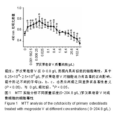
2.1 成骨细胞毒性分析结果 如图1所示,罗汉果皂苷V在0-0.8 g/L范围内显示出低细胞毒性,在6.25×10-3- 2.5×10⁻² g/L范围内明显促进成骨细胞生长(P < 0.05),在25.6-204.8 g/L范围内对成骨细胞生长有抑制作用(P < 0.05)。因此,选择6.25×10-3,1.25×10-2,2.5×10-2 g/L罗汉果皂苷V进行下一步研究。"
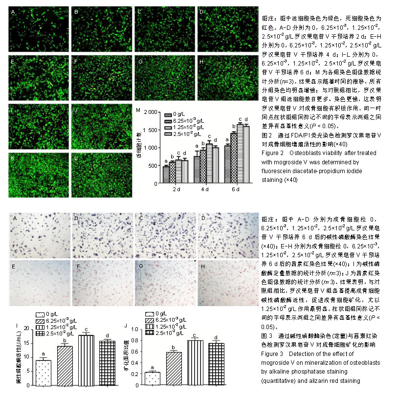
2.2 成骨细胞活性分析结果 如图2 FDA/PI荧光染色所示,活细胞染色为绿色,死细胞染色为红色。结果表明,罗汉果皂苷V对成骨细胞存活有明显影响,随着时间的推移,各组死细胞越来越多,但罗汉果皂苷V组活细胞数多于对照组(P < 0.05),这与细胞毒性实验结果一致。这表明罗汉果皂苷V对成骨细胞增殖有促进作用,尤其以1.25×10⁻² g/L最佳。 2.3 碱性磷酸酶活性分析结果 如图3A-D,I所示,与对照组相比,罗汉果皂苷V组以剂量依赖性显著提高成骨细胞碱性磷酸酶活性,特别是1.25×10⁻² g/L组提高碱性磷酸酶活性最为显著(P < 0.05)。 2.4 成骨细胞钙化结节染色 如图3E-H,J所示,罗汉果皂苷V对成骨细胞矿化有明显影响,以剂量依赖性显著促进成骨细胞矿化,特别是1.25×10⁻² g/L组效果最明显(P < 0.05)。"
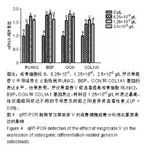
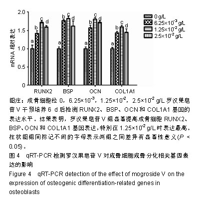
2.5 成骨细胞分化基因分析 如图4所示,与对照组相比,成骨分化相关基因RUNX2、BSP、OCN和COL1A1在罗汉果皂苷V干预6 d后表达水平显著升高,而且在浓度范围内呈剂量依赖性升高,以1.25×10⁻² g/L组表达量最高。RT-PCR结果与碱性磷酸酶活性和矿化结节实验结果相一致,均表明罗汉果皂苷V具有促进成骨细胞分化的作用。"
| [1]Cosman F, de Beur SJ, LeBoff MS, et al. Clinician's Guide to Prevention and Treatment of Osteoporosis. Osteoporos Int. 2014;25(10):2359-2381.[2]Bliuc D, Alarkawi D, Nguyen TV, et al. Risk of subsequent fractures and mortality in elderly women and men with fragility fractures with and without osteoporotic bone density: the Dubbo Osteoporosis Epidemiology Study. J Bone Miner Res. 2015;30(4):637-646.[3]Compston JE, McClung MR, Leslie WD. Osteoporosis. Lancet. 2019;393(10169):364-376.[4]Abrahamsen B, Osmond C, Cooper C. Life Expectancy in Patients Treated for Osteoporosis: Observational Cohort Study Using National Danish Prescription Data. J Bone Miner Res. 2015;30(9):1553-1559.[5]Siris ES, Adler R, Bilezikian J, et al. The clinical diagnosis of osteoporosis: a position statement from the National Bone Health Alliance Working Group. Osteoporos Int. 2014;25(5): 1439-1443.[6]Burge R, Dawson-Hughes B, Solomon DH, et al. Incidence and economic burden of osteoporosis-related fractures in the United States, 2005-2025.J Bone Miner Res. 2007;22(3): 465-475.[7]中华医学会骨质疏松和骨矿盐疾病分会.原发性骨质疏松症诊疗指南(2017)[J].中国全科医学,2017,20(32):3963-3982.[8]Marie PJ, Kassem M. Osteoblasts in osteoporosis: past, emerging, and future anabolic targets. Eur J Endocrinol. 2011;165(1):1-10.[9]曹建平,汤杰,刘合生,等.罗汉果皂苷生物活性研究进展[J].食品工业科技,2014,35(24):384-388,395.[10]Zhang M, Yang H, Zhang H, et al. Development of a process for separation of mogroside V from Siraitia grosvenorii by macroporous resins. Molecules. 2011;16(9):7288-7301.[11]Takasaki M, Konoshima T, Murata Y, et al. Anticarcinogenic activity of natural sweeteners, cucurbitane glycosides, from Momordica grosvenori. Cancer Lett. 2003;198(1):37-42.[12]Chen WJ, Wang J, Qi XY, et al. The antioxidant activities of natural sweeteners, mogrosides, from fruits of Siraitia grosvenori. Int J Food Sci Nutr. 2007;58(7):548-556.[13]Liu H, Qi X, Yu K, et al. AMPK activation is involved in hypoglycemic and hypolipidemic activities of mogroside-rich extract from Siraitia grosvenorii (Swingle) fruits on high-fat diet/streptozotocin-induced diabetic mice. Food Funct. 2019; 10(1):151-162.[14]Callaway DA, Jiang JX. Reactive oxygen species and oxidative stress in osteoclastogenesis, skeletal aging and bone diseases. J Bone Miner Metab. 2015;33(4):359-370.[15]Lin J, Zhu J, Wang Y, et al. Chinese single herbs and active ingredients for postmenopausal osteoporosis: From preclinical evidence to action mechanism. Biosci Trends. 2017;11(5):496-506.[16]Xiao W, Wang Y, Pacios S, et al. Cellular and Molecular Aspects of Bone Remodeling. Front Oral Biol. 2016;18:9-16.[17]Du L, Nong MN, Zhao JM, et al. Polygonatum sibiricum polysaccharide inhibits osteoporosis by promoting osteoblast formation and blocking osteoclastogenesis through Wnt/β-catenin signalling pathway. Sci Rep. 2016;6:32261.[18]Liang D, Ren H, Qiu T, et al. Extracts from plastrum testudinis reverse glucocorticoid-induced spinal osteoporosis of rats via targeting osteoblastic and osteoclastic markers. Biomed Pharmacother. 2016;82:151-160.[19]Pellegrini GG, Morales CC, Wallace TC, et al. Avenanthramides Prevent Osteoblast and Osteocyte Apoptosis and Induce Osteoclast Apoptosis in Vitro in an Nrf2-Independent Manner. Nutrients. 2016;8(7): E423.[20]Kalyanaraman H, Ramdani G, Joshua J, et al. A Novel, Direct NO Donor Regulates Osteoblast and Osteoclast Functions and Increases Bone Mass in Ovariectomized Mice. J Bone Miner Res. 2017;32(1):46-59.[21]Kassem A, Lindholm C, Lerner UH. Toll-Like Receptor 2 Stimulation of Osteoblasts Mediates Staphylococcus Aureus Induced Bone Resorption and Osteoclastogenesis through Enhanced RANKL. PLoS One. 2016;11(6):e0156708.[22]Zhang D, Fong C, Jia Z, et al. Icariin Stimulates Differentiation and Suppresses Adipocytic Transdifferentiation of Primary Osteoblasts Through Estrogen Receptor-Mediated Pathway. Calcif Tissue Int. 2016;99(2):187-198.[23]Choi BY, Park CH, Na YH, et al. Inhibition of RANKL-induced osteoclast differentiation through the downregulation of c-Fos and NFATc1 by Eremochloa ophiuroides (centipedegrass) extract. Mol Med Rep. 2016;13(5):4014-4022.[24]Baek JM, Kim JY, Yoon KH, et al. Ebselen Is a Potential Anti-Osteoporosis Agent by Suppressing Receptor Activator of Nuclear Factor Kappa-B Ligand-Induced Osteoclast Differentiation In vitro and Lipopolysaccharide-Induced Inflammatory Bone Destruction In vivo. Int J Biol Sci. 2016;12(5):478-488.[25]Sims NA, Martin TJ. Coupling Signals between the Osteoclast and Osteoblast: How are Messages Transmitted between These Temporary Visitors to the Bone Surface. Front Endocrinol (Lausanne). 2015;6:41.[26]Lassen NE, Andersen TL, Pløen GG, et al. Coupling of Bone Resorption and Formation in Real Time: New Knowledge Gained From Human Haversian BMUs.J Bone Miner Res. 2017;32(7):1395-1405.[27]Long F. Building strong bones: molecular regulation of the osteoblast lineage. Nat Rev Mol Cell Biol. 2011;13(1):27-38.[28]Liu Y, Lin Z, Guo J, et al. Notoginsenoside R1 significantly promotes in vitro osteoblastogenesis. Int J Mol Med. 2016; 38(2):537-544.[29]Huang L, Jin P, Lin X, et al. Beneficial effects of sulfonamide?based gallates on osteoblasts in vitro. Mol Med Rep. 2017; 15(3):1149-1156.[30]闫海锋,李林轩,覃金萍, 等.罗汉果研究综述[J].南方农业学报, 2011,42(11):1387-1391. [31]高媛,李慧敏,赵彤彤,等.罗汉果饮料的研制研究[J].食品安全质量检测学报,2018,9(14):3760-3764.[32]万艳娟,吴军林,吴清平.功能性甜味剂罗汉果甜苷的生理功能及食品应用研究进展[J].食品与发酵科技,2015,51(5):51-56.[33]Golub EE, Boesze-Battaglia K. The role of alkaline phosphatase in mineralization. Current Opinion in Orthopaedics. 2007;18(5):444-448.[34]Koromila T, Baniwal SK, Song YS, et al. Glucocorticoids antagonize RUNX2 during osteoblast differentiation in cultures of ST2 pluripotent mesenchymal cells. J Cell Biochem. 2014;115(1):27-33.[35]张弘,姚宇,孙佳栋,等.NELL-1调控RUNX2的P1启动子诱导成骨分化[J].中华口腔医学研究杂志(电子版),2017,11(4):197-203.[36]陶周善,周皖舒,江云云,等.骨形成蛋白联合雷奈酸锶对成骨细胞增殖和分化的影响[J].中国骨质疏松杂志,2018,24(2):165-169.[37]Foster BL, Ao M, Salmon CR, et al. Osteopontin regulates dentin and alveolar bone development and mineralization. Bone. 2018;107:196-207.[38]Hosseini S, Naderi-Manesh H, Vali H, et al. Contribution of osteocalcin-mimetic peptide enhances osteogenic activity and extracellular matrix mineralization of human osteoblast-like cells. Colloids Surf B Biointerfaces. 2019;173:662-671.[39]Zhang W, Chen J, Tao J, et al. The use of type 1 collagen scaffold containing stromal cell-derived factor-1 to create a matrix environment conducive to partial-thickness cartilage defects repair. Biomaterials. 2013;34(3):713-723. |
| [1] | Tang Hui, Yao Zhihao, Luo Daowen, Peng Shuanglin, Yang Shuanglin, Wang Lang, Xiao Jingang. High fat and high sugar diet combined with streptozotocin to establish a rat model of type 2 diabetic osteoporosis [J]. Chinese Journal of Tissue Engineering Research, 2021, 25(8): 1207-1211. |
| [2] | Li Zhongfeng, Chen Minghai, Fan Yinuo, Wei Qiushi, He Wei, Chen Zhenqiu. Mechanism of Yougui Yin for steroid-induced femoral head necrosis based on network pharmacology [J]. Chinese Journal of Tissue Engineering Research, 2021, 25(8): 1256-1263. |
| [3] | Liu Cong, Liu Su. Molecular mechanism of miR-17-5p regulation of hypoxia inducible factor-1α mediated adipocyte differentiation and angiogenesis [J]. Chinese Journal of Tissue Engineering Research, 2021, 25(7): 1069-1074. |
| [4] | Li Cai, Zhao Ting, Tan Ge, Zheng Yulin, Zhang Ruonan, Wu Yan, Tang Junming. Platelet-derived growth factor-BB promotes proliferation, differentiation and migration of skeletal muscle myoblast [J]. Chinese Journal of Tissue Engineering Research, 2021, 25(7): 1050-1055. |
| [5] | Hou Guangyuan, Zhang Jixue, Zhang Zhijun, Meng Xianghui, Duan Wen, Gao Weilu. Bone cement pedicle screw fixation and fusion in the treatment of degenerative spinal disease with osteoporosis: one-year follow-up [J]. Chinese Journal of Tissue Engineering Research, 2021, 25(6): 878-883. |
| [6] | Li Shibin, Lai Yu, Zhou Yi, Liao Jianzhao, Zhang Xiaoyun, Zhang Xuan. Pathogenesis of hormonal osteonecrosis of the femoral head and the target effect of related signaling pathways [J]. Chinese Journal of Tissue Engineering Research, 2021, 25(6): 935-941. |
| [7] | Zheng Xiaolong, He Xiaoming, Gong Shuidi, Pang Fengxiang, Yang Fan, He Wei, Liu Shaojun, Wei Qiushi. Bone turnover characteristics in patients with alcohol-induced osteonecrosis of the femoral head [J]. Chinese Journal of Tissue Engineering Research, 2021, 25(5): 657-661. |
| [8] | Ma Zetao, Zeng Hui, Wang Deli, Weng Jian, Feng Song. MicroRNA-138-5p regulates chondrocyte proliferation and autophagy [J]. Chinese Journal of Tissue Engineering Research, 2021, 25(5): 674-678. |
| [9] | Xiao Fangjun, Chen Shudong, Luan Jiyao, Hou Yu, He Kun, Lin Dingkun. An insight into the mechanism of Salvia miltiorrhiza intervention on osteoporosis based on network pharmacology [J]. Chinese Journal of Tissue Engineering Research, 2021, 25(5): 772-778. |
| [10] | Liu Bo, Chen Xianghe, Yang Kang, Yu Huilin, Lu Pengcheng. Mechanism of DNA methylation in exercise intervention for osteoporosis [J]. Chinese Journal of Tissue Engineering Research, 2021, 25(5): 791-797. |
| [11] | Wang Yujiao, Liu Dan, Sun Song, Sun Yong. Biphasic calcium phosphate loaded with advanced platelet rich fibrin can promote the activity of rabbit bone marrow mesenchymal stem cells [J]. Chinese Journal of Tissue Engineering Research, 2021, 25(4): 504-509. |
| [12] | Zhou Jihui, Yao Meng, Wang Yansong, Li Xinzhi, Zhou You, Huang Wei, Chen Wenyao. Influence of novel nanoscaffolds on biological behaviors of neural stem cells and the related gene expression [J]. Chinese Journal of Tissue Engineering Research, 2021, 25(4): 532-536. |
| [13] | Zhong Yuanming, Wan Tong, Zhong Xifeng, Wu Zhuotan, He Bingkun, Wu Sixian. Meta-analysis of the efficacy and safety of percutaneous curved vertebroplasty and unilateral pedicle approach percutaneous vertebroplasty in the treatment of osteoporotic vertebral compression fracture [J]. Chinese Journal of Tissue Engineering Research, 2021, 25(3): 456-462. |
| [14] | Nie Shaobo, Li Jiantao, Sun Jien, Zhao Zhe, Zhao Yanpeng, Zhang Licheng, Tang Peifu. Mechanical stability of medial support nail in treatment of severe osteoporotic intertrochanteric fracture [J]. Chinese Journal of Tissue Engineering Research, 2021, 25(3): 329-333. |
| [15] | Chen Yang, Huang Denggao, Gao Yuanhui, Wang Shunlan, Cao Hui, Zheng Linlin, He Haowei, Luo Siqin, Xiao Jingchuan, Zhang Yingai, Zhang Shufang. Low-intensity pulsed ultrasound promotes the proliferation and adhesion of human adipose-derived mesenchymal stem cells [J]. Chinese Journal of Tissue Engineering Research, 2021, 25(25): 3949-3955. |
| Viewed | ||||||
|
Full text |
|
|||||
|
Abstract |
|
|||||