Chinese Journal of Tissue Engineering Research ›› 2019, Vol. 23 ›› Issue (21): 3364-3370.doi: 10.3969/j.issn.2095-4344.1756
Previous Articles Next Articles
Isolation, identification and chondrogenic differentiation of rat articular cartilage-derived progenitor cells
Shen Xu1, Lu Yingjie1, Lu Dongdong1, Fang Yuepeng1, Zhou Naihui2, Zhu Xuesong1, Zhu Yueqian2
- 1Department of Orthopedics, 2Department of Dermatology, the First Affiliated Hospital of Soochow University, Suzhou 215006, Jiangsu Province, China
-
Revised:2019-03-10Online:2019-07-28Published:2019-07-28 -
Contact:Zhu Yueqian, Associate chief physician, Department of Dermatology, the First Affiliated Hospital of Soochow University, Suzhou 215006, Jiangsu Province, China; Zhu Xuesong, Researcher, Department of Orthopedics, the First Affiliated Hospital of Soochow University, Suzhou 215006, Jiangsu Province, China -
About author:Shen Xu, Master candidate, Department of Orthopedics, the First Affiliated Hospital of Soochow University, Suzhou 215006, Jiangsu Province, China. Lu Yingjie, Master, Physician, Department of Orthopedics, the First Affiliated Hospital of Soochow University, Suzhou 215006, Jiangsu Province, China. Shen Xu and Lu Yingjie contributed equally to this work. -
Supported by:the National Natural Science Foundation of China, No. 81772358 (to ZXS) and 81703144 (to ZNH); the Natural Science Foundation of Jiangsu Province (the Youth Program), No. BK20160350 (to ZYQ); the National Key R&D Program of China, No. SQ2018YFA010178 (to ZXS)
CLC Number:
Cite this article
Shen Xu, Lu Yingjie, Lu Dongdong, Fang Yuepeng, Zhou Naihui, Zhu Xuesong, Zhu Yueqian. Isolation, identification and chondrogenic differentiation of rat articular cartilage-derived progenitor cells[J]. Chinese Journal of Tissue Engineering Research, 2019, 23(21): 3364-3370.
share this article
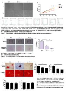
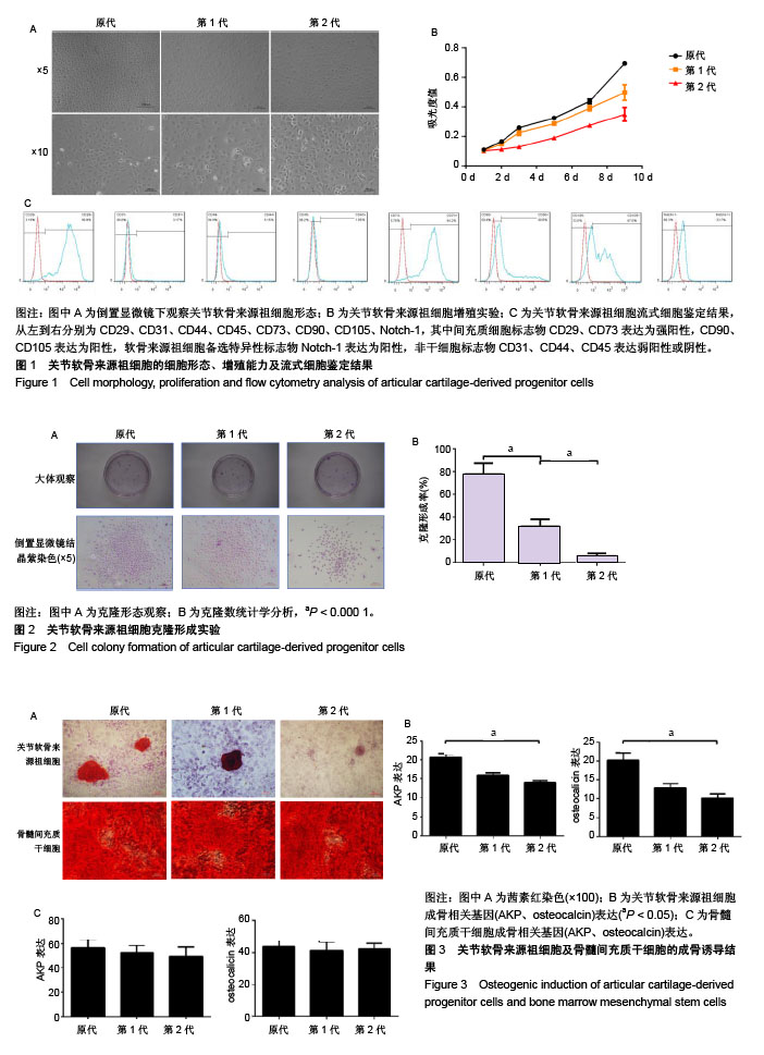
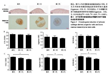
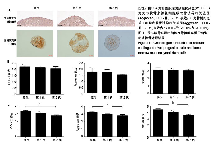
2.1 ACPCs的细胞形态和细胞活性 在倒置显微镜下观察,经过纤连蛋白分选后的ACPCs均为中间宽两边细长的短梭形,与间充质细胞的典型细胞形态类似。随着细胞代次的增加,在相同时间内ACPCs的细胞密度逐渐降低,但在细胞形态上未有明显改变,见图1A。 CCK-8结果显示,同一代次ACPCs的吸光度值随着培养时间延长持续升高;同一时间ACPCs的吸光度值在原代最高,其次是第1代,最后是第2代,培养1,3,5,7,9 d时原代与第1代,第1代与第2代差异均有显著性意义(P < 0.05),见图1B。 2.2 ACPCs的流式细胞鉴定结果 纤连蛋白分选后获得的大鼠ACPCs进行流式细胞鉴定,流式细胞仪计数结果显示:间充质细胞标志物CD29、CD73表达为强阳性,分别为96.8%和94.2%,CD90、CD105表达为阳性,分别为40.6%和67.0%,软骨来源祖细胞备选特异性标志物Notch-1表达为阳性,为33.7%;非干细胞标志物CD31、CD44、CD45表达弱阳性或阴性,分别为3.17%,5.15%和1.85%,见图1C。 2.3 ACPCs克隆形成实验结果 在各个代次细胞培养皿底部均有多个ACPCs单细胞克隆形成,见图2A。随后对不同代次细胞形成的克隆数进行比较,可见随着细胞代次的增加,ACPCs克隆数明显下降。对直径大于2 mm的克隆进行计数,不同代次之间差异有显著性意义(P < 0.000 1),见图2B。 2.4 成骨诱导结果 在肉眼下观察,ACPCs成骨诱导组橙红色范围随着代次增加逐渐减少。原代橙红色范围为孔中心及周围,颜色较深;第1代仅孔周围有部分橙红色区域,中心为蓝色区域;第2代孔内基本为浅蓝色区域。BMSCs成骨诱导组在原代、第1代和第2代均可见孔内区域为橙红色,橙红色范围随着细胞代次增加未见明显改变,见图3A。 在倒置显微镜下观察,ACPCs成骨诱导组在原代可见多个钙结节,第1代1个视野中仅可见1个钙结节,第2代无钙结节形成。BMSCs成骨诱导组在原代、第1代和第2代视野内基本均为橙红色钙结节,3个代次之间的差异不明显,见图3A。 ACPCs的RT-PCR结果显示,成骨诱导组的成骨相关基因AKP、osteocalcin均明显高于非诱导组,差异有显著性意义(P < 0.05),成骨相关基因在不同代次之间是逐渐降低的,在原代与第1代之间的降低幅度尤为明显,AKP、osteocalcin的表达在原代与第1代之间差异无显著性意义(P > 0.05),在第1代与第2代之间差异无显著性意义(P > 0.05),但原代与第2代之间差异有显著性意义(P < 0.05),见图3B。 BMSCs的RT-PCR结果显示,AKP、osteocalcin的表达在原代与第1代、第1代与第2代之间差异无显著性意义(P > 0.05),原代与第2代之间差异也无显著性意义(P > 0.05),见图3C。 2.5 成软骨诱导结果 在成软骨诱导21 d后,对细胞团块进行组织学染色分析,ACPCs成软骨诱导组细胞团块中有软骨组织典型结构,即软骨陷窝产生;Ⅱ型胶原染色可见ACPCs构建的团块全部为棕色阳性染色,不同代次之间阳性染色没有明显差异。BMSCs成软骨诱导组细胞团块中仅有部分区域有软骨陷窝产生;Ⅱ型胶原染色可见BMSCs构建的团块大部分组织为棕色阳性染色,且阳性染色区域随着细胞代次的增加而减少,到第2代时Ⅱ型胶原染色明显降低,见图4A。 ACPCs的RT-PCR结果显示,成软骨诱导组成软骨相关基因Aggrecan、COLⅡ、SOX9均明显高于非诱导组,差异有显著性意义(P < 0.05),Aggrecan、COL-Ⅱ、SOX9的表达在原代与第1代之间及第1代与第2代之间表达有减低,但基因表达差异无显著性意义(P > 0.05),原代与第2代之间差异也无显著性意义(P > 0.05),见图4B。 BMSCs的RT-PCR结果显示,成软骨诱导组成软骨相关基因Aggrecan、COL-Ⅱ、SOX9均明显高于非诱导组,差异有显著性意义(P < 0.05),Aggrecan、COL-Ⅱ、SOX9的表达在原代与第1代及第1代与第2代之间表达有减低,基因表达差异均无显著性意义(P > 0.05),但在原代与第2代之间差异有显著性意义(P < 0.05),见图4C。"
"
| [1] Jiang Y, Tuan RS. Origin and function of cartilage stem/progenitor cells in osteoarthritis. Nat Rev Rheumatol. 2015;11(4):206-212.[2] Centers for Disease Control and Prevention (CDC). Prevalence of doctor-diagnosed arthritis and arthritis-attributable activity limitation- United States, 2010-2012. MMWR Morb Mortal Wkly Rep. 2013; 62(44): 869-873.[3] Felson DT. An update on the pathogenesis and epidemiology of osteoarthritis. Radiol Clin North Am. 2004;42(1):1-9.[4] Guilbert JJ. The world health report 2002 - reducing risks, promoting healthy life. Educ Health (Abingdon). 2003;16(2):230.[5] 张爽,刘石,汪宇峰,等.间充质干细胞修复骨关节炎软骨损伤的临床应用意义[J].中国组织工程研究, 2018,22(33):5379-5385.[6] Medvedeva EV, Grebenik EA, Gornostaeva SN, et al. Repair of Damaged Articular Cartilage:Current Approaches and Future Directions. Int J Mol Sci. 2018;19(8): E2366.[7] Mithoefer K, McAdams T, Williams RJ, et al. Clinical efficacy of the microfracture technique for articular cartilage repair in the knee: an evidence-based systematic analysis. Am J Sports Med. 2009; 37(10):2053-2063.[8] Nakagawa Y, Mukai S, Yabumoto H, et al. Serial Changes of the Cartilage in Recipient Sites and Their Mirror Sites on Second-Look Imaging After Mosaicplasty. Am J Sports Med. 2016;44(5): 1243-1248.[9] Solheim E, Hegna J, Øyen J, et al. Results at 10 to 14 years after osteochondral autografting (mosaicplasty) in articular cartilage defects in the knee. Knee. 2013;20(4):287-290.[10] Hangody L, Dobos J, Baló E, et al. Clinical experiences with autologous osteochondral mosaicplasty in an athletic population: a 17-year prospective multicenter study. Am J Sports Med. 2010; 38(6):1125-1133.[11] Goyal D, Goyal A, Keyhani S, et al. Evidence-based status of second- and third-generation autologous chondrocyte implantation over first generation: a systematic review of level I and II studies. Arthroscopy. 2013;29(11):1872-1878.[12] Derrett S, Stokes EA, James M, et al. Cost and health status analysis after autologous chondrocyte implantation and mosaicplasty: a retrospective comparison. Int J Technol Assess Health Care. 2005; 21(3): 359-367.[13] Mistry H, Connock M, Pink J, et al. Autologous chondrocyte implantation in the knee: systematic review and economic evaluation. Health Technol Assess. 2017;21(6):1-294.[14] Park YB, Ha CW, Rhim JH, et al. Stem Cell Therapy for Articular Cartilage Repair: Review of the Entity of Cell Populations Used and the Result of the Clinical Application of Each Entity. Am J Sports Med. 2018;46(10):2540-2552.[15] 李乔乔,吴振强,张丽君,等.骨髓间充质干细胞的定向分化潜能[J].中国组织工程研究,2017,21(25): 4082-4087.[16] Williams R, Khan IM, Richardson K, et al. Identification and clonal characterisation of a progenitor cell sub-population in normal human articular cartilage. PLoS One. 2010;5(10):e13246.[17] Pelttari K, Winter A, Steck E, et al. Premature induction of hypertrophy during in vitro chondrogenesis of human mesenchymal stem cells correlates with calcification and vascular invasion after ectopic transplantation in SCID mice. Arthritis Rheum. 2006;54(10): 3254-3266.[18] 赵文慧,皮洪涛,冯万文,等.关节软骨细胞和骨髓间充质干细胞不同共培养方式对细胞增殖与分化的影响[J].中国组织工程研究,2019,23(1): 24-29.[19] Marcus P, De Bari C, Dell'Accio F, et al. Articular Chondroprogenitor Cells Maintain Chondrogenic Potential but Fail to Form a Functional Matrix When Implanted Into Muscles of SCID Mice. Cartilage. 2014; 5(4):231-240.[20] Dowthwaite GP, Bishop JC, Redman SN, et al. The surface of articular cartilage contains a progenitor cell population. J Cell Sci. 2004;117(Pt 6):889-897.[21] Ustunel I, Ozenci AM, Sahin Z, et al. The immunohistochemical localization of notch receptors and ligands in human articular cartilage, chondroprogenitor culture and ultrastructural characteristics of these progenitor cells. Acta Histochem. 2008;110(5):397-407.[22] Melero-Martin JM, Dowling MA, Smith M, et al. Expansion of chondroprogenitor cells on macroporous microcarriers as an alternative to conventional monolayer systems.Biomaterials. 2006;27(15): 2970-2979.[23] Melero-Martin JM, Dowling MA, Smith M, et al. Optimal in-vitro expansion of chondroprogenitor cells in monolayer culture. Biotechnol Bioeng.2006;93(3):519-533.[24] Martin JM, Smith M, Al-Rubeai M. Cryopreservation and in vitro expansion of chondroprogenitor cells isolated from the superficial zone of articular cartilage. Biotechnol Prog. 2005;21(1):168-177.[25] Sandrasaigaran P, Algraittee SJR, Ahmad AR, et al. Characterisation and immunosuppressive activity of human cartilage-derived mesenchymal stem cells. Cytotechnology. 2018;70(3):1037-1050.[26] Fellows CR, Williams R, Davies IR, et al. Characterisation of a divergent progenitor cell sub-populations in human osteoarthritic cartilage: the role of telomere erosion and replicative senescence. Sci Rep. 2017;7:41421.[27] Correa D, Lietman SA. Articular cartilage repair: Current needs, methods and research directions. Semin Cell Dev Biol. 2017;62: 67-77.[28] Levato R, Webb WR, Otto IA, et al. The bio in the ink: cartilage regeneration with bioprintable hydrogels and articular cartilage- derived progenitor cells. Acta Biomater. 2017;61:41-53.[29] Khan IM, Bishop JC, Gilbert S, et al. Clonal chondroprogenitors maintain telomerase activity and Sox9 expression during extended monolayer culture and retain chondrogenic potential. Osteoarthritis Cartilage. 2009;17(4):518-528. |
| [1] | Hou Jingying, Yu Menglei, Guo Tianzhu, Long Huibao, Wu Hao. Hypoxia preconditioning promotes bone marrow mesenchymal stem cells survival and vascularization through the activation of HIF-1α/MALAT1/VEGFA pathway [J]. Chinese Journal of Tissue Engineering Research, 2021, 25(7): 985-990. |
| [2] | Liang Xueqi, Guo Lijiao, Chen Hejie, Wu Jie, Sun Yaqi, Xing Zhikun, Zou Hailiang, Chen Xueling, Wu Xiangwei. Alveolar echinococcosis protoscolices inhibits the differentiation of bone marrow mesenchymal stem cells into fibroblasts [J]. Chinese Journal of Tissue Engineering Research, 2021, 25(7): 996-1001. |
| [3] | Geng Yao, Yin Zhiliang, Li Xingping, Xiao Dongqin, Hou Weiguang. Role of hsa-miRNA-223-3p in regulating osteogenic differentiation of human bone marrow mesenchymal stem cells [J]. Chinese Journal of Tissue Engineering Research, 2021, 25(7): 1008-1013. |
| [4] | Lun Zhigang, Jin Jing, Wang Tianyan, Li Aimin. Effect of peroxiredoxin 6 on proliferation and differentiation of bone marrow mesenchymal stem cells into neural lineage in vitro [J]. Chinese Journal of Tissue Engineering Research, 2021, 25(7): 1014-1018. |
| [5] | Zhu Xuefen, Huang Cheng, Ding Jian, Dai Yongping, Liu Yuanbing, Le Lixiang, Wang Liangliang, Yang Jiandong. Mechanism of bone marrow mesenchymal stem cells differentiation into functional neurons induced by glial cell line derived neurotrophic factor [J]. Chinese Journal of Tissue Engineering Research, 2021, 25(7): 1019-1025. |
| [6] | Pei Lili, Sun Guicai, Wang Di. Salvianolic acid B inhibits oxidative damage of bone marrow mesenchymal stem cells and promotes differentiation into cardiomyocytes [J]. Chinese Journal of Tissue Engineering Research, 2021, 25(7): 1032-1036. |
| [7] | Li Cai, Zhao Ting, Tan Ge, Zheng Yulin, Zhang Ruonan, Wu Yan, Tang Junming. Platelet-derived growth factor-BB promotes proliferation, differentiation and migration of skeletal muscle myoblast [J]. Chinese Journal of Tissue Engineering Research, 2021, 25(7): 1050-1055. |
| [8] | Liu Cong, Liu Su. Molecular mechanism of miR-17-5p regulation of hypoxia inducible factor-1α mediated adipocyte differentiation and angiogenesis [J]. Chinese Journal of Tissue Engineering Research, 2021, 25(7): 1069-1074. |
| [9] | Wang Shiqi, Zhang Jinsheng. Effects of Chinese medicine on proliferation, differentiation and aging of bone marrow mesenchymal stem cells regulating ischemia-hypoxia microenvironment [J]. Chinese Journal of Tissue Engineering Research, 2021, 25(7): 1129-1134. |
| [10] | Ma Zetao, Zeng Hui, Wang Deli, Weng Jian, Feng Song. MicroRNA-138-5p regulates chondrocyte proliferation and autophagy [J]. Chinese Journal of Tissue Engineering Research, 2021, 25(5): 674-678. |
| [11] | Wang Yujiao, Liu Dan, Sun Song, Sun Yong. Biphasic calcium phosphate loaded with advanced platelet rich fibrin can promote the activity of rabbit bone marrow mesenchymal stem cells [J]. Chinese Journal of Tissue Engineering Research, 2021, 25(4): 504-509. |
| [12] | Chen Junyi, Wang Ning, Peng Chengfei, Zhu Lunjing, Duan Jiangtao, Wang Ye, Bei Chaoyong. Decalcified bone matrix and lentivirus-mediated silencing of P75 neurotrophin receptor transfected bone marrow mesenchymal stem cells to construct tissue-engineered bone [J]. Chinese Journal of Tissue Engineering Research, 2021, 25(4): 510-515. |
| [13] | Zhou Jihui, Yao Meng, Wang Yansong, Li Xinzhi, Zhou You, Huang Wei, Chen Wenyao. Influence of novel nanoscaffolds on biological behaviors of neural stem cells and the related gene expression [J]. Chinese Journal of Tissue Engineering Research, 2021, 25(4): 532-536. |
| [14] | Jiang Tao, Ma Lei, Li Zhiqiang, Shou Xi, Duan Mingjun, Wu Shuo, Ma Chuang, Wei Qin. Platelet-derived growth factor BB induces bone marrow mesenchymal stem cells to differentiate into vascular endothelial cells [J]. Chinese Journal of Tissue Engineering Research, 2021, 25(25): 3937-3942. |
| [15] | Chen Yang, Huang Denggao, Gao Yuanhui, Wang Shunlan, Cao Hui, Zheng Linlin, He Haowei, Luo Siqin, Xiao Jingchuan, Zhang Yingai, Zhang Shufang. Low-intensity pulsed ultrasound promotes the proliferation and adhesion of human adipose-derived mesenchymal stem cells [J]. Chinese Journal of Tissue Engineering Research, 2021, 25(25): 3949-3955. |
| Viewed | ||||||
|
Full text |
|
|||||
|
Abstract |
|
|||||