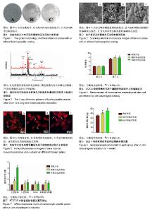
| [1] Gao Y, Liu S, Huang J, et al. The ECM-cell interaction of cartilage extracellular matrix on chondrocytes.Biomed Res Int. 2014;2014:648459. [2] Azuma Y, Ito M, Harada Y, et al. Low-intensity pulsed ultrasound accelerates rat femoral fracture healing by acting on the various cellular reactions in the fracture callus.J Bone Miner Res. 2001;16(4):671-680. [3] Li J, Pei M.Cell senescence: a challenge in cartilage engineering and regeneration.Tissue Eng Part B Rev. 2012; 18(4):270-287. [4] Jackson RW, Dieterichs C.The results of arthroscopic lavage and debridement of osteoarthritic knees based on the severity of degeneration: a 4- to 6-year symptomatic follow-up. Arthroscopy. 2003;19(1):13-20. [5] Dewan AK, Gibson MA, Elisseeff JH, et al. Evolution of autologous chondrocyte repair and comparison to other cartilage repair techniques.Biomed Res Int. 2014;2014: 272481. [6] 郑荣宗,吴伟东,应锦河.关节镜下自体骨软骨移植治疗膝关节软骨缺损[J].中国骨与关节损伤杂志,2014,29(10):1050-1051.[7] Chiang H, Liao CJ, Wang YH, et al. Comparison of articular cartilage repair by autologous chondrocytes with and without in vitro cultivation.Tissue Eng Part C Methods. 2010;16(2): 291-300. [8] Levi B, Hyun JS, Montoro DT, et al. In vivo directed differentiation of pluripotent stem cells for skeletal regeneration.Proc Natl AcadSci U S A. 2012;109(50): 20379-20384. [9] Handschel J, Berr K, Depprich RA, et al. Induction of osteogenic markers in differentially treated cultures of embryonic stem cells.Head Face Med. 2008;4:10. [10] Hashimoto J, Kariya Y, Miyazaki K.Regulation of proliferation and chondrogenic differentiation of human mesenchymal stem cells by laminin-5 (laminin-332).Stem Cells. 2006; 24(11):2346-2354. [11] Wong SP, Rowley JE, Redpath AN, et al. Pericytes, mesenchymal stem cells and their contributions to tissue repair.PharmacolTher. 2015;151:107-120. [12] Ahmed TA, Dare EV, Hincke M.Fibrin: a versatile scaffold for tissue engineering applications.Tissue Eng Part B Rev. 2008; 14(2):199-215. [13] Aoyama E, Hattori T, Hoshijima M, et al. N-terminal domains of CCN family 2/connective tissue growth factor bind to aggrecan.Biochem J. 2009;420(3):413-420. [14] Grande DA, Mason J, Light E, et al. Stem cells as platforms for delivery of genes to enhance cartilage repair.J Bone Joint Surg Am. 2003;85-A Suppl 2:111-116. [15] Kim MS, Hwang NS, Lee J, et al. Musculoskeletal differentiation of cells derived from human embryonic germ cells.Stem Cells. 2005;23(1):113-123. [16] Prosecká E, Buzgo M, Rampichová M, et al. Thin-layer hydroxyapatite deposition on a nanofiber surface stimulates mesenchymal stem cell proliferation and their differentiation into osteoblasts.J Biomed Biotechnol. 2012;2012:428503. [17] Lin L, Chow KL, Leng Y.Study of hydroxyapatite osteoinductivity with an osteogenic differentiation of mesenchymal stem cells.J Biomed Mater Res A. 2009;89(2): 326-335. [18] Be?karde? IG, Demirta? TT, Durukan MD, et al. Microwave-assisted fabrication of chitosan-hydroxyapatite superporous hydrogel composites as bone scaffolds.J Tissue Eng Regen Med. 2015;9(11):1233-1246. [19] Doi K, Kubo T, Takeshita R,et al. Inorganic polyphosphate adsorbed onto hydroxyapatite for guided bone regeneration: an animal study.Dent Mater J. 2014;33(2):179-186. [20] Wang Y, Yu X, Baker C, et al. Mineral particles modulateosteo-chondrogenic differentiation of embryonic stem cell aggregates.Acta Biomater. 2016;29:42-51. [21] Shibuya I, Yoshimura K, Miyamoto Y, et al. Octacalcium phosphate suppresses chondrogenic differentiation of ATDC5 cells.Cell Tissue Res. 2013;352(2):401-412. [22] Hung KY, Lo SC, Shih CS, et al. Titanium surface modified by hydroxyapatite coating for dental implants.Surface & Coatings Technology. 2013;231(9):337-345. [23] Bayram C, Demirbilek M, Cali?kan N, et al. Osteoblast activity on anodized titania nanotubes: effect of simulated body fluid soaking time.J Biomed Nanotechnol. 2012;8(3):482-490. [24] LinDY, WangXX. Preparation of hydroxyapatite coating on smooth implant surface by electrodeposition.Ceramics International. 2011;37(1):403-406. [25] Albayrak O, El-Atwani O,Altintas S. Hydroxyapatite coating on titanium substrate by electrophoretic deposition method: Effects of titanium dioxide inner layer on adhesion strength and hydroxyapatite decomposition.Surface & Coatings Technology. 2008;202(11):2482-2487. [26] Yang GL, He FM, Hu JA, et al. Effects of biomimetically and electrochemically deposited nano-hydroxyapatite coatings on osseointegration of porous titanium implants.Oral Surg Oral Med Oral Pathol Oral Radiol Endod. 2009;107(6):782-789. [27] Zhao SF, Dong WJ, Jiang QH, et al. Effects of zinc-substituted nano-hydroxyapatite coatings on bone integration with implant surfaces.J Zhejiang UnivSci B. 2013;14(6):518-525. [28] Yang GL, He FM, Hu JA, et al. Biomechanical comparison of biomimetically and electrochemically deposited hydroxyapatite-coated porous titanium implants.J Oral Maxillofac Surg. 2010;68(2):420-427. [29] Zhao JJ, Dong XC, Bian MM, et al. Solution combustion method for synthesis of nanostructured hydroxyapatite, fluorapatite and chlorapatite.Applied Surface Science. 2014;314(10):1026-1033. [30] Costa DO, Prowse PD, Chrones T, et al. The differential regulation of osteoblast and osteoclast activity by surface topography of hydroxyapatite coatings.Biomaterials. 2013; 34(30):7215-7226. [31] Rupp F, Scheideler L, Olshanska N, et al. Enhancing surface free energy and hydrophilicity through chemical modification of microstructured titanium implant surfaces.J Biomed Mater Res A. 2006;76(2):323-334. |

