Chinese Journal of Tissue Engineering Research ›› 2019, Vol. 23 ›› Issue (32): 5221-5226.doi: 10.3969/j.issn.2095-4344.1494
Previous Articles Next Articles
Application and progress of finite element analysis in scoliosis biomechanical research
Sun Fengyuan1, Li Zongyuan1, He Xi1, Yang Zongde2
- 1College of Basic Medical Sciences, Naval Medical University, Shanghai 200433, China; 2Department of Spinal Surgery, Changhai Hospital, Naval Medical University, Shanghai 200433, China
-
Online:2019-11-18Published:2019-11-18 -
About author:Sun Fengyuan, College of Basic Medical Sciences, Naval Medical University, Shanghai 200433, China Li Zongyuan, College of Basic Medical Sciences, Naval Medical University, Shanghai 200433, China Sun Fengyuan and Li Zongyuan contributed equally to this paper.
CLC Number:
Cite this article
Sun Fengyuan, Li Zongyuan, He Xi, Yang Zongde. Application and progress of finite element analysis in scoliosis biomechanical research[J]. Chinese Journal of Tissue Engineering Research, 2019, 23(32): 5221-5226.
share this article

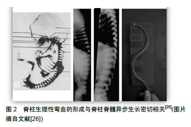
有限元分析在特发性脊柱侧凸的生物力学研究领域已经有了大量的尝试,取得了不少的进展,并愈发受到关注[1,4-7]。目前已经有不少的有限元分析在脊柱侧凸领域的应用报道,且大多集中在外固定矫正设备的设计与应用模拟[8-10]、优化手术方案以及预估临床干预的效果[11],但是青少年特发性脊柱侧凸完整的有限元分析模型报道较少[1],在脊柱侧凸病因学的应用报道也不多。有限元分析在脊柱侧凸生物力学领域应用的可行性已经得到充分的验证,研究领域主要集中在手术和支具模拟、病因学研究、并发症研究和有限元分析模型优化几个方面: 2.1 手术和支具模拟 有限元分析可以解决传统手术和支具模拟中尸体标本不足、可重复性不强、模拟数据采集困难等问题,展现出了不可取代的优势。有限元分析法在脊柱侧凸手术和支具模拟中已经有了大量应用。1986年Viviani等[12]最早将有限元分析应用在脊柱侧凸手术的模拟上,指导和优化手术矫正过程,他们建立的模型为二维线性模型,故存在还原性差的问题。2000年Gignac等[13]建立的有限元分析模型包含了胸廓的部分结构,而且研究了Boston支具的生物力学原理,实现了对其的改良。但是他们尚未构建全脊柱模型,故试验误差依然较大。2004年Perie等[14]建立了3例有肋骨、骨盆的青少年特发性脊柱侧凸的有限元分析模型,探索了躯体内部作用力的相互关系。自此,通过有限元分析研究了解支具生物力学原理,进行支具改良和个性化设计的可行性得到了验证。 2.1.1 生物力学关系研究 有限元分析用于手术和支具的模拟,可以直观的得到支具内部、机体内部以及支具与机体之间的生物力学关系。近年来出现许多具有代表性的支具生物力学关系研究。2010年Lafon等[6]构建了10例重度特发性脊柱侧凸患者的个体化有限元分析模型,证明了原位弯棒与CD旋棒技术的矫正效果相似,还证明了双侧棒的弯曲对融合区内外的脊柱具有互补的效果。2013年Zhang等[15]建立了Lenke5型特发性脊柱侧凸患者有限元分析模型,研究4种手术方式的去旋转和压缩序贯矫正过程,得到4种手术方式矫正下患者脊柱Cobb角及顶椎旋转角度的变化,有助于指导特发性脊柱侧凸手术的选择和预估预后。2016年Sattout等[16]评估了Providence夜间支具治疗青少年特发性脊柱侧凸的生物力学效果,指出仰卧位发挥了冠状位弯曲矫形的主要效果,而支具只发挥了次要效果。由支具产生的弯曲力矩可以在生长板上产生再平衡作用,有助于抑制椎体的不平衡生长。2018年Aubin等[17]利用6个青少年特发性脊柱侧凸病例的有限元分析模型对cv和ant系绳开展生物力学评估,他们的生物力学研究发现了cv和ant系绳之间的差异,并指出了因患者特定特征而产生的变异性。同年Agarwal等[18]通过改变椎间盘的材料特性,改变脊柱侧凸模型的轴向刚度,进行参数化研究,发现随着脊柱轴向刚度的增加,杆上的应力增加。他们的研究建立了仅受重力作用的在Cobb角度中的百分比校正与将生长棒上的最大Von Mises应力限制为 255 MPa所需的干扰间隔之间的关系,指出了将应力限制在选定的标称值所需的干扰间隔随着脊柱刚度的增加而减小,并且随着时间的推移,脊柱变得更硬(自动融合),每个模型的适当干扰间隔也会减少。这表明最佳的干扰频率是一个时间相关的变量,必须实现以保持最大冯·米塞斯应力下的指定安全系数。 2.1.2 设计和改良支具 支具的设计和改良是复杂的和需要实验证明的过程,有限元分析因为其独有的优势,在这一领域里发挥着越来越大的作用。2007年Liao等[9]通过有限元分析建模改进Boston支具,使其质量降低了12.4%-18%,而预后相同,减少了患者的不耐受。2015年Weiss等[19]考虑到支具矫正(和顺应性)决定了支具治疗脊柱侧弯的最终结果,指出基于分类方法的脊柱侧弯支具优于基于有限元分析模型分析的方法。2016年Cobetto等[20]的随机对照研究希望能够通过数值模拟和有限元分析模型与计算机辅助设计与制造系统相结合的方法,在所有3个平面上设计更高效更轻便可以3D改进的支具,并且指出这种新方法可能改善青少年特发性脊柱侧凸的远期疗效。另外,通过使用该仿真平台可以在3D模式下改进青少年特发性脊柱侧凸的支具。2017年Hachem等[21]建立了含骨生长板的猪脊椎和肋骨的有限元分析模型,用以开发治疗小儿脊柱侧弯的无融合设备。他们的成果经过开发和验证后或许可以作为评估无融合装置的替代平台,并可能以此节约动物试验的昂贵经济成本和时间成本。 2.1.3 改善手术效果 有限元分析被用于手术模拟以改良手术方法、改善预后效果、评估手术结果。2013年Little等[5]构建了8例具有韧带、肋骨和椎体的特发性脊柱侧凸患者有限元分析模型,研究钉棒内固定矫正,证明矫正力和预后有关。2014年Agarwal等[22]的有限元分析研究发现适当缩小分散间隔可以在不妨碍预期脊柱生长的同时减少生长杆断裂的概率。2015年Galbusera等[23]提出了构建一种生物力学软件个性化模拟矫正患者的脊柱畸形。该方法基于从双平面放射照相图像获得的脊柱解剖结构构建的有限元分析模型。该法有望为患者提供更加个性化的治疗策略,但是目前距离实际应用仍有较大距离。2017年海涌等[24]研究各种生长棒固定的选择与早发性脊柱侧凸相邻节段生物力学的影响,得知在生长棒手术治疗早发性脊柱侧凸患者中,双生长棒及头侧的椎板钩固定可以更好地降低相邻节段的力。李明等[25]在2018年利用有限元分析法研究不同材料矫形棒在青少年特发性脊柱侧凸矢状面重建中的作用,提示采用强度较大的钴铬合金棒及适当加大矫形棒预弯弧度有助于矢状面力线的恢复,但也会导致螺钉拔出应力增加。 2.2 病因学研究 目前青少年特发性脊柱侧凸的发病机制仍不清楚,很多学者将生长发育作为研究青少年特发性脊柱侧凸发病机制的切入点,其中脊柱脊髓异步生长假说是其主要假说之一,它的内容为青少年时期脊柱和脊髓生长不同步,脊柱生长速度快于脊髓,导致脊髓对脊柱产生了牵拉作用(图2)[26]。脊柱脊髓异步生长假说即认为此作用力导致了青少年特发性脊柱侧凸的发生,目前脊柱脊髓异步生长作用引起青少年特发性脊柱侧凸发病的具体机制仍未阐明。此外人们还普遍认识到,青少年特发性脊柱侧凸是在脊柱不对称负荷和椎体生长调节的生物力学过程中进展的。由于其突出的力学模拟优势,有限元分析在青少年特发性脊柱侧凸生物力学的病因学研究中展现出了很大的潜力。1998年Azegami等[27]建立了完整的有限元分析模型,有限元分析模型以八面体网格分元,再以有限元分析法根据压缩屈曲学说引发T4-T10青少年特发性脊柱侧凸,得到同实际测量一模一样的青少年特发性脊柱侧凸进展过程,这是较早构建真正的有限元分析非线性模型的研究之一。"

2.2.1 脊柱异常生长 2002年Villemure等[28]的研究建立了一个包含椎体生长和生长调节的有限元分析模型,以代表脊柱侧弯畸形的进展。总体而言,其模型充分代表了椎体和脊柱侧弯畸形的自我维持进展。他们的研究证明了该模型方法的可行性,并与当时脊柱侧弯的其他生物力学研究相比,实现了更完整的脊柱侧弯的脊柱模拟。既往的有限元分析模型往往都是静态的,而他们的模型能够模拟脊柱的生长,这是有限元分析脊柱侧凸生物力学研究的又一进步。2011年Shi等[29]构建了包涵生长动力学在的脊柱有限元分析模型,模拟了不同的冠状和矢状弯曲,并且利用青少年特发性脊柱侧凸和正常生长剖面模拟了10年的脊柱生长过程。他们的研究结果验证了加速生长可能会加快脊柱侧弯的进展,因此脊柱生长过快可能会成为一个渐进的危险因素。他们的研究有可能是脊柱脊髓异步生长作用在有限元分析生物力学研究的证据支撑。 2.2.2 解剖、受力和生物力学性质不对称的影响 2003年Goto等[30]构建青少年特发性脊柱侧凸的有限元分析形成过程模型,证明了骨形成作用抑制青少年特发性脊柱侧凸的发生,骨吸收作用促进青少年特发性脊柱侧凸的发生。2016年Wang等[31]应用有限元分析验证了在不对称负荷的情况下,小关节会出现小关节压缩性变形,小关节限制椎体的旋转和摆位的能力减小,小关节接触力的不对称负载加速了腰椎的不对称。同年贾少薇等[32]利用有限元分析研究指出了脊柱侧凸与腰骶部结构特点之间具有关联性。2017年有学者利用一个简单的生物力学模型指出,脊柱形态的差异和相应的脊柱弹性变化可能与特发性脊柱侧凸有关。同年Li等[33]为探讨不对称张力对特发性脊柱侧弯的影响,建立了啮齿类动物的脊柱侧弯模型。他们的研究认为,不对称张力导致椎体上的蛋白表达和骨质量不对称,最终导致解剖不对称。此外解剖不对称加剧了张力不对称,这是首次研究表明特发性脊柱侧凸存在不对称的恶性循环。Wang等[34]在系统和持续的低骨密度这一领域进行了包括有限元分析的病例对照研究,是在这一问题上开展的首次综合性研究,为青少年特发性脊柱侧凸不同骨层次的未定义独特病理变化提供直接证据,指出系统和持续的低骨密度是侧凸进展的独立预后因素,但是青少年特发性脊柱侧凸中骨质量是如何受到影响的根本问题仍然存在争议。2018年Song等[35]建立了不同骨矿状态的特发性脊柱有限元分析模型,并进行轴向加载模拟。他们的研究指出,在青少年特发性脊柱侧弯患者中,低骨量可能会加重侧凸进展,诱发更严重的腰椎代偿性脊柱侧弯。同时体质量增加也是曲线进展的一个危险因素。2018年Schlager等[36]根据无视力畸形患者的CT数据,建立了胸膜、胸部和脊柱的有限元分析模型,研究证实了不对称的胸膜腔内压与脊柱生物力学和病理学的潜在相关性。 2.3 并发症研究 2015年Pasha等[37]评估了脊柱侧弯融合对脊柱和骨盆之间转移负荷的影响,指出骶骨终板的生物力学负荷与术后姿势平衡和脊柱侧凸手术后脊柱骨盆矫正的代偿性变化有关。这为术后姿势平衡和代偿提供了理论依据。2016年Henao等[38]建立有限元分析特异性混合模型,成功复制了脊柱侧弯矫正操作中神经损伤的生物力学模型。此生物力学模型有望能够作为研究避免术后神经损伤并发症的实验平台。2017年Xu等[39]的有限元分析研究为成人退变性脊柱侧凸会严重增加脊髓对环形振动的反应提供了实验性证据,并指出这有可能导致脊柱侧凸畸形的加重。同年Agarwal等[40]采用多种代表性的脊柱侧凸有限元分析模型,分析了干扰力和频率对多种代表性脊柱侧弯曲线的影响, 并建立了高牵张力与螺钉松动之间的关系,证实了在具有最佳干扰力的螺钉-骨界面下载荷的有限元分析结果。这项研究还将第二常见的并发症螺钉松动与高干扰力联系起来,由他们的研究结果可以看出,优化双生长棒的生物力学环境可以明显减少与生长棒相关的生物力学并发症。在脊柱侧弯矫正手术中应用的力量对神经结构施加压力,可能会导致神经并发症。在矫正过程中,不同类型的应力对神经结构的生物力学影响尚不清楚。2018年Henao等[41]利用有限元分析,通过数值模拟,对脊柱侧弯矫正过程中的脊髓和神经进行生物力学分析,评价脊柱侧弯矫正动作和应力对脊柱神经结构的生物力学影响。他们的研究采用了新的混合患者特异性模型,对不同矫正术对脊髓和脊髓神经的相对应激进行了量化,指出哈灵顿牵拉动作是诱发胸腰椎区域神经并发症最重要的应力。 2.4 优化有限元分析模型 2.4.1 数据提取和建模方法 2018年Pea等[42]的研究显示了基于一种新的单冠状位X射线片和表面形貌建模方法设计和模拟支架矫正的可行性,这种创新的方法可以用于改善支具设计,对患者的辐射剂量更小。2018年Hadaqali等[43]论证了一种从现有的多块规范性椎骨上半自动化构建脊柱侧凸椎体几何结构的方法,以减少繁琐庞大的建模工作量。 2.4.2 有限元分析件的优化 2016年Vergari等[44]的42例青少年特发性脊柱侧凸回顾性和前瞻性研究指出,个性化脊柱功能单位的机械性能的可以提高模拟的准确性。2017年Xu等[45]的研究结果表明,几种不同有限元分析模型的综合预测(中值或平均值)可改善在相同载荷和边界条件下人体腰椎行为的预测。他们进行了广泛的验证及网格收敛和材料敏感性分析,证明了他们开发的有限元分析模型结果与现有文献中的实验数据和仿真结果是一致的。他们介绍的验证建模方法可用于功能失调的脊柱的建模,如椎间盘退变和脊柱侧弯。 2.4.3 软组织的有限元分析构建 2015年Little等[46]为研究脊柱椎间盘及周围软组织对脊柱侧凸柔韧性的影响,建立了脊柱侧凸有限元分析模型,发现脊柱侧凸中对柔韧性影响最大的解剖结构上椎间盘。有限元分析模型中的软组织的构建是目前有限元分析主要的挑战和发展方向之一,而他们构建了软组织的有限元分析模拟,虽仍然存在还原度的问题,但是有限元分析进步的重要尝试。"

| [1]黄盛佳,霍洪军,杨学军,等. PUMCⅡd1型青少年特发性脊柱侧凸三维有限元模型的建立[J]. 中国组织工程研究, 2014,18(26): 4219-4223.[2]黄盛佳,霍洪军,杨学军,等. PUMCⅡd1型青少年特发性脊柱侧凸后路三维矫形手术有限元研究[J]. 国际骨科学杂志, 2016,37(1): 46-52.[3]李晔.王以朋.贾少薇.等. 三维有限元法分析先天性脊柱侧凸冠状面腰骶段的柔韧性[J]. 中国组织工程研究,2017,21(27): 4366-4372.[4]Salmingo R, Tadano S, Fujisaki K, et al. Corrective force analysis for scoliosis from implant rod deformation. Clin Biomech (Bristol, Avon). 2012;27(6): 545-550.[5]Little JP, Izatt MT, Labrom RD, et al. An FE investigation simulating intra-operative corrective forces applied to correct scoliosis deformity. Scoliosis. 2013;8(1): 9.[6]Lafon Y, Steib JP, Skalli W. Intraoperative three dimensional correction during in situ contouring surgery by using a numerical model. Spine (Phila Pa 1976). 2010;35(4):453-459.[7]Gardner-Morse M, Stokes IA. Three-dimensional simulations of the scoliosis derotation maneuver with Cotrel-Dubousset instrumentation. J Biomech. 1994;27(2): 177-181.[8]盛林,汪学松,吴志宏,等, 不同矫形力矫正特发性脊柱侧凸的三维有限元分析[J]. 中国组织工程研究与临床康复,2009,13(30): 5972-5976.[9]Liao YC, Feng CK, Tsai MW, et al. Shape modification of the Boston brace using a finite-element method with topology optimization. Spine (Phila Pa 1976). 2007;32(26): 3014-3019.[10]Kessler JI. Efficacy of a new computer-aided design/computer-aided manufacture orthosis in the treatment of adolescent idiopathic scoliosis. J Pediatr Orthop. 2008;17(4): 207.[11]刘伟强,张振军,孙艺萄,等. 有限元法在腰椎融合术与置换术生物力学研究中应用进展[J]. 医用生物力学, 2018,33(1)): 82-88.[12]Viviani GR, Ghista DN, Lozada PJ, et al. Biomechanical analysis and simulation of scoliosis surgical correction. Clin Orthop Relat Res. 1986; (208): 40-47.[13]Gignac D, Aubin CE, Dansereau J, et al. Optimization method for 3D bracing correction of scoliosis using a finite element model. Eur Spine J. 2000; 9(3): 185-190.[14]Perie D, Aubin CE, Petit Y, et al. Personalized biomechanical simulations of orthotic treatment in idiopathic scoliosis. Clin Biomech (Bristol, Avon). 2004;19(2): 190-195.[15]Zhang H, Hu X, Wang Y, et al. Use of finite element analysis of a Lenke type 5 adolescent idiopathic scoliosis case to assess possible surgical outcomes. Comput Aided Surg. 2013;18(3-4): 84-92.[16]Sattout A, Clin J, Cobetto N, et al. Biomechanical assessment of providence nighttime brace for the treatment of adolescent idiopathic scoliosis. Spine Deform. 2016;4(4):253-260.[17]Aubin C, Clin J, Rawlinson J. Biomechanical simulations of costo-vertebral and anterior vertebral body tethers for the fusionless treatment of pediatric scoliosis. J Orthop Res. 2018;36(1): 254-264.[18]Agarwal A, Jayaswal A, Goel VK, et al. Patient-specific distraction regimen to avoid growth-rod failure. Spine. 2017; 43(4): E221-E226.[19]Weiss H, Kleban A. Development of CAD/CAM based brace models for the treatment of patients with scoliosis-classification based approach versus finite element modelling. Asian Spine J. 2015;9(5): 661.[20]Cobetto N, Aubin CÉ, Parent S, et al. 3D correction of AIS in braces designed using CAD/CAM and FEM: a randomized controlled trial. Scoliosis Spinal Disord. 2017;12(1):24.[21]Hachem B, Aubin C, Parent S. Porcine spine finite element model: a complementary tool to experimental scoliosis fusionless instrumentation. Eur Spine J. 2017;26(6):1610-1617.[22]Agarwal A, Agarwal AK, Jayaswal A, et al. Smaller interval distractions may reduce chances of growth rod breakage without impeding desired spinal growth: a finite element study. Spine Deform. 2014; 2(6): 430-436.[23]Galbusera F, Bassani T, La Barbera L, et al. Planning the surgical correction of spinal deformities: toward the identification of the biomechanical principles by means of numerical simulation. Front Bioeng Biotechnol. 2015;3:178.[24]海涌,潘爱星,李永刚,等. 早发性脊柱侧凸不同生长棒固定方式对邻近节段生物力学的影响[J]. 中华医学杂志, 2017,97(48):3768-3773.[25]李明,范建平,赵检等.不同材料矫形棒在青少年特发性脊柱侧凸矢状面重建中作用的有限元研究[J]. 脊柱外科杂志,2018,16(6):358-362.[26]Yang Z, Xie Y, Li M. Three-dimensional spring model: A new hypothesis of pathogenesis of adolescent idiopathic scoliosis. Med Hypotheses. 2009;73(5):709-713.[27]Azegami H, Murachi S, Kitoh J, et al. Etiology of idiopathic scoliosis. Computational study. Clin Orthop Relat Res, 1998;(357): 229-236.[28]Villemure I, Aubin CE, Dansereau J, et al. Simulation of progressive deformities in adolescent idiopathic scoliosis using a biomechanical model integrating vertebral growth modulation. J Biomech Eng. 2002;124(6): 784-790.[29]Shi L, Wang D, Driscoll M, et al. Biomechanical analysis and modeling of different vertebral growth patterns in adolescent idiopathic scoliosis and healthy subjects. Scoliosis. 2011; 6: 11.[30]Goto M, Kawakami N, Azegami H, et al. Buckling and bone modeling as factors in the development of idiopathic scoliosis. Spine (Phila Pa 1976). 2003;28(4): 364-370; discussion 371.[31]Wang L, Zhang B, Chen S, et al. A validated finite element analysis of facet joint stress in degenerative lumbar scoliosis. World Neurosurgery. 2016; 95: 126-133.[32]贾少薇,张顺心,范顺成,等. 脊柱侧凸腰骶椎结构的有限元分析及其变形趋势[J]. 医用生物力学, 2017,32(3):235-241.[33]Li QY, Zhong GB, Liu ZD, et al. Effect of asymmetric tension on biomechanics and metabolism of vertebral epiphyseal plate in a rodent model of scoliosis. Orthop Surg. 2017;9(3):311-318.[34]Wang Z, Chen H, Yu YE, et al. Unique local bone tissue characteristics in iliac crest bone biopsy from adolescent idiopathic scoliosis with severe spinal deformity. Sci Rep. 2017;7(1):40265.[35]Song XX, Jin LY, Li XF, et al. Effects of low bone mineral status on biomechanical characteristics in idiopathic scoliotic spinal deformity. World Neurosurg. 2018;110:e321-e329.[36]Schlager B, Niemeyer F, Galbusera F, et al. Asymmetrical intrapleural pressure distribution: a cause for scoliosis? A computational analysis. Eur J Appl Physiol. 2018.[37]Pasha S, Aubin CE, Labelle H, et al. The biomechanical effects of spinal fusion on the sacral loading in adolescent idiopathic scoliosis. Clin Biomech. 2015;30(9):981-987.[38]Henao J, Aubin CE, Labelle H, et al. Patient-specific finite element model of the spine and spinal cord to assess the neurological impact of scoliosis correction: preliminary application on two cases with and without intraoperative neurological complications. Comput Methods Biomech Biomed Engin. 2015;19(8):901-910.[39]Xu M, Yang J, Lieberman I, et al. Finite element method-based study for effect of adult degenerative scoliosis on the spinal vibration characteristics. Comput Biol Med. 2017;84:53-58.[40]Agarwal A, Agarwal AK, Jayaswal A, et al. Outcomes of optimal distraction forces and frequencies in growth rod surgery for different types of scoliotic curves: an in silico and in vitro study. Spine Deform. 2017; 5(1): 18-26.[41]Henao J, Labelle H, Arnoux PJ, et al. Biomechanical simulation of stresses and strains exerted on the spinal cord and nerves during scoliosis correction maneuvers. Spine Deform. 2018;6(1): 12-19.[42]Pea R, Dansereau J, Caouette C, et al. Computer-assisted design and finite element simulation of braces for the treatment of adolescent idiopathic scoliosis using a coronal plane radiograph and surface topography. Clin Biomech. 2018;54:86-91.[43]Hadagali P, Peters JR, Balasubramanian S. Morphing the feature-based multi-blocks of normative/healthy vertebral geometries to scoliosis vertebral geometries: development of personalized finite element models. Comput Methods Biomech Biomed Engin. 2018; 21(4):297-324.[44]Vergari C, Courtois I, Ebermeyer E, et al. Experimental validation of a patient-specific model of orthotic action in adolescent idiopathic scoliosis. Eur Spine J. 2016;25(10):3049-3055.[45]Xu M, Yang J, Lieberman IH, et al. Lumbar spine finite element model for healthy subjects: development and validation. Comput Methods Biomech Biomed Engin. 2017;20(1):1-15. [46]Little JP, Adam CJ. The effect of soft tissue properties on spinal flexibility in scoliosis: biomechanical simulation of fulcrum bending. Spine (Phila Pa 1976). 2009;34(2):E76-82.[47]Cegoñino J, Calvo-Echenique A, Pérez-del Palomar A. Influence of different fusion techniques in lumbar spine over the adjacent segments: A 3D finite element study. J Orthop Res. 2015;33(7):993-1000.[48]Wang H, Wang X, Chen W, et al. Biomechanical comparison of interspinous distraction device and facet screw fixation system on the motion of lumbar spine: a finite element analysis. Chin Med J (Engl). 2014;127(11):2078-2084. |
| [1] | Xu Feng, Kang Hui, Wei Tanjun, Xi Jintao. Biomechanical analysis of different fixation methods of pedicle screws for thoracolumbar fracture [J]. Chinese Journal of Tissue Engineering Research, 2021, 25(9): 1313-1317. |
| [2] | Lu Dezhi, Mei Zhao, Li Xianglei, Wang Caiping, Sun Xin, Wang Xiaowen, Wang Jinwu. Digital design and effect evaluation of three-dimensional printing scoliosis orthosis [J]. Chinese Journal of Tissue Engineering Research, 2021, 25(9): 1329-1334. |
| [3] | Chen Xinmin, Li Wenbiao, Xiong Kaikai, Xiong Xiaoyan, Zheng Liqin, Li Musheng, Zheng Yongze, Lin Ziling. Type A3.3 femoral intertrochanteric fracture with augmented proximal femoral nail anti-rotation in the elderly: finite element analysis of the optimal amount of bone cement [J]. Chinese Journal of Tissue Engineering Research, 2021, 25(9): 1404-1409. |
| [4] | Liang Yan, Zhao Yongfei, Xu Shuai, Zhu Zhenqi, Wang Kaifeng, Liu Haiying, Mao Keya. Imaging evaluation of short-segment fixation and fusion for degenerative lumbar scoliosis assisted by highly selective nerve root block [J]. Chinese Journal of Tissue Engineering Research, 2021, 25(9): 1423-1427. |
| [5] | Zhou Jihui, Li Xinzhi, Zhou You, Huang Wei, Chen Wenyao. Multiple problems in the selection of implants for patellar fracture [J]. Chinese Journal of Tissue Engineering Research, 2021, 25(9): 1440-1445. |
| [6] | Xu Yulin, Shen Shi, Zhuo Naiqiang, Yang Huilin, Yang Chao, Li Yang, Zhao Heng, Zhao Lu. Biomechanical comparison of three different plate fixation methods for acetabular posterior column fractures in standing and sitting positions [J]. Chinese Journal of Tissue Engineering Research, 2021, 25(6): 826-830. |
| [7] | Cai Qunbin, Zou Xia, Hu Jiantao, Chen Xinmin, Zheng Liqin, Huang Peizhen, Lin Ziling, Jiang Ziwei. Relationship between tip-apex distance and stability of intertrochanteric femoral fractures with proximal femoral anti-rotation nail: a finite element analysis [J]. Chinese Journal of Tissue Engineering Research, 2021, 25(6): 831-836. |
| [8] | Song Chengjie, Chang Hengrui, Shi Mingxin, Meng Xianzhong. Research progress in biomechanical stability of lateral lumbar interbody fusion [J]. Chinese Journal of Tissue Engineering Research, 2021, 25(6): 923-928. |
| [9] | Liu Zhao, Xu Xilin, Shen Yiwei, Zhang Xiaofeng, Lü Hang, Zhao Jun, Wang Zhengchun, Liu Xuzhuo, Wang Haitao. Guiding role and prospect of staging and classification combined collapse prediction method for osteonecrosis of femoral head [J]. Chinese Journal of Tissue Engineering Research, 2021, 25(6): 929-934. |
| [10] | Xie Chongxin, Zhang Lei. Comparison of knee degeneration after anterior cruciate ligament reconstruction with or without remnant preservation [J]. Chinese Journal of Tissue Engineering Research, 2021, 25(5): 735-740. |
| [11] | Liang Yan, Zhao Yongfei, Zhu Zhenqi, Liu Haiying, Mao Keya. Minimally invasive transforaminal lumbar interbody fusion in the treatment of sciatic scoliosis caused by lumbar disc herniation: a 2-year follow-up of coronal and sagittal balance [J]. Chinese Journal of Tissue Engineering Research, 2021, 25(3): 409-413. |
| [12] | Zhou Jihui, Li Xinzhi, Zhou You, Huang Wei, Chen Wenyao. Comparison of the advantages and disadvantages of multiple implants in treatment of traumatic dislocation of sternoclavicular joint [J]. Chinese Journal of Tissue Engineering Research, 2021, 25(3): 443-448. |
| [13] | Nie Shaobo, Li Jiantao, Sun Jien, Zhao Zhe, Zhao Yanpeng, Zhang Licheng, Tang Peifu. Mechanical stability of medial support nail in treatment of severe osteoporotic intertrochanteric fracture [J]. Chinese Journal of Tissue Engineering Research, 2021, 25(3): 329-333. |
| [14] | Tan Jiachang, Yuan Zhenchao, Wu Zhenjie, Liu Bin, Zhao Jinmin. Biomechanical analysis of elastic nail combined with end caps and wire fixation for long oblique femoral shaft fractures [J]. Chinese Journal of Tissue Engineering Research, 2021, 25(3): 334-338. |
| [15] | Chen Lu, Zhang Jianguang, Deng Changgong, Yan Caiping, Zhang Wei, Zhang Yuan. Finite element analysis of locking screw assisted acetabular cup fixation [J]. Chinese Journal of Tissue Engineering Research, 2021, 25(3): 356-361. |
| Viewed | ||||||
|
Full text |
|
|||||
|
Abstract |
|
|||||