Chinese Journal of Tissue Engineering Research ›› 2019, Vol. 23 ›› Issue (34): 5454-5461.doi: 10.3969/j.issn.2095-4344.1445
Previous Articles Next Articles
Collagen/heparin sulfate scaffold combined with neural stem cells promote motor function recovery after spinal cord injury
- 1Department of Orthopedics, First Affiliated Hospital of Chengdu Medical College, Chengdu 610500, Sichuan Province, China; 2Second Affiliated Hospital of Kunming Medical University, Kunming 650101, Yunan Province, China; 3Department of Orthopedics, Affiliated Union Hospital of Tongji Medical College, Huazhong University of Science and Technology, Wuhan 430022, Hubei Province, China
-
Received:2019-05-22Online:2019-12-08Published:2019-12-08 -
Contact:Jiang Tao, Chief physician, Associate professor, Department of Orthopedics, First Affiliated Hospital of Chengdu Medical College, Chengdu 610500, Sichuan Province, China -
About author:Cao Zongrui, Attending physician, Lecturer, Department of Orthopedics, First Affiliated Hospital of Chengdu Medical College, Chengdu 610500, Sichuan Province, China -
Supported by:Sichuan Provincial Department of Education-Supported Project, No. 15AZ0265 (to JT)
CLC Number:
Cite this article
Cao Zongrui, Zheng Bo, Zhong Lin, Hu Liangcong, Zhang Xiuli, Qu bo, Jiang Tao. Collagen/heparin sulfate scaffold combined with neural stem cells promote motor function recovery after spinal cord injury[J]. Chinese Journal of Tissue Engineering Research, 2019, 23(34): 5454-5461.
share this article

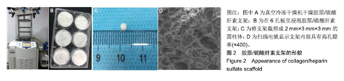
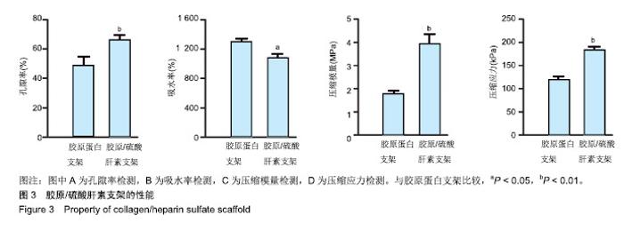
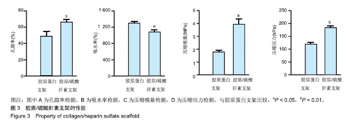
2.1 胶原/硫酸肝素的特性 胶原/硫酸肝素支架需要在真空冷冻干燥机中冷冻干燥成型,见图2A。将支架浸入1%NaOH溶液中浸泡12 h、去离子水中浸泡48 h,见图2B。然后裁剪成2 mm×3 mm×3 mm圆柱体,见图2C。扫描电镜显示胶原/硫酸肝素支架内部具有高孔隙率,见图2D,这符合神经网络,促进细胞存活,促进轴突再生。 胶原/硫酸肝素支架的孔隙率高于胶原蛋白支架(P < 0.05),见图3A,促进了营养液的扩散和组织形成。胶原/硫酸肝素支架的吸水率低于胶原蛋白支架(P < 0.05),见图3B。胶原/硫酸肝素支架的压缩模量高于胶原蛋白支架[(3.93±0.26),(1.77±0.24) MPa,P < 0.01],见图3C。胶原/硫酸肝素支架的压缩应力高于胶原蛋白支架[(184.95± 23.89),(118.47±12.68) kPa,P < 0.01],见图3D。"
"
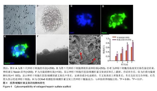
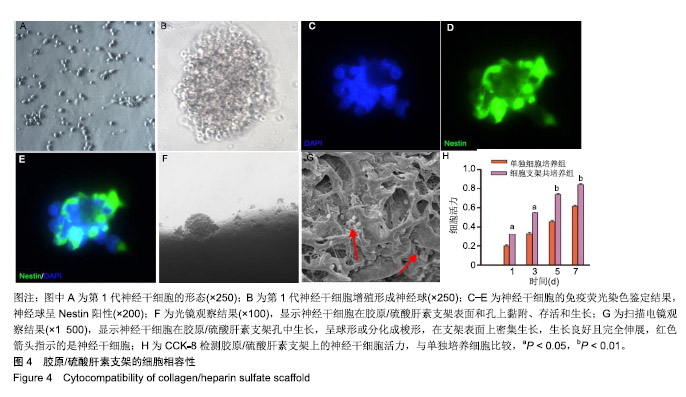
2.2 支架与神经干细胞的相容性 光学显微镜下分离的第1代神经干细胞增殖并形成漂浮球,见图4A,B。应用免疫荧光染色检测神经球呈Nestin阳性,见图4C-E。将神经干细胞种植到胶原/硫酸肝素支架上7 d后,光学显微镜下可见神经干细胞在胶原/硫酸肝素支架表面和孔上黏附、存活和生长,表明胶原/硫酸肝素具有良好的生物相容性,见图4F。扫描电镜显示许多神经干细胞在胶原/硫酸肝素支架孔中生长,细胞呈球形或分化成梭形,在胶原/硫酸肝素支架表面上密集生长,生长良好且完全伸展,见图4G。结果表明胶原/硫酸肝素支架可为神经干细胞提供微环境,促进细胞的黏附、伸长和分化,具有良好的细胞相容性。 2.3 共培养期间神经干细胞的存活率 随着共培养时间的延长,两组吸光度值均逐渐增加,共培养组各个时间点的吸光度值均高于单独细胞培养组(P < 0.05),见图4H。结果表明胶原硫酸肝素支架组没有细胞毒性,具有良好的细胞相容性。"
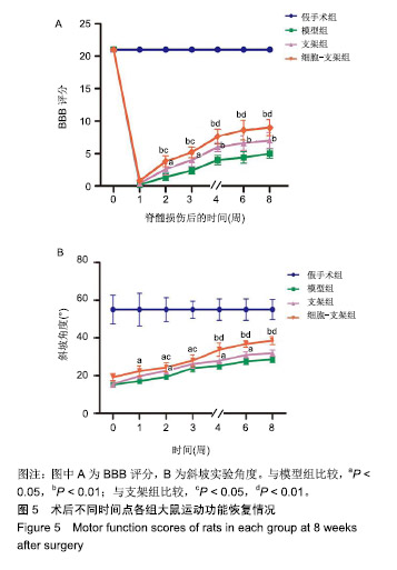
2.4 动物体内实验结果 2.4.1 实验动物数量分析 造模过程中无动物死亡,纳入的60只大鼠均进入结果分析。 2.4.2 行为学观察结果 模型组、支架组、细胞-支架组术后1周的BBB评分几乎为0,随着时间的推移后肢运动功能有所恢复,模型组的恢复速度明显慢于支架组、细胞-支架组,从术后2周开始,细胞-支架组的BBB评分明显高于模型组支架组(P < 0.05),见图5A。斜板实验中,模型组、支架组、细胞-支架组均可见后肢运动功能的恢复,细胞-支架组和支架组的角度变化趋势均优于模型组(P < 0.05),并且从术后2周开始,细胞-支架组斜坡角度明显大于支架组(P < 0.05),见图5B。"
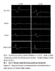
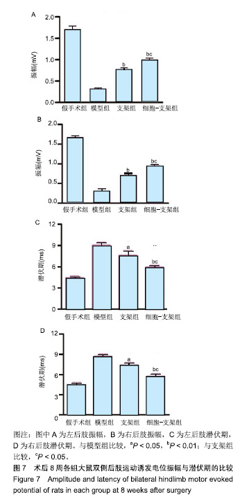
2.4.3 电生理检测结果 术后8周,代表性的运动诱发电位峰在模型组中几乎消失,细胞-支架组、支架组的运动诱发电位峰值或多或少地恢复,特别是在细胞-支架组恢复非常突出,见图6;①左侧后肢运动诱发电位:模型组、支架组、细胞-支架组的振幅分别为(0.30±0.04),(0.76±0.04),(0.97±0.07) mV,模型组的振幅低于支架组、细胞-支架组(P < 0.01),支架组低于细胞-支架组(P < 0.05);模型组、支架组、细胞-支架组的潜伏期分别为(9.02±0.44),(7.58±0.74),(5.96±0.29) ms,模型组的潜伏期长于支架组、细胞-支架组(P < 0.01),支架组的潜伏期长于细胞-支架组(P < 0.05);②右侧后肢的运动诱发电位:模型组、支架组、细胞-支架组的振幅分别为(0.30±0.06),(0.70±0.05),(0.90±0.07) mV,模型组的振幅低于支架组、细胞-支架组(P < 0.01),支架组低于细胞-支架组(P < 0.05);模型组、支架组、细胞-支架组的潜伏期分别为(8.70±0.35),(7.36±0.47),(5.74±0.43) ms,模型组的潜伏期长于支架组、细胞-支架组(P < 0.01),支架组的潜伏期长于细胞-支架组(P < 0.05),见图7。"

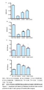
"
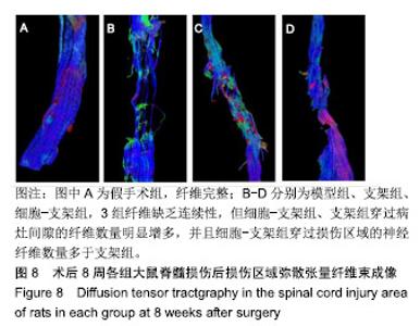
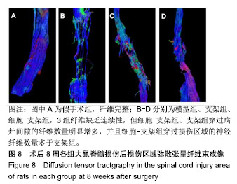
2.4.4 弥散张量成像结果 通过处理弥散张量成像数据得到弥散张量纤维束成像图像,可见假手术组纤维完整;脊髓横断后,模型组病变部位周围脊髓纤维缺乏不同方向的连续性;与模型组相比,细胞-支架组、支架组穿过病灶间隙的纤维数量明显增多,并且细胞-支架组穿过损伤区域的神经纤维数量多于支架组,见图8。弥散张量成像图像结果与运动功能结果一致。 "
| [1]Singh A,Tetreault L,Kalsiryan S,et al.Global prevalence and incidence of traumatic spinal cord injury. Clin Epidemiol.2014; 6:309-331.[2]Organization WH.International perspectives on spinal cord injury. Weed Res. 2013;11(4): 314-316.[3]Hagg T,Oudega M.Degenerative and spontaneous regenerative processes after spinal cord injury. J Neurotrauma. 2006;23(3-4):264-280.[4]Gentleman E,Lay AN,Dickerson DA,et al.Mechanical characterization of collagen fibers and scaffolds for tissue engineering. Biomaterials. 2003;24(21):3805-3813.[5]Yamane K,Mazaki T,Shiozaki Y,et al.Collagen-Binding Hepatocyte Growth Factor (HGF) alone or with a Gelatin- furfurylamine Hydrogel Enhances Functional Recovery in Mice after Spinal Cord Injury. Sci Rep.2018;8(1):917.[6]Shi Q,Gao W,Han X,et al.Collagen scaffolds modified with collagen-binding bFGF promotes the neural regeneration in a rat hemisected spinal cord injury model.Sci China Life Sci. 2014;57(2):232-240.[7]Chen X,Zhao Y,Li X,et al.Functional Multichannel Poly(Propylene Fumarate)-Collagen Scaffold with Collagen-Binding Neurotrophic Factor 3 Promotes Neural Regeneration After Transected Spinal Cord Injury.Adv Healthc Mater.2018;7(14):e1800315.[8]Iozzo RV.Matrix proteoglycans: from molecular design to cellular function.Annu Rev Biochem.1998.67(1):609-652.[9]Shrestha B,Coykendall K,Li Y,et al.Repair of injured spinal cord using biomaterial scaffolds and stem cells.Stem Cell Res Ther.2014;5(4):91.[10]Li G,Che MT,Zhang K,et al.Graft of the NT-3 persistent delivery gelatin sponge scaffold promotes axon regeneration, attenuates inflammation, and induces cell migration in rat and canine with spinal cord injury. Biomaterials.2016;83:233-248.[11]Rao JS,Zhao C,Zhang A,et al.NT3-chitosan enables de novo regeneration and functional recovery in monkeys after spinal cord injury.Proc Natl Acad Sci U S A. 2018;115(24): E5595-E5604.[12]Li G,Che MT,Zeng X,et al.Neurotrophin-3 released from implant of tissue-engineered fibroin scaffolds inhibits inflammation, enhances nerve fiber regeneration, and improves motor function in canine spinal cord injury.J Biomed Mater Res A.2018;106(8):2158-2170.[13]Yang Z,Zhang A,Duan H,et al.NT3-chitosan elicits robust endogenous neurogenesis to enable functional recovery after spinal cord injury.Proc Natl Acad Sci U S A. 2015;112(43): 13354-13359.[14]Liu S,Schackel T,Weidner N,et al.Biomaterial-Supported Cell Transplantation Treatments for Spinal Cord Injury: Challenges and Perspectives.Front Cell Neurosci.2017;11:430.[15]Ma YH,Zeng X,Qiu XC,et al.Perineurium-like sheath derived from long-term surviving mesenchymal stem cells confers nerve protection to the injured spinal cord.Biomaterials. 2018; 160:37.[16]Duan H,Li X,Wang C,et al.Functional hyaluronate collagen scaffolds induce NSCs differentiation into functional neurons in repairing the traumatic brain injury.Acta Biomater.2016;45: 182-195.[17]Lai BQ,Feng B,Che MT,et al.A Modular Assembly of Spinal Cord-Like Tissue Allows Targeted Tissue Repair in the Transected Spinal Cord.Adv Sci (Weinh).2018;5(9):1800261.[18]Zhang J,Lu X,Feng G,et al.Chitosan scaffolds induce human dental pulp stem cells to neural differentiation: potential roles for spinal cord injury therapy. Cell Tissue Res. 2016; 366(1): 129-142.[19]Kaneko A,Matsushita A,Sankai Y.A 3D nanofibrous hydrogel and collagen sponge scaffold promotes locomotor functional recovery, spinal repair, and neuronal regeneration after complete transection of the spinal cord in adult rats.Biomed Mater.2015;10(1):015008.[20]Zhang W,Yan Q,Zeng YS,et al.Implantation of adult bone marrow-derived mesenchymal stem cells transfected with the neurotrophin-3 gene and pretreated with retinoic acid in completely transected spinal cord. Brain Res. 2010;1359: 256-2571.[21]Wang JM, Zeng YS, Wu JL,et al.Cograft of neural stem cells and schwann cells overexpressing TrkC and neurotrophin-3 respectively after rat spinal cord transection. Biomaterials. 2011;32(30): 7454-7468.[22]Kelley BJ,Harel NY,Kim CY,et al.Diffusion tensor imaging as a predictor of locomotor function after experimental spinal cord injury and recovery.J Neurotrauma.2014;31(15):1362-1373.[23]Wu GH,Shi HJ,Che MT,et al.Recovery of paralyzed limb motor function in canine with complete spinal cord injury following implantation of MSC-derived neural network tissue. Biomaterials.2018;181:15-34.[24]Parker D.Functional changes after spinal lesions: implications for interventions.Neural Regen Res. 2018;13(5):811-812.[25]Wang Q,Zhang H,Xu H,et al.Novel multi-drug delivery hydrogel using scar-homing liposomes improves spinal cord injury repair. Theranostics.2018;8(16):4429-4446.[26]Madigan NN, McMahon S, O'Brien T, et al.Current tissue engineering and novel therapeutic approaches to axonal regeneration following spinal cord injury using polymer scaffolds.Respir Physiol Neurobiol. 2009;169(2):183-199.[27]Vigani B, Rossi S, Sandri G, et al.Design and criteria of electrospun fibrous scaffolds for the treatment of spinal cord injury.Neural Regen Res. 2017;12(11):1786-1790.[28]Ricard-Blum S,Beraud M,Raynal N,et al.Structural requirements for heparin/heparan sulfate binding to type V collagen.J Biol Chem. 2006;281(35):25195-25204.[29]Chen Y Scully M,Dawson G,et al.Perturbation of the heparin/heparin-sulfate interactome of human breast cancer cells modulates pro-tumourigenic effects associated with PI3K/Akt and MAPK/ERK signalling.Thromb Haemost.2013; 109(6):1148-1157.[30]Lu Q,Zhang S,Hu K,et al.Cytocompatibility and blood compatibility of multifunctional fibroin/collagen/heparin scaffolds. Biomaterials. 2007;28(14):2306-2313.[31]甘晓,南吴力.硫酸肝素/胶原蛋白神经组织工程支架修复周围神经损伤[J].中国组织工程研究,2016,20(25): 3744-3749.[32]张仁坤,涂悦,赵明亮,等.3-D打印胶原蛋白-硫酸肝素仿生脊髓支架的研究[J].中国修复重建外科杂志, 2015,29(8):1022-1027.[33]曹雄彬,戴军,宫丽,等.胶原蛋白-硫酸肝素生物支架在猪脑内的生物相容性分析[J].中国组织工程研究, 2015,19(21): 3361-3365.[34]Leong NL,Arshi A,Kabir N,et al.In vitro and in vivo evaluation of heparin mediated growth factor release from tissue- engineered constructs for anterior cruciate ligament reconstruction.J Orthop Res. 2015;33(2):229-236.[35]Ogle BM,Bursac N,Domian I,et al.Distilling complexity to advance cardiac tissue engineering. Sci Transl Med. 2016; 8(342):342ps13.[36]Jang SH, Lee J, Yeo SS.Central post-stroke pain due to injury of the spinothalamic tract in patients with cerebral infarction: a diffusion tensor tractography imaging study.Neural Regen Res. 2017;12(12):2021-2024. |
| [1] | Min Youjiang, Yao Haihua, Sun Jie, Zhou Xuan, Yu Hang, Sun Qianpu, Hong Ensi. Effect of “three-tong acupuncture” on brain function of patients with spinal cord injury based on magnetic resonance technology [J]. Chinese Journal of Tissue Engineering Research, 2021, 25(在线): 1-8. |
| [2] | Jiang Hongying, Zhu Liang, Yu Xi, Huang Jing, Xiang Xiaona, Lan Zhengyan, He Hongchen. Effect of platelet-rich plasma on pressure ulcers after spinal cord injury [J]. Chinese Journal of Tissue Engineering Research, 2021, 25(8): 1149-1153. |
| [3] | Shen Jinbo, Zhang Lin. Micro-injury of the Achilles tendon caused by acute exhaustive exercise in rats: ultrastructural changes and mechanism [J]. Chinese Journal of Tissue Engineering Research, 2021, 25(8): 1190-1195. |
| [4] | Wan Ran, Shi Xu, Liu Jingsong, Wang Yansong. Research progress in the treatment of spinal cord injury with mesenchymal stem cell secretome [J]. Chinese Journal of Tissue Engineering Research, 2021, 25(7): 1088-1095. |
| [5] | Zeng Yanhua, Hao Yanlei. In vitro culture and purification of Schwann cells: a systematic review [J]. Chinese Journal of Tissue Engineering Research, 2021, 25(7): 1135-1141. |
| [6] | Kong Desheng, He Jingjing, Feng Baofeng, Guo Ruiyun, Asiamah Ernest Amponsah, Lü Fei, Zhang Shuhan, Zhang Xiaolin, Ma Jun, Cui Huixian. Efficacy of mesenchymal stem cells in the spinal cord injury of large animal models: a meta-analysis [J]. Chinese Journal of Tissue Engineering Research, 2021, 25(7): 1142-1148. |
| [7] | Liang Xueqi, Guo Lijiao, Chen Hejie, Wu Jie, Sun Yaqi, Xing Zhikun, Zou Hailiang, Chen Xueling, Wu Xiangwei. Alveolar echinococcosis protoscolices inhibits the differentiation of bone marrow mesenchymal stem cells into fibroblasts [J]. Chinese Journal of Tissue Engineering Research, 2021, 25(7): 996-1001. |
| [8] | Duan Liyun, Cao Xiaocang. Human placenta mesenchymal stem cells-derived extracellular vesicles regulate collagen deposition in intestinal mucosa of mice with colitis [J]. Chinese Journal of Tissue Engineering Research, 2021, 25(7): 1026-1031. |
| [9] | Guan Qian, Luan Zuo, Ye Dou, Yang Yinxiang, Wang Zhaoyan, Wang Qian, Yao Ruiqin. Morphological changes in human oligodendrocyte progenitor cells during passage [J]. Chinese Journal of Tissue Engineering Research, 2021, 25(7): 1045-1049. |
| [10] | Ma Binxiang, He Wanqing, Zhou Guangchao, Guan Yonglin. Triptolide improves motor dysfunction in rats following spinal cord injury [J]. Chinese Journal of Tissue Engineering Research, 2021, 25(5): 701-706. |
| [11] | Liu Liu, Zhou Qingzhu, Gong Zhuo, Liu Boyan, Yang Bin, Zhao Xian. Characteristics and manufacturing techniques of collagen/inorganic materials for constructing tissue-engineered bone [J]. Chinese Journal of Tissue Engineering Research, 2021, 25(4): 607-613. |
| [12] | Li Jie, Ma Yuewen, Kang Nan, Zhang Jing, Zhang Yu. Radial extracorporeal shock wave therapy promotes the proliferation of neural stem cells in hippocampus of cerebral infarction rats and inhibits miR-124 expression [J]. Chinese Journal of Tissue Engineering Research, 2021, 25(31): 4981-4987. |
| [13] | Sun Jianwei, Yang Xinming, Zhang Ying. Effect of montelukast combined with bone marrow mesenchymal stem cell transplantation on spinal cord injury in rat models [J]. Chinese Journal of Tissue Engineering Research, 2021, 25(25): 3962-3969. |
| [14] | Xu Xiaoming, Chen Yan, Song Qian, Yuan Lu, Gu Jiaming, Zhang Lijuan, Geng Jie, Dong Jian. Human placenta derived mesenchymal stem cell gel promotes the healing of radiation skin damage in SD rats [J]. Chinese Journal of Tissue Engineering Research, 2021, 25(25): 3976-3980. |
| [15] | Xu Weilong, Zuo Yuan, Xin Daqi, He Chenyang, Zhao Peng, Shi Ming, Zhou Boyuan, Liu Yating, Zhao Yan. Selection of modeling methods for acute compressive spinal cord injury: a network Meta-analysis [J]. Chinese Journal of Tissue Engineering Research, 2021, 25(23): 3767-3772. |
| Viewed | ||||||
|
Full text |
|
|||||
|
Abstract |
|
|||||