Chinese Journal of Tissue Engineering Research ›› 2018, Vol. 22 ›› Issue (10): 1618-1624.doi: 10.3969/j.issn.2095-4344.0725
Previous Articles Next Articles
Biological scaffolds and mesenchymal stem cells in bone tissue engineering
- The First Affiliated Hospital of Harbin Medical University, Harbin 150001, Heilongjiang Province, China
-
Received:2017-11-30Online:2018-04-08Published:2018-04-08 -
Contact:Song Ke-guan, M.D., Chief physician, Master’s supervisor, the First Affiliated Hospital of Harbin Medical University, Harbin 150001, Heilongjiang Province, China -
About author:Liu Xiang-jie, Master candidate, the First Affiliated Hospital of Harbin Medical University, Harbin 150001, Heilongjiang Province, China
CLC Number:
Cite this article
Liu Xiang-jie, Song Ke-guan. Biological scaffolds and mesenchymal stem cells in bone tissue engineering[J]. Chinese Journal of Tissue Engineering Research, 2018, 22(10): 1618-1624.
share this article
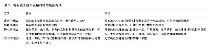
2.1 生物支架材料 生物支架材料目前主要有两种,分别是人工合成的支架材料和天然的支架材料[8]。 2.1.1 人工合成支架材料 人工合成有机支架材料:人工合成支架材料的种类复杂,其中大部分人工合成支架材料几乎是有机高分子化合物,它们具有共同的理化特性:机械性能好,拉伸强度强,生物相容性优良,生物降解性能强;同时也具有很大的缺点:亲水性能较差,吸附率较低,降解的产物酸性较大,炎症反应发生率较高[9]。人工合成支架材料目前大多数有聚乙烯醇、聚乳酸、聚羟基乙酸、聚乳酸羟基乙酸[10-12],其中聚乳酸、聚羟基乙酸受到越来越多的科研工作者的关注。聚乳酸具有明显的优越性能:一方面物理机械性能较强,另一方面加工技术较容易。更重要的是聚乳酸具有较强的降解性能[13]。有人应用聚乳酸修复兔全层软骨缺损,96%的兔关节缺损部位可观察到光滑、稳固的新生软骨,这个实验证明聚乳酸能促进软骨修复组织的再次生长。羟基乙酸可由聚羟基乙酸分解而产生,尽管聚羟基乙酸能很好地提高细胞黏附、增殖及分化,但是聚羟基乙酸也具有致命的缺陷,就是降解的速度太快,而且容易在体内大量的积聚,使细胞中毒而裂解,最严重的是使细胞死亡[14]。El Sayed等[15]将鼻中隔软骨细胞接种在聚羟基乙酸上,发现聚羟基乙酸具有修复软骨的功能,可作为组织工程的支架材料进行深入研究。聚乙烯醇主要用于治疗人工软骨假体修复、整形修复手术、创伤骨再生的治疗及制备缓释药物载体等。Kobayashi等[16]用聚乙烯醇进行重力压缩实验和应力力学检测实验,结果证明聚乙烯醇与人半月板在生物力学方面具有一致的黏弹性。实验结论证明聚乙烯醇与半月板组织工程化具有代偿半月板的功能,并且已在临床中得到了应用。Cohen等[17]将聚乳酸羟基乙酸与藻酸盐与软骨细胞复合,最终在兔体内植入此复合物,12周后发现兔体内有新生软骨生成。 人工合成无机支架材料:羟基磷灰石硬度大,骨传导性强,承重耐受力好,在自然界中可从骨无机盐中提取,与天然骨中的无机盐成分非常接近。羟基磷灰石可被复合到其他支架材料中,这样可使复合后的支架材料具有更加优越的性能,比如明显提高力学机械强度,但羟基磷灰石同样具有不可回避的缺陷,例如骨诱导活性较其他材料明显较低,脆性较大,塑形能力较弱,降解时间长等[18]。周立伟等[19]采用纳米羟基磷灰石/聚酰胺66复合人工骨修复兔颅骨缺损,结果表明支架材料具有良好的生物相容性,未对兔颅内代谢及功能产生负面影响,修复12周后,发现复合支架材料与颅骨之间的界线较模糊,再次证明此复合支架材料具有明显的骨诱导作用。段宏等[20]使用纳米羟基磷灰石/聚酰胺66复合人工骨支架材料修复45例肿瘤患者的胫骨、股骨、指骨、肱骨骨缺损,平愈合周期大约为2.8个月,实验结果证明纳米羟基磷灰石/聚酰胺66复合人工骨的生物相容性较为优越。Sotome等[21]将胶原、纳米羟基磷灰石与海藻酸钠复合在一起,将此复合支架材料植入到大鼠体内,5周后发现大鼠体内有新生骨组织生成。Bhumiratana等[22]将蚕丝蛋白复合到羟基磷灰石上制成一种新的支架模型,复合支架的硬度和机械性能显著提高,复合支架材料较复合前的支架材料具有更好的骨传导性,而且此复合支架模型中的羟基磷灰石对于新生骨形成具有很大的促进作用。有实验表明将羟基磷灰石与松质骨胶原复合也可获得良好的传导性和降解性、骨引导及骨诱导特性[23]。 2.1.2 天然生物支架材料 胶原:胶原在自然界中主要存在5种,它们分别是Ⅰ、Ⅱ、Ⅲ、Ⅳ和Ⅴ型,研究最多的是Ⅰ型胶原,其功能主要是连接组织和器官,保护机体等,由于胶原具有亲水特性,可促进细胞的黏附,并对细胞能够产生趋化作用,对细胞的生长能够起到很好的诱导作用。胶原构成了细胞外基质的主要骨架,胶原可与多种支架材料复合在一起,复合后的支架材料可用于软骨组织修复、细胞分化等[24]。 目前有研究证明Ⅰ、Ⅱ型胶原均可诱导软骨组织的生成,并且可促进软骨细胞增殖、分化,但Ⅰ型胶原可同时诱导软骨细胞的去分化[25]。目前医用胶原主要是从牛肌腱和牛皮中提取出来的。胶原具有很多优点,包括无毒性、良好的生物相容性等。但胶原也有很多致命缺点,比如缺乏一定的机械强度。在细胞培养过程中,胶原与其他材料相比也具有很大优点,比如可促进细胞的生长、分化等,因此被广泛研究,其中在医用材料中颇受青睐。胶原在治疗急性骨折、创伤骨缺损及软骨组织工程等方面也得到广泛应用[26]。目前在市场上销售的胶原膜主要有Bio-Gide® Alloderm®BioMendCytoplast® 等[27]。商品胶原膜在术后18周内可完全被吸收。实验结果表明胶原对成纤维细胞的诱导分化作用更强[28]。 壳聚糖:壳聚糖已被广泛应用于医学领域、水处理、金属提取及回收,壳聚糖具有良好的生物相容性,微生物降解性能好,毒性弱,具有一定的免疫调节活性,降解产物毒性较低,无免疫原性,无致癌性[29]。壳聚糖具有高化学反应活性,容易被一些化学试剂修饰,可用于生长因子传递的载体[30]。但壳聚糖也存在致命的缺点,比如机械性能较差,缺乏一定的骨传导性,但可复合到其他仿生材料中,如金属材料、生物陶瓷材料,这样二者优势互补,可在骨组织工程中发挥更大的作用[31]。壳聚糖为带阳离子的高分子碱性化合物,而细胞膜往往带负电荷,这样使得二者之间能够产生静电引力,有利于细胞黏附在壳聚糖表面[32]。但Montembaul等[33]认为壳聚糖是由细胞外基质分泌的诱导剂,使细胞容易黏附在壳聚糖表面,但壳聚糖本身却不起支架作用。Fakhry 等[34]通过实验发现成骨细胞在壳聚糖的黏附和播散更有优势。有实验将骨髓间充质干细胞黏附到壳聚糖表面,结果证明可诱导骨组织细胞的修复。并且被现在越来越多地用于非病毒基因载体[35-37]。Cruz等[38]将骨髓基质干细胞复合到壳聚糖支架上,结果发现细胞可均匀分布在微孔道内,并且在4周内一直保持增殖,骨髓基质干细胞可向破骨细胞及成骨细胞分化。由于壳聚糖的溶解速率较缓慢,因此可把壳聚糖制作为药物缓释载体,可显著减少患者的给药频率[39]。 明胶:明胶是由胶原水解而成,具有加工容易、价格便宜、免疫原性低等优点[40]。但明胶也有很多缺点,例如不能应用于支架材料,主要是因为明胶生物相容性较差,降解速率较快等,但可将明胶与其他支架材料复合在一起,有研究证明将明胶与聚磷酸钙纤维复合在一起,可制备出明胶/聚磷酸钙纤维复合支架材料,明胶/聚磷酸钙纤维复合支架材料的降解率随时间延长逐渐降低,2周后逐渐趋于平缓[41]。明胶亲水性能较好,属于两性电解质,已被广泛用于各种研究[42]。 丝素蛋白:丝素蛋白主要由蚕丝组成,蚕丝在力学方面表现优势明显,这可能是因为蚕丝在某些方面具有丝素蛋白的分级结构。目前为止,丝素蛋白由于有众多优点,已被广泛制成各种产品,例如三维支架、无纺网等[43]。纯丝素蛋白的抗拉强度较大,为610-690 MPa,抗拉伸强度系数为15-17 GPa,断裂伸长率为4%- 16%。自然界中的丝素蛋白具有以下特征:良好的机械性能和理化特性,如透气透湿性和缓释性等;无毒性;在外科领域的应用具备很长的历史;在体内外降解率可被控制;有独特的力学性能[44]。自然界中的丝素蛋白主要由氨基酸组成,大约有18种氨基酸,其中甘氨酸、丝氨酸、丙氨酸和色氨酸的含量最多,其中由一条重链(325 ku)和一条轻链(25 ku)组成,二者之间通过S-S键结合组成。丝素蛋白作为天然生物高分子材料,由于自身具有很大优点,受到众多材料专家的广泛关注,目前被广泛应用于人工神经、皮肤、骨骼、血管、肌键、韧带和角膜等领域。有些丝素蛋白本身不含丝胶,这样的丝素蛋白有很多优点,例如无免疫原性。无论是丝素蛋白还是丝胶蛋白,在机械性能上和拉伸力度上明显优于其他生物支架材料。但丝素蛋白本身有很多不同的形式,不同的形式在力学性能方面却相差不大,在力学方面具有明显优势的是以纤维模行式存在的丝素蛋白。 2.2 支架材料的制备方法 支架材料的制备方法主要有冷冻干燥法、熔融成型法、静电纺丝法、3D打印技术(表1)[45]。冷冻干燥法是指将含有水的化合物冻结到零度以下,使化合物冻结成冰,然后再将冰转变为蒸气的干燥方法。该制备方法的优点是支架具有较高的孔隙率,操作简单、方便,缺点是需要加入一定的交联剂才能固定高分子网络结构,因此可能导致溶剂残留[46]。熔融成型法是将聚合物粉末与可沥混合均匀,倒入模具中加热到一定的温度,成形后在水中浸泡,除去可沥去相,得到二维空间支架。该方法优点是孔隙高度规则,缺点是需对材料进行加热,可能引起聚合物性质的改变[47]。静电纺丝技术是将聚合物带上高压静电,当外界电场力足够大时,聚合物液滴可克服表面张力形成喷射细流,沉积于基布上形成纳米纤维膜。静电纺丝法优点是操作简单、成本低,制备的支架材料具有大的比表面积,可促进细胞的增殖与黏附,为细胞提供适宜的生长空间,但同样具有很多缺点,比如得到的纳米纤维彼此粘连在一起,不易分离,效率低下,纳米纤维材料抗拉伸强度低,支架结构稳定性不好,强度、刚度较低,易导致种植细胞损伤[48]。3D打印技术可通过计算机、CT等仪器精确扫描计算支架材料的各种比例需求,实现支架个体 化[49]。3D打印技术可解决传统支架制备方法存在的一系列问题[50]。但3D打印技术对确定多种复合材料的打印参数还存在一定的不足[51]。 "
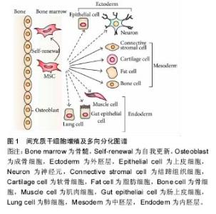
2.3 间充质干细胞 2.3.1 间充质干细胞的增殖与分化 骨组织工程中,种子细胞的选择面临一个重大的难题,早期成骨细胞是大多数研究的焦点,但要想获取一定量的成骨细胞非常困难,并且成骨细胞在体外的扩增能力有限。随着研究的不断深入,间充质干细胞成为研究的热点,因为间充质干细胞具有很强的自我复制及多向分化能力,因此被认为是相对较为理想的种子细胞[52]。有实验证明不同种类小鼠的骨髓往往具有不同的分化能力、增殖能力。有实验表明胎儿骨髓间充质干细胞比成人骨髓间充质干细胞更具有优势,胎儿骨髓比成人有更强的增殖和分化潜能;但在造血功能及恢复方面,成人比胎儿强。2003年Romanov等[53]从人脐血中分离出间充质干细胞,被命名为人脐血间充干细胞,实验证明脐血间充质干细胞在细胞增殖能力及成骨分化潜力方面都明显较骨髓间充质干细胞强,而且脐血间充干细胞具有培养简单、来源广泛等优点[54]。目前有研究者应用胰蛋白酶、胶原酶来分离基质细胞,但这些酶解方法都不太理想。胶原酶法的主要缺点是使组织过度消化,导致细胞活力降低,降解细胞表面的受体,降低了细胞的功能[55],但优点是可在短时间内获得大量贴壁生长的间充质干细胞,这样显著提高了原代培养的效率。也可通过组织培养方法获得,但存在原代细胞培养周期较长的缺点,优点是操作简单、容易获得、经济方便。在细胞培养中,细胞球培养是间充质干细胞体外分化研究最常用的培养方式之一。细胞球培养可使细胞充分互相接触,构建完美的三维环境,这样有利于细胞基质之间的连接、空间结构的形成、细胞间信号的传递[56]。目前骨髓间充质干细胞在软骨损伤修复及再生方面具有很大优势[57]。近年来脂肪干细胞的研究得到了广泛关注,因为脂肪干细胞来源较为广泛,创伤较小,再生能力较强,这样可有效解决间充质干细胞取材问题[58]。目前关于间充质干细胞-3D支架研究大多数还停留在动物实验阶段,临床治疗效果也没有取得突破性进展。尽管目前在骨不连的治疗中取得了令人满意的疗效,但还是以传统的切开清创、取自体骨植骨效果最佳,在植骨的同时可复合间充质干细胞移植来加快骨折的愈合过程。随着骨组织工程的发展,在不久的未来,间充质干细胞的一些难题都将可能得到解决,到时候,移植间充质干细胞来治疗骨不连必将得到广泛的临床应用。间充质干细胞的多向分化图谱见图1[59]。 与间充质干细胞具有相似功能的是内皮祖细胞,内皮祖细胞是由Asahara于1997年首次发现,其可分化为血管内皮细胞,在特定微环境下可促进间充质干细胞成骨[60]。内皮祖细胞不仅可分化成血管内皮细胞,而且还能分化为成骨细胞。当机体受到外伤缺血时,内皮祖细胞可迅速进入缺血部位,促进新生血管形成,诱导成骨细胞快速增殖。当机体发生骨折破坏骨组织血供及骨的完整性时,骨折部位可释放炎性因子等炎症递质,炎性细胞因子动员内皮祖细胞向外周循环迁移,到达缺血部位,促进骨折部位血供和骨折愈合[61-62]。内皮祖细胞容易从机体中获得和分离,并且对机体创伤小,而骨髓间充质干细胞只能在麻醉下通过手术从骨髓中提取,但内皮祖细胞对骨折的治疗目前仍处于临床前研究阶段,临床应用报道较少[63]。 2.3.2 间充质干细胞结合生物支架材料复合物研究 随着生物支架材料的不断发展,间充质干细胞结合生物支架材料的研究也越来越受到广大科研工作者的青睐,将间充质干细胞结合生物支架材料上可以形成一个细胞-材料移植复合物,形成的移植复合物可替代自体骨来治疗临床中的骨缺损,这种方法具有很大的优势,与自体骨移植相比,最重要的优势就是减少患者二次受伤,且适合于大面积骨缺损,这样给患者自身的损伤降到最低。随着骨组织工程的高速发展,将间充质干细胞与生物支架材料复合,优势就在于可根据骨缺损的形态任意改变形状来适应骨缺损的填充,促进骨缺损的再生与修复,但不足之处在于机械硬度达不到自体骨的程度,降解时间较长[64]。有研究将人脐带血间充质干细胞和人骨髓间充质干细胞分别种植在磷酸钙石灰泥上,再将人脐带血间充质干细胞-磷酸钙石灰泥复合物和人骨髓间充质干细胞-磷酸钙石灰泥复合物植入到骨缺损动物模型中,空白组为单一的磷酸钙石灰泥,在第12,24周末分别采用影像学和组织学观察骨缺损的修复再生程度,结果发现与单纯的磷酸钙石灰泥相比,实验组新生骨组织和细胞及再生血管均明显高于空白组[65]。骨组织工程目前在临床没有得到广泛应用,最主要的原因为组织工程骨的血管化程度低。血管化与骨缺损的修复有着密切关系。血供是影响骨缺损修复的一个重要因素,如果植入复合支架材料附近的血供不足,将会直接影响骨缺损的修复。在骨缺损修复的骨组织工程中,最常使用的种子细胞通常包括间充质干细胞、内皮细胞、内皮祖细胞,其中血管化的关键步骤是内皮细胞的增殖与分化。目前内皮祖细胞成为复合支架材料移植中研究的热点之一,其主要原因在于内皮祖细胞可分化成为内皮细胞,促进血管的形成[66]。 2.3.3 间充质干细胞结合细胞因子研究 间充质干细胞常被用来充当骨组织工程中的种子细胞,不但具有干细胞的功能,而且还利于外源性基因的导入和表达。目前大多数的研究模式是将体外培养的间充质干细胞导入到目的基因,然后再移植到缺损的动物模型当中,促进机体组织损伤的修复与再生。这种方法的优点在于将细胞与基因紧密结合在一起,为临床中骨缺损治疗提供了一个新的策略。间充质干细胞与细胞因子是骨组织工程中不可或缺的2个重要因素,二者密不可分,但二者之间的作用机制还有待于研究。目前为止,比较明朗的作用机制是细胞因子可通过促进和调节间充质干细胞的增殖、分化和趋化过程来提高间充质干细胞的诱导能力。目前研究较多的细胞因子有骨形态发生蛋白2、血管内皮生长因子、粒细胞集落刺激因子、成纤维细胞生长因子、胰岛素样生长因子、转化生长因子β1。骨形态发生蛋白2是转化生长因子β超家族一员,是此家族中骨诱导作用最强的[67]。骨形态发生蛋白最早是由Urist发现的,Uris从皮质骨中提取物中获得了骨形态发生蛋白。到目前为止,在 转化生长因子β超家族中共有43个成员,骨形态发生蛋白能够诱导间充质干细胞分化为骨、软骨、肌腱、韧带、神经组织。骨形态发生蛋白2在骨母细胞向矿化细胞的转化过程中发挥着重要调节作 用[68]。血管内皮生长因子主要在血管生长调节方面发挥着重要作用,其应用最多的是诱导血管再生。血管内皮生长因子能够诱导内皮细胞迁移、增殖、分化,并且可构建新生血管,还能活化血管内皮细胞,并增加其功能。血管内皮生长因子主要在血管再生与血循环中起着不可替代的作用[69]。但骨形态发生蛋白2和血管内皮生长因子在骨修复与血管再生的不同阶段常发挥不同的作用,并且大量国内外文献报道联合多种因子的同步应用可显著提高二者对骨组织再生与修复的能力,因此可将二者联合在一起共同作用于间充质干细胞,使其共同促进成骨再生与修复及血管化。粒细胞集落刺激因子的主要来源是血管内皮细胞、成纤维细胞、血小板、间皮细胞。粒细胞集落刺激因子是一种糖蛋白,相对分子质量约为20 000,大约有174个氨基酸组成,其受体包含813个氨基酸残基。粒细胞集落刺激因子可促进间充质干细胞的增殖与分化[70-71]。成纤维细胞生长因子对血管内皮细胞具有很好的保护作用,并且可与血管内皮生长因子共同作用,二者联合在一起可发挥放大效应。成纤维细胞生长因子还可与间充质干细胞联合在一起,形成移植复合物,在临床中治疗骨缺损,而且该法明显优于单独使用骨髓间充质干细胞移植[72]。 "

| [1]Calori GM,Mazza E,Colombo M,et al.The use of bone-graft substitutes in large bone defects: any specific needs.Injury. 2011; 42(S2):S56-63.[2]Yang Y,Niu X,Zhang Q,et al.A comparative study of calcium sulfate artificial bone graft versus allograft in the reconstruction of bone defect after tumor curettage.Chin Med J (Engl).2014;127(17):3092-3097.[3]Wang L,Ma XY,Zhang Y,et al.Repair of segmental bone defect using Totally Vitalized tissue engineered bone graft by a combined perfusion seeding and culture system.PLoS One.2014;9(4):e94276[4]Hogan MV,Bagayoko N,James R,et al.Tissue engineering solutions for tendon repair.J Am Acad Orthop Surg. 2011;19(3):134.[5]Oryan A,Alidadi S,Moshiri A,et al.Bone regenerative medicine: classic options, novel strategies, and future directions.J Orthop Surg Res. 2014;9(1):18. [6]Sathy BN, Watson BM, Kinard LA, et al.Bone Tissue Engineering with Multilayered Scaffolds-Part II: Combining Vascularization with Bone Formation in Critical-Sized Bone Defect.Tissue Eng Part A. 2015;21 (19-20):2495-2503. [7]Cunniffe GM,Vinardell T,Murphy JM,et al.Porous decellularized tissue engineered hypertrophic cartilage as a scaffold for large bone defect healing.Acta Biomater. 2015;23:82-90.[8]Kosuge D,Khan WS,Haddad B,et al.Biomaterials and scaffolds in bone and musculoskeletal engineering.Curr Stem Cell Res Ther. 2013;8(3): 185-191.[9]Liao J,Shi K,Ding Q,et al.Recent developments in scaffold-guided cartilage tissue regeneration.J Biomed Nanotechnol. 2014;10(10): 3085-3104. [10]Sabrina M,Simona S,Jani H,et al.Osteogenic and osteoclastogenic differentiation of co-cultured cells in polylactic acid– nanohydroxyapatite fiber scaffolds.J Biotechnol.2015;204(6):53-62. [11]Boontharika C,Thanaphum O,Nunthawan N,et al.The efficacy of polycaprolactone/hydroxyapatite scaffold in combination with mesenchymal stem cells for bone tissue engineering.J Biomed Mater Res A.2016;104(1):264-271. [12]Tang CM,Tian YH,Shan HH.Poly (vinyl alcohol) nanocomposites reinforced with bamboo charcoal nanoparticles: mineralization behavior and characterization.Materials.2015;8(7):4895-4911. [13]Zhou R,Xu W,Chen F,et al.Amorphous calcium phosphate nanospheres/polylactide composite coated tantalum scaffold: facile preparation, fast biomineralization and subchondral bone defect repair application.Colloids Surf B Biointerfaces.2014;123:236-245.[14]石宗利,杜心康,王彦平.聚乳酸钙/左旋聚乳酸软骨组织工程支架复合材料的分析[J].中国临床床康复,2004,8(17):3373-3375[15]El Sayed K,Marzahn U,John T,et al.PGA-associated heterotopic chondrocyte cocultures: implications of nasoseptal and auricular chondrocytes in articular cartilage repair.J Tissue Eng Regen Med. 2013;7(1):61-72.[16]Kobayashi M,Toguchida J,Oka M.Preliminary study of polyvinyl alcohol-hydrogel(PVA-H) artificial meniscus.Biomaterials. 2003; 24(4):639-647.[17]Cohen SB,Meirisch CM,Wilson HA,et al.The use of absorbable copolymer pads with alginate and cells for articular cartilage repair in rabbits.Biomaterials. 2003;24(15):2653-2660.[18]Hu JX,Zhu YJ,Tong H,et al.A detailed study of homogeneous agarose/hydroxyapatite nanocomposites for load-bearing bone tissue. Int J Biol Macromol.2016;82(1):134-143.[19]周立伟,魏世成,李玉宝,等.纳米羟基磷灰石/聚酰胺66复合人工骨修复颅骨缺损的动物实验研究[J].口腔医学, 2009,29(11):561-563.[20]段宏,张开伟,闵理,等.纳米羟基磷灰石聚酰胺66骨填充材料修复肢体良性肿瘤术后骨缺损的疗效分析[J].中国骨与关节外科, 2009,2(5): 341-346.[21]Sotome S,Uemure T,Kjkuchi M,et al.Synthesis and in vivo evaluation of a novel hydroxyapatite/collagen as a bone filler and a drug delivery carrier of bone morphogenetic protein. Materials Science and Engineering C. 2004;24(18):341-349.[22]Bhumiratana S,Grayson WL,Castaneda A,et al.Nucleation and growth of mineralized bone matrix on silk-hydroxyapatite composite scaffolds. Biomaterials.2011;32(11):2812-2820. [23]Arahira T,Todo M.Variation of mechanical behavior of beta-TCP/ collagen two phase composite scaffold with mesenchymal stem cell in vitro.J Mech Behav Biomed Mater.2016;61:464-474.[24]Levingstone TJ,Thompson E,Matsiko A,et al.Multi-layered collagen-based scaffolds for osteochondral defect repair in Rabbits. Acta Biomaterialia.2016;32(3):149-160.[25]Zhang L,Yuan T,Guo L,et al.An in vitro study of collagen hydrogel to induce the chondrogenic differentiation of mesenchymal stem cells.J Biomed Mater Res A. 2012;100(10):2717-2725.[26]Lee CH,Singla A,Lee Y.Biomedical applications of collagen.Int J Pharm, 2001;221(1-2):1-22.[27]Bottino MC,Thomas V,Schmidt G,et al.Recent advances in the development of GTR/GBR membranes for periodontal regenera-tion—a materials perspective.Dent Mater. 2012;28(7):703-721.[28]Ghanaati S.Non-cross-linked porcine-based collagen I–III mem-branes do not require high vascularization rates for their integra-tion within the implantation bed: A paradigm shift.Acta Biomaterialia.2012;8(8):3061-3072.[29]Ray SD.Potential aspects of chitosan as pharmaceutical excipient.Acta Pol Pharm.2011;68(5):619-622.[30]Rami L,Malaise S,Delmond S,et al.Physicochemical modulation of chitosan-based hydrogels induces different biological responses: interest for tissue engineering.J Biomed Mater Res A. 2014;102(10): 3666-3676[31]Costa-Pinto AR,Reis RL,Neves NM.Scaffolds based bone tissue engineering: the role of chitosan. Tissue Eng Part B Rev. 2011;17(5): 331-347.[32]Kumar MN,Muzzarelli RA,Muzzarelli C,et al. Chitosan chemistry and pharmaceutical perspectives. Chem Rev.2004;104(12):6017-6084.[33]Montembault A,Tahiri K,Korwin-Zmijowska C,et al.A material decoy of biological media based on chitosan physical hydrogels: application to cartilage tissue engineering.Biochimie.2006;88(5): 551-564.[34]Fakhry A,Schneider GB,Zaharias R,et al.Chitosan supports the initial attachment and spreading of osteoblasts preferentially over fibroblasts. Biomaterials. 2004;25(11):2075-2079.[35]Hirschmann MT,Keller L,Hirschmann A,et al.One-year clinical and MR imaging outcome after partial meniscal replacement in stabilized knees using a collagen meniscus implant.Knee Surg Sports Traumatol Arthrosc.2013;21(3):740-747.[36]Qi BW,Yu AX,Zhu SB,et al.Chitosan/poly (vinyl alcohol) hydrogel combined with Ad-hTGF-β1transfected mesenchymal stem cells to re-pair rabbit articular cartilage defects.Exp Biol Med (Maywood). 2013;238(1):23-30.[37]Eroglu E,Tiwari PM,Waffo AB,et al.A nonviral pHEMA+chitosan nanosphere-mediated high-efficiency gene delivery system.Int J Nano-medicine.2013;8:1403-1415.[38]Cruz DM,Gomes M,Reis RL,et al.Differentiation of mesenchymal stem cells in chitosan scaffolds with double micro and macroporosity.J Biomed Mater Res A. 2010;95(4):1182-1193.[39]李瑞欣,马小军,王卫明,等.复合万古霉素海藻酸钠/壳聚糖缓释载体的体内释放实验[J].中国组织工程研究与临床康复, 2009,13(38):7535-7538.[40]Lin WH,Yu JS,Chen GP,et al.Fabrication of multi-biofunctional gelatin-based electrospun fibrous scaffolds for enhancement of osteogenesis of mesenchymal stem cells. Colloids Surf B Biointerfaces. 2016;138(2):26-31. [41]朱凌云,王彦平,石宗利,等.构建球磨碳酸钙/聚磷酸钙纤维/聚乳酸组织工程支架复合材料[J].中国组织工程研究与临床康复, 2009,13(47):9265-9268.[42]Draghi L,Resta S,Pirozzolo MG,et al.Microspheres leaching for scaffold porosity control.Mater Sci Mater Med. 2005;16(12):1093-1097.[43]Lin JH,Chen CK,Wen SP,et al.Poly-L-lactide/sodium alginate/chitosan microsphere hybrid scaffolds made with braiding manufacture and adhesion technique: Solution to the incongruence between porosity and compressive strength.Mater Sci Eng C Mater Biol Appl. 2015;52: 111-120. [44]Lai GJ,Shalumon KT,Chen SH,et al.Composite chitosan/silk fibroin nanofibers for modulation of osteogenic differentiation and proliferation of human mesenchymal stem cells.Carbohydr Polym.2014;111:288-297.[45]Tang D,Tare RS,Yang LY,et al.Biofabrication of bone tissue: approaches, challenges and translation for bone regeneration. Biomaterials.2016;83(3):363-382.[46]Mohammad A,Abbas T,Ghasem M.Preparation, characterization and biocompatible properties of b-chitin/silk fibroin/ nanohydroxyapatite composite scaffolds prepared using a freeze-drying method.RSC Adv.2016;6(3):7048-7060.[47]Ze inI,Hu tmacher DW,Tan KC,etal.Fusedde position Modeling of novel scaffol darchitectu rest or tissue engineering application.Biomaterials. 2002;23(4):1169-1185.[48]Chakraborty S,Liao IA.Electrohydrodynamics: A facile technique to fabricate drug delivery systems.Adv Drug Deliv Rev. 2009;61(12): 1043-1054.[49]LDu MC,Chen B,Meng QY,et al.3D bioprinting of BMSC-laden methacrylamide gelatin scaffolds with CBD-BMP2-collagen microfibers.Biofabrication.2015;7(4):1-10.[50]Liu FH.Fabrication of bioceramic bone scaffolds for tissue engineering.J Mater Eng Perform.2014;23(10):3762-3769.[51]Yoon YJ,Moon SK,Hwang JH.3D printing as an ei cient way for comparative study of biomimetic structures-trabecular bone and honeycomb.J Mech Sci Technol. 2014;28(11):4635-4640.[52]Liu Y,Shu XZ,Prestwich GD.Osteochondral defect repair with autologous bone marrow-derived mesenchymal stem cells in an injectable, in situ, cross-linked synthetic extracellular matrix.Tissue Eng.2006;12(12):3405-3416.[53]Romanov YA,Svintsitskaya VA,Smirnov VN.Searching for alternativesources of postnatal human mesenchymal stem cells: candidate MSC-like cells from umbilical cord.Stem Cells.2003;21(1): 105-108.[54]Baksh D,Yao R,Tuan RS.Comparison of proliferative and multilineage differentiation potential of human mesenchymal stem cells derived from umbilical cord and bone marrow.Stem Cells.2007; 25(6): 1384-1392.[55]Hung CT, Mauck RL. Biological assays: cellular level.In: Biomedical Technology and Devices Handbook. Moore JE,Zouridakis G,eds.CRS Press,London.2004:151-153.[56]Musumeci G,Mobasheri A,Trovato FM,et al. Biosynthesis of colla-genⅠ,Ⅱ,RUNX2 and lubricin at different time points of chondro-genic differentiation in a 3D in vitro model of human mesenchymal stem cells derived from adipose tissue.Acta Histochem.2014;116(8):1407-1417.[57]Mendez-Ferrer S,Scadden DT,Sanchez-Aguilera A.Bone marrow stem cells:current and emerging concepts.Ann N Y Acad Sci. 2015;1335: 32-44.[58]Jiang H,Zhang J,Zhang Z,et al.Effect of transplanted adipose de-rived stem cells in mice exhibiting idiopathic pulmonary fibrosis.Mol Med Rep.2015;12(4):5933-5938.[59]Yousefi AM,James PF,Akbarzadeh R,et al.Prospect of Stem Cells in Bone Tissue Engineering: A Review.Stem Cells Int. 2016;2016(10): 6180487.[60]Rioja AY,Tiruvannamalai Annamalai R,Paris S,et al. Endothelial sprouting and network formation in collagen- and fibrin-based modular microbeads.Acta Biomater.2016;29:33-41.[61]Zigdon-Giladi H,Rudich U,Michaeli-Geller G,et al.Recentadvances in bone regeneration using adult stem cells.World J Stem Cells. 2015; 7(3):630-640.[62]Ayata RE,Chabaud S,Auger M,et al.Behaviour of endothelial cells in a tridimensional in vitro environment.Biomed Res Int.2015;2015:630461.[63]63.Knight MN,Hankenson KD.Mesenchymal stem cells in bone regeneration.Adv Wound Care (New Rochelle). 2013;2(6):306-316.[64]Lin WH,Yu JS,Chen GP,et al.Fabrication of multi-biofunctional gelatin-based electrospun fibrous scaffolds for enhancement of osteogenesis of mesenchymal stem cells.Colloids Surf B Biointerfaces. 2016;138(2):26-31.[65]Chen W,Liu J,Manuchehrabadi N,et al.Umbilical cord and bone marrow mesenchymal stem cell seeding on macroporous calcium phosphate for bone regeneration in rat cranial defects.Biomaterials. 2013; 34(38):9917-9925.[66].Xing W,Mu D,Wang Q,et al.Improvement of fat graft survival with autologous bone marrow aspirate and bone marrow concentrate: A one-step method.Plast Reconstr Surg. 2016;137(4):676-668.[67]Li L,Zhou G,Wang Y1,et al.Controlled dual delivery of BMP-2 and dexamethasone by nanoparticle-embedded electrospun nanofibers for the efficient repair of critical-sized rat calvarial defect.Biomaterials. 2015;37:218-229.[68]Sun J,Li J,Li C,et al.Role of bone morphogenetic protein- 2 in osteogenic differentiation of mesenchymal stem cells.Mol Med Rep. 2015;12(3):4230-4237.[69]Wei X,Mao Z,Hou Y,et al.Local administration of TGF-1 /VEGF165 gene-transduced bone mesenchymal stem cells for Achillesallograft replacement of the anterior cruciate ligament in rabbits.Biochem Biophys Res Commun. 2011;406(2):204-210.[70]Liu J,Walker NM,Cook MT,et al.Function Cftr in crypt epitheliumof organotypic enteroid cultures from murine small intestine.Am J Physiol Cell Physiol. 2012;302(10):C1492-C1503.[71]Shyu WC,Lin SZ,Yang HI,et al.Functional recovery of stroke ratsinduced by granulocyte colony-stimulating factor-stimulated stem cells.Circulation. 2004;110(13):1847-1854.[72]Wang JL,Sun JF,Tang YX,et al.Basic fibroblast growth factor attenuates the degeneration of injured spinal cord motor endplates. Neural Regen Res.2013;8(24):2213-2224.[1]Calori GM,Mazza E,Colombo M,et al.The use of bone-graft substitutes in large bone defects: any specific needs.Injury. 2011; 42(S2):S56-63.[2]Yang Y,Niu X,Zhang Q,et al.A comparative study of calcium sulfate artificial bone graft versus allograft in the reconstruction of bone defect after tumor curettage.Chin Med J (Engl).2014;127(17):3092-3097.[3]Wang L,Ma XY,Zhang Y,et al.Repair of segmental bone defect using Totally Vitalized tissue engineered bone graft by a combined perfusion seeding and culture system.PLoS One.2014;9(4):e94276[4]Hogan MV,Bagayoko N,James R,et al.Tissue engineering solutions for tendon repair.J Am Acad Orthop Surg. 2011;19(3):134.[5]Oryan A,Alidadi S,Moshiri A,et al.Bone regenerative medicine: classic options, novel strategies, and future directions.J Orthop Surg Res. 2014;9(1):18. [6]Sathy BN, Watson BM, Kinard LA, et al.Bone Tissue Engineering with Multilayered Scaffolds-Part II: Combining Vascularization with Bone Formation in Critical-Sized Bone Defect.Tissue Eng Part A. 2015;21 (19-20):2495-2503. [7]Cunniffe GM,Vinardell T,Murphy JM,et al.Porous decellularized tissue engineered hypertrophic cartilage as a scaffold for large bone defect healing.Acta Biomater. 2015;23:82-90.[8]Kosuge D,Khan WS,Haddad B,et al.Biomaterials and scaffolds in bone and musculoskeletal engineering.Curr Stem Cell Res Ther. 2013;8(3): 185-191.[9]Liao J,Shi K,Ding Q,et al.Recent developments in scaffold-guided cartilage tissue regeneration.J Biomed Nanotechnol. 2014;10(10): 3085-3104. [10]Sabrina M,Simona S,Jani H,et al.Osteogenic and osteoclastogenic differentiation of co-cultured cells in polylactic acid– nanohydroxyapatite fiber scaffolds.J Biotechnol.2015;204(6):53-62. [11]Boontharika C,Thanaphum O,Nunthawan N,et al.The efficacy of polycaprolactone/hydroxyapatite scaffold in combination with mesenchymal stem cells for bone tissue engineering.J Biomed Mater Res A.2016;104(1):264-271. [12]Tang CM,Tian YH,Shan HH.Poly (vinyl alcohol) nanocomposites reinforced with bamboo charcoal nanoparticles: mineralization behavior and characterization.Materials.2015;8(7):4895-4911. [13]Zhou R,Xu W,Chen F,et al.Amorphous calcium phosphate nanospheres/polylactide composite coated tantalum scaffold: facile preparation, fast biomineralization and subchondral bone defect repair application.Colloids Surf B Biointerfaces.2014;123:236-245.[14]石宗利,杜心康,王彦平.聚乳酸钙/左旋聚乳酸软骨组织工程支架复合材料的分析[J].中国临床床康复,2004,8(17):3373-3375[15]El Sayed K,Marzahn U,John T,et al.PGA-associated heterotopic chondrocyte cocultures: implications of nasoseptal and auricular chondrocytes in articular cartilage repair.J Tissue Eng Regen Med. 2013;7(1):61-72.[16]Kobayashi M,Toguchida J,Oka M.Preliminary study of polyvinyl alcohol-hydrogel(PVA-H) artificial meniscus.Biomaterials. 2003; 24(4):639-647.[17]Cohen SB,Meirisch CM,Wilson HA,et al.The use of absorbable copolymer pads with alginate and cells for articular cartilage repair in rabbits.Biomaterials. 2003;24(15):2653-2660.[18]Hu JX,Zhu YJ,Tong H,et al.A detailed study of homogeneous agarose/hydroxyapatite nanocomposites for load-bearing bone tissue. Int J Biol Macromol.2016;82(1):134-143.[19]周立伟,魏世成,李玉宝,等.纳米羟基磷灰石/聚酰胺66复合人工骨修复颅骨缺损的动物实验研究[J].口腔医学, 2009,29(11):561-563.[20]段宏,张开伟,闵理,等.纳米羟基磷灰石聚酰胺66骨填充材料修复肢体良性肿瘤术后骨缺损的疗效分析[J].中国骨与关节外科, 2009,2(5): 341-346.[21]Sotome S,Uemure T,Kjkuchi M,et al.Synthesis and in vivo evaluation of a novel hydroxyapatite/collagen as a bone filler and a drug delivery carrier of bone morphogenetic protein. Materials Science and Engineering C. 2004;24(18):341-349.[22]Bhumiratana S,Grayson WL,Castaneda A,et al.Nucleation and growth of mineralized bone matrix on silk-hydroxyapatite composite scaffolds. Biomaterials.2011;32(11):2812-2820. [23]Arahira T,Todo M.Variation of mechanical behavior of beta-TCP/ collagen two phase composite scaffold with mesenchymal stem cell in vitro.J Mech Behav Biomed Mater.2016;61:464-474.[24]Levingstone TJ,Thompson E,Matsiko A,et al.Multi-layered collagen-based scaffolds for osteochondral defect repair in Rabbits. Acta Biomaterialia.2016;32(3):149-160.[25]Zhang L,Yuan T,Guo L,et al.An in vitro study of collagen hydrogel to induce the chondrogenic differentiation of mesenchymal stem cells.J Biomed Mater Res A. 2012;100(10):2717-2725.[26]Lee CH,Singla A,Lee Y.Biomedical applications of collagen.Int J Pharm, 2001;221(1-2):1-22.[27]Bottino MC,Thomas V,Schmidt G,et al.Recent advances in the development of GTR/GBR membranes for periodontal regenera-tion—a materials perspective.Dent Mater. 2012;28(7):703-721.[28]Ghanaati S.Non-cross-linked porcine-based collagen I–III mem-branes do not require high vascularization rates for their integra-tion within the implantation bed: A paradigm shift.Acta Biomaterialia.2012;8(8):3061-3072.[29]Ray SD.Potential aspects of chitosan as pharmaceutical excipient.Acta Pol Pharm.2011;68(5):619-622.[30]Rami L,Malaise S,Delmond S,et al.Physicochemical modulation of chitosan-based hydrogels induces different biological responses: interest for tissue engineering.J Biomed Mater Res A. 2014;102(10): 3666-3676[31]Costa-Pinto AR,Reis RL,Neves NM.Scaffolds based bone tissue engineering: the role of chitosan. Tissue Eng Part B Rev. 2011;17(5): 331-347.[32]Kumar MN,Muzzarelli RA,Muzzarelli C,et al. Chitosan chemistry and pharmaceutical perspectives. Chem Rev.2004;104(12):6017-6084.[33]Montembault A,Tahiri K,Korwin-Zmijowska C,et al.A material decoy of biological media based on chitosan physical hydrogels: application to cartilage tissue engineering.Biochimie.2006;88(5): 551-564.[34]Fakhry A,Schneider GB,Zaharias R,et al.Chitosan supports the initial attachment and spreading of osteoblasts preferentially over fibroblasts. Biomaterials. 2004;25(11):2075-2079.[35]Hirschmann MT,Keller L,Hirschmann A,et al.One-year clinical and MR imaging outcome after partial meniscal replacement in stabilized knees using a collagen meniscus implant.Knee Surg Sports Traumatol Arthrosc.2013;21(3):740-747.[36]Qi BW,Yu AX,Zhu SB,et al.Chitosan/poly (vinyl alcohol) hydrogel combined with Ad-hTGF-β1transfected mesenchymal stem cells to re-pair rabbit articular cartilage defects.Exp Biol Med (Maywood). 2013;238(1):23-30.[37]Eroglu E,Tiwari PM,Waffo AB,et al.A nonviral pHEMA+chitosan nanosphere-mediated high-efficiency gene delivery system.Int J Nano-medicine.2013;8:1403-1415.[38]Cruz DM,Gomes M,Reis RL,et al.Differentiation of mesenchymal stem cells in chitosan scaffolds with double micro and macroporosity.J Biomed Mater Res A. 2010;95(4):1182-1193.[39]李瑞欣,马小军,王卫明,等.复合万古霉素海藻酸钠/壳聚糖缓释载体的体内释放实验[J].中国组织工程研究与临床康复, 2009,13(38):7535-7538.[40]Lin WH,Yu JS,Chen GP,et al.Fabrication of multi-biofunctional gelatin-based electrospun fibrous scaffolds for enhancement of osteogenesis of mesenchymal stem cells. Colloids Surf B Biointerfaces. 2016;138(2):26-31. [41]朱凌云,王彦平,石宗利,等.构建球磨碳酸钙/聚磷酸钙纤维/聚乳酸组织工程支架复合材料[J].中国组织工程研究与临床康复, 2009,13(47):9265-9268.[42]Draghi L,Resta S,Pirozzolo MG,et al.Microspheres leaching for scaffold porosity control.Mater Sci Mater Med. 2005;16(12):1093-1097.[43]Lin JH,Chen CK,Wen SP,et al.Poly-L-lactide/sodium alginate/chitosan microsphere hybrid scaffolds made with braiding manufacture and adhesion technique: Solution to the incongruence between porosity and compressive strength.Mater Sci Eng C Mater Biol Appl. 2015;52: 111-120. [44]Lai GJ,Shalumon KT,Chen SH,et al.Composite chitosan/silk fibroin nanofibers for modulation of osteogenic differentiation and proliferation of human mesenchymal stem cells.Carbohydr Polym.2014;111:288-297.[45]Tang D,Tare RS,Yang LY,et al.Biofabrication of bone tissue: approaches, challenges and translation for bone regeneration. Biomaterials.2016;83(3):363-382.[46]Mohammad A,Abbas T,Ghasem M.Preparation, characterization and biocompatible properties of b-chitin/silk fibroin/ nanohydroxyapatite composite scaffolds prepared using a freeze-drying method.RSC Adv.2016;6(3):7048-7060.[47]Ze inI,Hu tmacher DW,Tan KC,etal.Fusedde position Modeling of novel scaffol darchitectu rest or tissue engineering application.Biomaterials. 2002;23(4):1169-1185.[48]Chakraborty S,Liao IA.Electrohydrodynamics: A facile technique to fabricate drug delivery systems.Adv Drug Deliv Rev. 2009;61(12): 1043-1054.[49]LDu MC,Chen B,Meng QY,et al.3D bioprinting of BMSC-laden methacrylamide gelatin scaffolds with CBD-BMP2-collagen microfibers.Biofabrication.2015;7(4):1-10.[50]Liu FH.Fabrication of bioceramic bone scaffolds for tissue engineering.J Mater Eng Perform.2014;23(10):3762-3769.[51]Yoon YJ,Moon SK,Hwang JH.3D printing as an ei cient way for comparative study of biomimetic structures-trabecular bone and honeycomb.J Mech Sci Technol. 2014;28(11):4635-4640.[52]Liu Y,Shu XZ,Prestwich GD.Osteochondral defect repair with autologous bone marrow-derived mesenchymal stem cells in an injectable, in situ, cross-linked synthetic extracellular matrix.Tissue Eng.2006;12(12):3405-3416.[53]Romanov YA,Svintsitskaya VA,Smirnov VN.Searching for alternativesources of postnatal human mesenchymal stem cells: candidate MSC-like cells from umbilical cord.Stem Cells.2003;21(1): 105-108.[54]Baksh D,Yao R,Tuan RS.Comparison of proliferative and multilineage differentiation potential of human mesenchymal stem cells derived from umbilical cord and bone marrow.Stem Cells.2007; 25(6): 1384-1392.[55]Hung CT, Mauck RL. Biological assays: cellular level.In: Biomedical Technology and Devices Handbook. Moore JE,Zouridakis G,eds.CRS Press,London.2004:151-153.[56]Musumeci G,Mobasheri A,Trovato FM,et al. Biosynthesis of colla-genⅠ,Ⅱ,RUNX2 and lubricin at different time points of chondro-genic differentiation in a 3D in vitro model of human mesenchymal stem cells derived from adipose tissue.Acta Histochem.2014;116(8):1407-1417.[57]Mendez-Ferrer S,Scadden DT,Sanchez-Aguilera A.Bone marrow stem cells:current and emerging concepts.Ann N Y Acad Sci. 2015;1335: 32-44.[58]Jiang H,Zhang J,Zhang Z,et al.Effect of transplanted adipose de-rived stem cells in mice exhibiting idiopathic pulmonary fibrosis.Mol Med Rep.2015;12(4):5933-5938.[59]Yousefi AM,James PF,Akbarzadeh R,et al.Prospect of Stem Cells in Bone Tissue Engineering: A Review.Stem Cells Int. 2016;2016(10): 6180487.[60]Rioja AY,Tiruvannamalai Annamalai R,Paris S,et al. Endothelial sprouting and network formation in collagen- and fibrin-based modular microbeads.Acta Biomater.2016;29:33-41.[61]Zigdon-Giladi H,Rudich U,Michaeli-Geller G,et al.Recentadvances in bone regeneration using adult stem cells.World J Stem Cells. 2015; 7(3):630-640.[62]Ayata RE,Chabaud S,Auger M,et al.Behaviour of endothelial cells in a tridimensional in vitro environment.Biomed Res Int.2015;2015:630461.[63]63.Knight MN,Hankenson KD.Mesenchymal stem cells in bone regeneration.Adv Wound Care (New Rochelle). 2013;2(6):306-316.[64]Lin WH,Yu JS,Chen GP,et al.Fabrication of multi-biofunctional gelatin-based electrospun fibrous scaffolds for enhancement of osteogenesis of mesenchymal stem cells.Colloids Surf B Biointerfaces. 2016;138(2):26-31.[65]Chen W,Liu J,Manuchehrabadi N,et al.Umbilical cord and bone marrow mesenchymal stem cell seeding on macroporous calcium phosphate for bone regeneration in rat cranial defects.Biomaterials. 2013; 34(38):9917-9925.[66].Xing W,Mu D,Wang Q,et al.Improvement of fat graft survival with autologous bone marrow aspirate and bone marrow concentrate: A one-step method.Plast Reconstr Surg. 2016;137(4):676-668.[67]Li L,Zhou G,Wang Y1,et al.Controlled dual delivery of BMP-2 and dexamethasone by nanoparticle-embedded electrospun nanofibers for the efficient repair of critical-sized rat calvarial defect.Biomaterials. 2015;37:218-229.[68]Sun J,Li J,Li C,et al.Role of bone morphogenetic protein- 2 in osteogenic differentiation of mesenchymal stem cells.Mol Med Rep. 2015;12(3):4230-4237.[69]Wei X,Mao Z,Hou Y,et al.Local administration of TGF-1 /VEGF165 gene-transduced bone mesenchymal stem cells for Achillesallograft replacement of the anterior cruciate ligament in rabbits.Biochem Biophys Res Commun. 2011;406(2):204-210.[70]Liu J,Walker NM,Cook MT,et al.Function Cftr in crypt epitheliumof organotypic enteroid cultures from murine small intestine.Am J Physiol Cell Physiol. 2012;302(10):C1492-C1503.[71]Shyu WC,Lin SZ,Yang HI,et al.Functional recovery of stroke ratsinduced by granulocyte colony-stimulating factor-stimulated stem cells.Circulation. 2004;110(13):1847-1854.[72]Wang JL,Sun JF,Tang YX,et al.Basic fibroblast growth factor attenuates the degeneration of injured spinal cord motor endplates. Neural Regen Res.2013;8(24):2213-2224. |
| [1] | Pu Rui, Chen Ziyang, Yuan Lingyan. Characteristics and effects of exosomes from different cell sources in cardioprotection [J]. Chinese Journal of Tissue Engineering Research, 2021, 25(在线): 1-. |
| [2] | Lin Qingfan, Xie Yixin, Chen Wanqing, Ye Zhenzhong, Chen Youfang. Human placenta-derived mesenchymal stem cell conditioned medium can upregulate BeWo cell viability and zonula occludens expression under hypoxia [J]. Chinese Journal of Tissue Engineering Research, 2021, 25(在线): 4970-4975. |
| [3] | Zhang Tongtong, Wang Zhonghua, Wen Jie, Song Yuxin, Liu Lin. Application of three-dimensional printing model in surgical resection and reconstruction of cervical tumor [J]. Chinese Journal of Tissue Engineering Research, 2021, 25(9): 1335-1339. |
| [4] | Hou Jingying, Yu Menglei, Guo Tianzhu, Long Huibao, Wu Hao. Hypoxia preconditioning promotes bone marrow mesenchymal stem cells survival and vascularization through the activation of HIF-1α/MALAT1/VEGFA pathway [J]. Chinese Journal of Tissue Engineering Research, 2021, 25(7): 985-990. |
| [5] | Shi Yangyang, Qin Yingfei, Wu Fuling, He Xiao, Zhang Xuejing. Pretreatment of placental mesenchymal stem cells to prevent bronchiolitis in mice [J]. Chinese Journal of Tissue Engineering Research, 2021, 25(7): 991-995. |
| [6] | Liang Xueqi, Guo Lijiao, Chen Hejie, Wu Jie, Sun Yaqi, Xing Zhikun, Zou Hailiang, Chen Xueling, Wu Xiangwei. Alveolar echinococcosis protoscolices inhibits the differentiation of bone marrow mesenchymal stem cells into fibroblasts [J]. Chinese Journal of Tissue Engineering Research, 2021, 25(7): 996-1001. |
| [7] | Fan Quanbao, Luo Huina, Wang Bingyun, Chen Shengfeng, Cui Lianxu, Jiang Wenkang, Zhao Mingming, Wang Jingjing, Luo Dongzhang, Chen Zhisheng, Bai Yinshan, Liu Canying, Zhang Hui. Biological characteristics of canine adipose-derived mesenchymal stem cells cultured in hypoxia [J]. Chinese Journal of Tissue Engineering Research, 2021, 25(7): 1002-1007. |
| [8] | Geng Yao, Yin Zhiliang, Li Xingping, Xiao Dongqin, Hou Weiguang. Role of hsa-miRNA-223-3p in regulating osteogenic differentiation of human bone marrow mesenchymal stem cells [J]. Chinese Journal of Tissue Engineering Research, 2021, 25(7): 1008-1013. |
| [9] | Lun Zhigang, Jin Jing, Wang Tianyan, Li Aimin. Effect of peroxiredoxin 6 on proliferation and differentiation of bone marrow mesenchymal stem cells into neural lineage in vitro [J]. Chinese Journal of Tissue Engineering Research, 2021, 25(7): 1014-1018. |
| [10] | Zhu Xuefen, Huang Cheng, Ding Jian, Dai Yongping, Liu Yuanbing, Le Lixiang, Wang Liangliang, Yang Jiandong. Mechanism of bone marrow mesenchymal stem cells differentiation into functional neurons induced by glial cell line derived neurotrophic factor [J]. Chinese Journal of Tissue Engineering Research, 2021, 25(7): 1019-1025. |
| [11] | Duan Liyun, Cao Xiaocang. Human placenta mesenchymal stem cells-derived extracellular vesicles regulate collagen deposition in intestinal mucosa of mice with colitis [J]. Chinese Journal of Tissue Engineering Research, 2021, 25(7): 1026-1031. |
| [12] | Pei Lili, Sun Guicai, Wang Di. Salvianolic acid B inhibits oxidative damage of bone marrow mesenchymal stem cells and promotes differentiation into cardiomyocytes [J]. Chinese Journal of Tissue Engineering Research, 2021, 25(7): 1032-1036. |
| [13] | Wang Xianyao, Guan Yalin, Liu Zhongshan. Strategies for improving the therapeutic efficacy of mesenchymal stem cells in the treatment of nonhealing wounds [J]. Chinese Journal of Tissue Engineering Research, 2021, 25(7): 1081-1087. |
| [14] | Wang Shiqi, Zhang Jinsheng. Effects of Chinese medicine on proliferation, differentiation and aging of bone marrow mesenchymal stem cells regulating ischemia-hypoxia microenvironment [J]. Chinese Journal of Tissue Engineering Research, 2021, 25(7): 1129-1134. |
| [15] | Zeng Yanhua, Hao Yanlei. In vitro culture and purification of Schwann cells: a systematic review [J]. Chinese Journal of Tissue Engineering Research, 2021, 25(7): 1135-1141. |
| Viewed | ||||||
|
Full text |
|
|||||
|
Abstract |
|
|||||