Chinese Journal of Tissue Engineering Research ›› 2018, Vol. 22 ›› Issue (31): 5021-5026.doi: 10.3969/j.issn.2095-4344.0565
Previous Articles Next Articles
Research progress of non-human primate models for bone defect implant materials
Yan Xia1, 2, Zhang Ya-nan1, 2, Meng Zeng-dong2, 3
- 1Medical School of Kunming University of Science and Technology, Kunming 650500, Yunnan Province, China; 2Department of Orthopedics, the Affiliated Hospital of Kunming University of Science and Technology, Kunming 650032, Yunnan Province, China; 3Department of Orthopedics, The First People’s Hospital of Yunnan Province, Kunming 650032, Yunnan Province, China
-
Online:2018-11-08Published:2018-11-08 -
Contact:Meng Zeng-dong, MD, Chief physician, Professor, Department of Orthopedics, the Affiliated Hospital of Kunming University of Science and Technology, Kunming 650032, Yunnan Province, China; Department of Orthopedics, The First People’s Hospital of Yunnan Province, Kunming 650032, Yunnan Province, China -
About author:Yan Xia, Master candidate, Medical School of Kunming University of Science and Technology, Kunming 650500, Yunnan Province, China; Department of Orthopedics, the Affiliated Hospital of Kunming University of Science and Technology, Kunming 650032, Yunnan Province, China -
Supported by:the National Natural Science Foundation of China, No. 31860264; the Basic Applied Research Project in Yunnan Province, No. 2015FB094
CLC Number:
Cite this article
Yan Xia, Zhang Ya-nan, Meng Zeng-dong. Research progress of non-human primate models for bone defect implant materials[J]. Chinese Journal of Tissue Engineering Research, 2018, 22(31): 5021-5026.
share this article
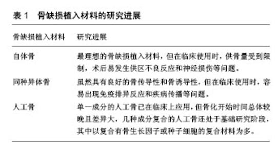
2.1 骨缺损植入材料的研究进展 骨缺损植入材料是在出现不能自愈的骨缺损问题后植入患处起修复、填充、固定和支撑等作用的医用材料,常见的骨缺损植入材料主要分为3类,包括:自体骨、同种异体骨和人工骨,其研究进展如表1所示。 2.1.1 自体骨 自体骨取自自体,取骨后对自体各方面无较大影响,没有疾病的传播而且几乎不会诱发机体的免疫反应,但是数量受到限制,且容易出现供区不良反应和神经损伤等问题[9-10]。如Baumhauer等[11]收集了130例做过自体骨移植手术的病例,在术后3,24,36,52周时与患者一起根据目测类比量表量化评估供骨区的疼痛度,最后所得疼痛值≥20便认定为有临床显著疼痛,在术后3周有46例(35%)发生疼痛,在术后24周有16例(12%)发生疼痛,在术后36周有8例(6.2%)发生疼痛,在术后52周有11例(8.5%)发生疼痛,表明在自体骨移植术后1年内供骨区易出现急性或持续性疼痛。 2.1.2 同种异体骨 同种异体骨即取自除自体外同物种个体的骨,由于是同种异体移植物,该移植物的特性受供体的影响,易出现移植排异反应、疾病传播等问 题[12]。如有研究统计了9例利用同种异体骨移植修复肿瘤切除后骨盆缺损的骨盆重建病例[13],在术后平均11.8个月的随访中,最短8个月,最长15个月,其中1例出现切口感染和腹胀,2例出现骨吸收,1例在8个月时出现骨折,说明同种异体骨用于治疗骨缺损疾病容易产生不良反应。 2.1.3 人工骨 人工骨则是起替代自体骨或修复骨缺损作用的人工材料,由单一成分或几种成分复合而成,不仅具有自体骨和同种异体骨的优点,而且不受供源量的限制,是目前最有前景的用于治疗骨缺损的骨植入材料,亦是目前骨缺损植入材料领域的研究热点[14-17]。临床上单一成分骨缺损植入材料已有应用,如有研究统计利用3D打印羟基磷灰石修复8个月到13岁儿童颅骨缺损19例,术后平均随访时间2.7年,所有患者的美容效果均令人满意,没有出现不良反应[18],脑血流量得到提高,但骨化开始时间差异较大,整体偏晚,平均时间为13个月,最早开始时间为术后3个月,最晚开始时间为术后22个月。 人工骨作为最有潜力应用于治疗骨缺损的骨植入材料,是医学和生物材料科学领域的研究热点。研究人员一直致力于开发出具有能够满足细胞及血管的生长要求的三维多孔立体结构,且具有一定的机械强度以便能够提供足够的机械支撑力,同时具有良好的生物相容性、良好的生物降解性、良好的骨传导性、良好的骨诱导性的骨缺损植入材料。但随着研究的深入,发现单一成分材料无法同时满足骨缺损植入材料的要求,如珊瑚虽然具有与人骨相似的孔隙结构,但其被机体吸收的速度快于骨新生的速度且无骨诱导性[19-22],所以几种特性互补成分的复合体材料应运而生,而且近年的研究基本都集中于复合体材料[23]。已经研制出的满足骨缺损植入材料要求的人工骨只有少部分用于临床,是因为大部分材料的研究还处于动物实验阶段,而且现阶段的实验动物多为鼠、兔、犬、猪、羊等,其实验结果还不能有力的说明该材料应用于临床治疗的有效性,仍需进一步研究证明。只有为数不多的材料建立了非人灵长类动物模型[24],其中以复合有骨生长因子或种子细胞的复合材料最多。 骨生长因子是骨组织的细胞产生并分泌的一些多肽,通过信号传导,对成骨细胞和破骨细胞的增殖及活性起促进或抑制作用,进而对骨新生起重要调节作用。分泌方式包括骨细胞自分泌和成骨细胞旁分泌两种。目前已经发现多种骨生长因子,如骨形态发生蛋白(bone morphogenetic protein,BMP)、转化生长因子β(transforming growth factor-β,TGF-β)、骨骼生长因子(skeletal growth factor,SGF)等[25],其中研究的较多的用于制备骨缺损植入复合材料的骨生长因子是骨形态发生蛋白2及调控因子,如转化生长因子β。 种子细胞是指组织工程中重建组织和器官时所用的细胞,具有再次分化能力。骨组织工程中可用的种子细胞有多种,如骨髓间充质干细胞、胚胎干细胞和牙髓干细胞等[26],但每种细胞都有其各自的缺点,如骨髓间充质干细胞的分化潜能随着成年供者的年龄增大而降低;对胚胎干细胞的分化调控机制研究不够透彻,并且涉及伦理问题;对牙髓干细胞的研究时间较短,其纯化、体外大量增殖等均是难题。现阶段,理想的种子细胞还未出现。研究人员在努力寻找理想的种子细胞的同时,也选用骨髓间充质干细胞和牙髓干细胞作为骨缺损植入复合材料的种子细胞。骨髓间充质干细胞是一种多功能干细胞,能分化成骨细胞、软骨细胞和脂肪细胞等,将其作为种子细胞应用于医学研究愈发受到关注。牙髓干细胞自2000年被Gronthos等[27]提出以来,因其具有与骨髓间充质干细胞相似的多向分化能力,被越来越多的应用在组织工程中。 2.2 骨缺损植入材料的非人灵长类动物模型研究现状 将动物实验结果外推至临床应用,究其可信度,主要取决于该实验动物与人的相似度,即“类人性”。非人灵长类动物模型作为基础研究和临床应用间的纽带,是不可替代的。但建立非人灵长类动物动物经费昂贵,动物资源紧张,致使许多研究停滞不前,因此在筛选近5年内骨缺损植入材料的非人灵长类动物模型时发现相关研究较少,而且文章之间关联性较低,研究内容不能构成一个系统,比较散乱。 2.2.1 复合有骨生长因子的复合材料修复骨缺损的非人灵长类动物模型研究现状 Chanchareonsook等[28]以具有碳酸酯取代的羟基磷灰石涂层的工程化的聚己内酯为支架,建立支架复合外源性重组人骨形态发生蛋白2组、支架复合自体骨髓间充质干细胞组和纯支架对照组,每组8个样品,修复猕猴右侧下颌骨15 mm缺损。实验过程中,由于钢板、螺钉的松动以及伤口的感染,最终支架复合外源性重组人骨形态发生蛋白2组获得6个样品、支架复合自体骨髓间充质干细胞组获得5个样品和纯支架对照组获得3个样品。总体结果显示,该实验设计的聚己内酯支架承载能力不足,并且在植入6个月后未实现完全的骨愈合。但是,通过组间对比发现载有重组人骨形态发生蛋白2的聚己内酯支架组显示出骨再生潜能。Zhou等[29]通过对比重组人骨形态发生蛋白2和脱钙冻干同种异体骨复合材料组、重组人骨形态发生蛋白2和珊瑚羟基磷灰石复合材料组、脱钙冻干同种异体骨组和珊瑚羟基磷灰石组修复猕猴下颌骨20 mm缺损。结果显示,含有重组人骨形态发生蛋白2的2个实验组表现出良好的骨诱导能力。Xie等[30]则采用可吸收胶原海绵(absorbable collagen sponge,ACS)作为支架材料,复合有anti-骨形态发生蛋白2的复合材料捕获内源性的骨形态发生蛋白2修复食蟹猴的下颌骨15 mm连续缺陷,发现其修复效果明显优于使用同型对照抗体的对照组。Ripamonti等[31]对比人转化生长因子β3+7%羟基磷灰石/珊瑚复合材料和7%羟基磷灰石/珊瑚复合材料修复25 mm直径孔的颅骨缺损,结果显示发现人转化生长因子β3具有调节骨形态发生蛋白2、成骨蛋白1等释放的作用,复合有人转化生长因子β3的复合材料具有骨重建的潜能。 2.2.2 复合有种子细胞的复合材料修复骨缺损的非人灵长类动物模型研究现状 Masaoka等[32]以β-磷酸三钙为支架材料,修复食蟹猴左侧股骨50 mm连续缺陷,分3次手术,第1次手术植入β-磷酸三钙支架材料、第2次手术在相同部位植入复合有骨髓间充质干细胞的β-磷酸三钙支架材料,第3次手术则在第2次手术后成功重建股骨的位置制造13 mm缺损,不植入任何材料,观察再生骨的愈合能力。对比第1,2次手术的结果发现复合有骨髓间充质干细胞的β-磷酸三钙支架材料具有良好的骨再生潜能,第3次手术结果则显示再生骨亦具有自愈能力。Fan等[33]通过对比骨髓间充质干细胞/β-磷酸三钙组、β-磷酸三钙组、空白对照组修复猕猴胫骨20 mm连续缺陷,在术后第4,8,12周进行取样观查,发现含骨髓间充质干细胞材料组在修复速率及修复效果等方面均优于无骨髓间充质干细胞组,表现出良好的骨再生潜能。Araki等[34]以胶原凝胶为支架材料,建立胶原凝胶/骨髓间充质干细胞组、胶原凝胶组、空白对照组,进行非人灵长类动物的髌骨沟软骨直径3 mm,深5 mm缺损的修复,在术后第6,12,24周进行取样观查,结果显示,胶原凝胶/骨髓间充质干细胞组的重建速率及重建效果均优于其他两组,同时移植骨髓间充质干细胞克服了软骨和自体软骨细胞移植的缺点,取得了良好的修复效果。Bachtiar等[35]使用不同配比(30/70/0,50/50/0,1/1/1)的羟基磷灰石、磷酸三钙和壳聚糖制作支架材料并复合牙髓干细胞用于非人灵长类动物的下颌骨10 mm× 20 mm缺损的重建,与空白对照组比较,发现基本无支架残留且骨再生情况良好。 2.2.3 其他材料修复骨缺损的非人灵长类动物模型研究现状 除去上述两类材料之外,亦有其他的材料建立了骨缺损非人灵长类动物模型。Goh等[36]采用聚己内 酯-磷酸三钙和自体骨移植修复猕猴拔牙后牙槽骨的缺损处,发现采用聚己内酯-磷酸三钙植入缺损处,能够很好的维持牙槽骨的轮廓,但骨再生情况却比自体骨移植组差很多。有研究采用聚丙烯、聚D、L-乳酸共聚物制作植入材料或在植入材料内填充无机牛骨矿物质用于维持猕猴拔牙后牙槽骨的形状、大小取得良好效果,有效地减少骨质的流失,为临床义齿修复方法提供新希望[37-39]。Jakus等[40]以质量比为9∶1的羟基磷灰石、聚己内酯通过3D打印制作植入材料并用于猕猴的颅骨缺损修复,发现有带血管蒂的软组织覆盖植入材料,且植入材料内部有新骨生成,说明该材料具有成骨能力。"

| [1] 岳飞翔,隋海明.骨缺损动物模型的研究进展[J].中国民族民间医药,2015,24(16):35-36.[2] Li Y, Chen SK, Li L, et al. Bone defect animal models for testing efficacy of bone substitute biomaterials. J Orthop Translat. 2015;3(3):95-104. [3] Leigh SR. Brain growth, life history, and cognition in primate and human evolution. Am J Primatol. 2004;62(3):139-164. [4] Xing J, Witherspoon DJ, Ray DA, et al. Mobile DNA elements in primate and human evolution. Am J Phys Anthropol. 2007; 134(S45):2-19. [5] Sato K, Sasaki E. Genetic engineering in nonhuman primates for human disease modeling. J Hum Genet. 2018;63(2): 125-131. [6] Estes JD, Wong SW, Brenchley JM. Nonhuman primate models of human viral infections. Nat Rev Immunol. 2018; 18(6):390-404. [7] Shen L, Chen CY, Huang D, et al. Pathogenic events in a nonhuman primate model of oral poliovirus infection leading to paralytic poliomyelitis. J Virol. 2017;91(14): 2310-2316. [8] Yao YG, Chen YB, Liang B. The 3rd Symposium on animal models of primates-the application of non-human primates to basic research and translational medicine. J Genet Genomics. 2015;42(6):339-341. [9] 袁冰,韦卓.骨缺损修复的研究进展[J].生物骨科材料与临床研究, 2014,11(3):38-41.[10] 王兴,刘洪臣.自体骨移植修复种植位点骨缺损的研究进展[J].口腔颌面修复学杂志,2016,17(1):49-52.[11] Baumhauer J, Pinzur MS, Donahue R, et al. Site selection and pain outcome after autologous bone graft harvest. Foot Ankle Int. 2014;35(2):104-107. [12] 镐英杰,于磊,李志磊,等.同种异体骨与自体骨在骨缺损治疗中的应用比较[J].实用骨科杂志,2015,21(7):592-566.[13] Wang W, Wang Y, Bi W, et al. Allogeneic bone transplantation for pelvic reconstruction of large skeletal defects after tumor resection. Zhongguo Xiu Fu Chong Jian Wai Ke Za Zhi. 2014; 28(3):331-334. [14] 郝强,赵丽,关继奎,等.人工骨材料在骨缺损修复中的应用[J].中国组织工程研究与临床康复,2009,13(34):6745-6748.[15] 辛雷,苏佳灿.人工骨修复材料的现状与展望[J].创伤外科杂志, 2011,13(3):272-284.[16] 王迎军,杜昶,赵娜如,等.仿生人工骨修复材料研究[J].华南理工大学学报(自然科学版),2012,40(10):51-58.[17] 许瑾,吴晶晶,王晓冬,等.羟基磷灰石复合骨组织工程支架的研究进展[J].生物骨科材料与临床研究,2016,13(2):63-66.[18] Beuriat PA, Szathmari A, Grassiot B, et al. Why a hydroxyapatite cranioplasty can be used to repair a cranial bone defect in children: experience of 19 cases. Neurochirurgie. 2016;62(5):251-257. [19] Guillemin G, Patat JL, Fournie J, et al. The use of coral as a bone graft substitute. J Biomed Mater Res. 2010;21(5): 557-567. [20] Guillemin G, Meunier A, Dallant P, et al. Comparison of coral resorption and bone apposition with two natural corals of different porosities. J Biomed Mater Res. 2010;23(7):765-779. [21] Vuola J, Böhling T, Kinnunen J, et al. Natural coral as bone-defect-filling material. J Biomed Mater Res. 2015;51(1): 117-122. [22] Green DW, Ben-Nissan B, Yoon KS, et al. Natural and synthetic coral biomineralization for human bone revitalization. Trends Biotechnol. 2016;35(1):43-54. [23] Siddiqui JA, Partridge NC. Physiological bone remodeling: systemic regulation and growth factor involvement. Physiology. 2016;31(3):233-245. [24] Ripamonti U, Duarte R, Ferretti C. Re-evaluating the induction of bone formation in primates. Biomaterials. 2014;35(35): 9407-9422. [25] Mendes LF, Katagiri H, Tam WL, et al. Advancing osteochondral tissue engineering: bone morphogenetic protein, transforming growth factor, and fibroblast growth factor signaling drive ordered differentiation of periosteal cells resulting in stable cartilage and bone formation in vivo. Stem Cell Res Ther. 2018;9(1):42. [26] 张治金,全仁夫,岳振双,等.软骨组织工程中种子细胞的研究进展[J].中国医药导报,2017,14(23):36-39.[27] Gronthos S, Mankani M, Brahim J, et al. Postnatal human dental pulp stem cells (DPSCs) in vitro and in vivo. Proc Natl Acad Sci U S A. 2000;97(25):13625-13630. [28] Chanchareonsook N, Tideman H, Feinberg SE, et al. Segmental mandibular bone reconstruction with a carbonate-substituted hydroxyapatite-coated modular endoprosthetic poly(ε-caprolactone) scaffold in Macaca fascicularis. J Biomed Mater Res B Appl Biomater. 2014; 102(5):962-976. [29] Zhou M, Peng X, Mao C, et al. The value of SPECT/CT in monitoring prefabricated tissue-engineered bone and orthotopic rhBMP-2 implants for mandibular reconstruction. PLoS One. 2015;10(9):e0137167. [30] Xie Y, Su Y, Min S, et al. Collagen sponge functionalized with chimeric anti-bmp-2 monoclonal antibody mediates repair of critical-size mandibular continuity defects in a nonhuman primate model. Biomed Res Int. 2017;2017(2):8094152. [31] Ripamonti U, Klar RM, Parak R, et al. Tissue segregation restores the induction of bone formation by the mammalian transforming growth factor-β3 in calvarial defects of the non-human primate Papio ursinus. Biomaterials. 2016;86: 21-32. [32] Masaoka T, Yoshii T, Yuasa M, et al. Bone defect regeneration by a combination of a β-tricalcium phosphate scaffold and bone marrow stromal cells in a non-human primate model. Open Biomed Eng J. 2016;10(1):2-11. [33] Fan H, Zeng X, Wang X, et al. Efficacy of prevascularization for segmental bone defect repair using β-tricalcium phosphate scaffold in rhesus monkey. Biomaterials. 2014;35(26): 7407-7415. [34] Araki S, Imai S, Ishigaki H, et al. Improved quality of cartilage repair by bone marrow mesenchymal stem cells for treatment of an osteochondral defect in a cynomolgus macaque model. Acta Orthop. 2015;86(1):119-126. [35] Bachtiar EW, Amir LR, Suhardi P, et al. Scaffold degradation during bone tissue reconstruction in Macaca nemestrina mandible. Interv Med Appl Sci. 2016;8(2):77-81. [36] Goh BT, Chanchareonsook N, Tideman H, et al. The use of a polycaprolactone-tricalcium phosphate scaffold for bone regeneration of tooth socket facial wall defects and simultaneous immediate dental implant placement in Macaca fascicularis. J Biomed Mater Res A. 2014;102(5):1379-1388. [37] Min S, Liu Y, Tang J, et al. Alveolar ridge dimensional changes following ridge preservation procedure with novel devices: part 1-CBCTlinear analysis in non-human primate model. Clin Oral Implants Res. 2016;27(1):97-105. [38] Omran M, Min S, Abdelhamid A, et al. Alveolar ridge dimensional changes following ridge preservation procedure: part-2-CBCT 3D analysis in non-human primate model. Clin Oral Implants Res. 2016;27(7):859-866. [39] Su Y, Tang J, Min S, et al. Alveolar ridge dimensional changes following ridge preservation procedure with novel devices: part 3 – histological analysis in non-human primate model. Clin Oral Implants Res. 2017;28(11):252-261. [40] Jakus AE, Rutz AL, Jordan SW, et al. Hyperelastic "bone": a highly versatile, growth factor-free, osteoregenerative, scalable, and surgically friendly biomaterial. Sci Transl Med. 2016;8(358):358ra127. |
| [1] | Zhang Chao, Lü Xin. Heterotopic ossification after acetabular fracture fixation: risk factors, prevention and treatment progress [J]. Chinese Journal of Tissue Engineering Research, 2021, 25(9): 1434-1439. |
| [2] | Chen Jiming, Wu Xiaojing, Liu Tianfeng, Chen Haicong, Huang Chengshuo. Effects of silymarin on liver injury and bone metabolism induced by carbon tetrachloride in mice [J]. Chinese Journal of Tissue Engineering Research, 2021, 25(8): 1224-1228. |
| [3] | Tan Jingyu, Liu Haiwen. Genome-wide identification, classification and phylogenetic analysis of Fasciclin gene family for osteoblast specific factor 2 [J]. Chinese Journal of Tissue Engineering Research, 2021, 25(8): 1243-1248. |
| [4] | Kong Desheng, He Jingjing, Feng Baofeng, Guo Ruiyun, Asiamah Ernest Amponsah, Lü Fei, Zhang Shuhan, Zhang Xiaolin, Ma Jun, Cui Huixian. Efficacy of mesenchymal stem cells in the spinal cord injury of large animal models: a meta-analysis [J]. Chinese Journal of Tissue Engineering Research, 2021, 25(7): 1142-1148. |
| [5] | Ma Binxiang, He Wanqing, Zhou Guangchao, Guan Yonglin. Triptolide improves motor dysfunction in rats following spinal cord injury [J]. Chinese Journal of Tissue Engineering Research, 2021, 25(5): 701-706. |
| [6] | Liu Qing, Wan Bijiang. Effect of acupotomy therapy on the expression of Bcl-2/Bax in synovial tissue of collagen-induced arthritis rats [J]. Chinese Journal of Tissue Engineering Research, 2021, 25(5): 729-734. |
| [7] | Zhang Liang, Ma Xiaoyan, Wang Jiahong. Regulatory mechanism of Shenshuai Yin on cell apoptosis in the kidney of chronic renal failure rats [J]. Chinese Journal of Tissue Engineering Research, 2021, 25(23): 3672-3677. |
| [8] | Xie Yang, Lü Zhiyu, Zhang Shujiang, Long Ting, Li Zuoxiao. Effects of recombinant adeno-associated virus mediated nerve growth factor gene transfection on oligodendrocyte apoptosis and myelination in experimental autoimmune encephalomyelitis mice [J]. Chinese Journal of Tissue Engineering Research, 2021, 25(23): 3678-3683. |
| [9] | Hu Guang, Guan Zhiyu, Zhang Kaiwei . Mechanism underlying the interventional effect of Panax Notoginsenosides on ovariectomized osteoporotic fracture rats [J]. Chinese Journal of Tissue Engineering Research, 2021, 25(2): 172-177. |
| [10] | Li Huye, Dou Zenghua, Kong Deyuan, Cai Jinlian, Li Zeqing. Cornuside I inhibits interleukin-6-mediated inflammation in knee osteoarthritis rats [J]. Chinese Journal of Tissue Engineering Research, 2021, 25(2): 211-215. |
| [11] | Chen Yutong, Li Chenchen, Liu Yang, Zheng Yaqin, Yang Xihua, An Meiwen. Establishment of an acute radioactive skin injury model in Wistar rats [J]. Chinese Journal of Tissue Engineering Research, 2021, 25(2): 237-241. |
| [12] | Xie Wenjie, Zhou Gang, Xie Jinmei, Liu Jiao, Li Pengfei, Yang Fan, Cui Di. Mechanism of myocardial oxidative damage in a rat model of one-time exhaustive exercise [J]. Chinese Journal of Tissue Engineering Research, 2021, 25(2): 247-252. |
| [13] | Mu Yufeng, Wei Lina, Wu Yong, Shao Anliang, Chen Liang, Qu Shuxin, Xu Liming. Development and evaluation of alpha-galactosyl antigen-deficient rabbit model [J]. Chinese Journal of Tissue Engineering Research, 2021, 25(2): 281-285. |
| [14] | Wang Yan, Dong Benchao, Wang Ying, Sun Lei, Lu Bin, Bai Haohao, Tian Aixian, Ma Jianxiong, Ma Xinlong. Animal models of osteonecrosis of the femoral head: modeling methods and characteristics [J]. Chinese Journal of Tissue Engineering Research, 2021, 25(2): 292-297. |
| [15] | Ma Dujun, Peng Liping, Chen Feng, Jiang Shunwan, Jiang Jinting, Gao Kun, Lin Zhanpeng. Research strategy of gene editing technology in the gene treatment of osteoarthritis [J]. Chinese Journal of Tissue Engineering Research, 2021, 25(2): 298-303. |
| Viewed | ||||||
|
Full text |
|
|||||
|
Abstract |
|
|||||