Chinese Journal of Tissue Engineering Research ›› 2026, Vol. 30 ›› Issue (33): 8761-8777.doi: 10.12307/2026.484
Previous Articles Next Articles
Causal relationships between immune cells and childhood asthma and the mediating role of inflammatory proteins
Gong Jiaxuan, Lin Lin, Wang Chun, Zhang Xingxing, Xiong Ying
- School of Acupuncture-Moxibustion and Tuina, and School of Health Preservation and Rehabilitation, Nanjing University of Chinese Medicine, Nanjing 210023, Jiangsu Province, China
-
Received:2025-12-06Revised:2026-04-01Online:2026-11-28Published:2026-06-16 -
Contact:Xiong Ying, PhD, Professor, School of Acupuncture-Moxibustion and Tuina, and School of Health Preservation and Rehabilitation, Nanjing University of Chinese Medicine, Nanjing 210023, Jiangsu Province, China -
About author:Gong Jiaxuan, MS candidate, School of Acupuncture-Moxibustion and Tuina, and School of Health Preservation and Rehabilitation, Nanjing University of Chinese Medicine, Nanjing 210023, Jiangsu Province, China -
Supported by:the National Natural Science Foundation of China, No. 81973970 (to XY)

CLC Number:
Cite this article
Gong Jiaxuan, Lin Lin, Wang Chun, Zhang Xingxing, Xiong Ying. Causal relationships between immune cells and childhood asthma and the mediating role of inflammatory proteins[J]. Chinese Journal of Tissue Engineering Research, 2026, 30(33): 8761-8777.
share this article
Add to citation manager EndNote|Reference Manager|ProCite|BibTeX|RefWorks
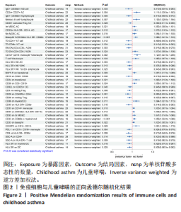
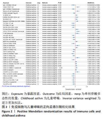
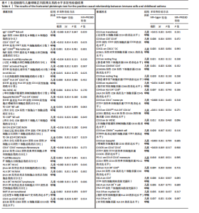
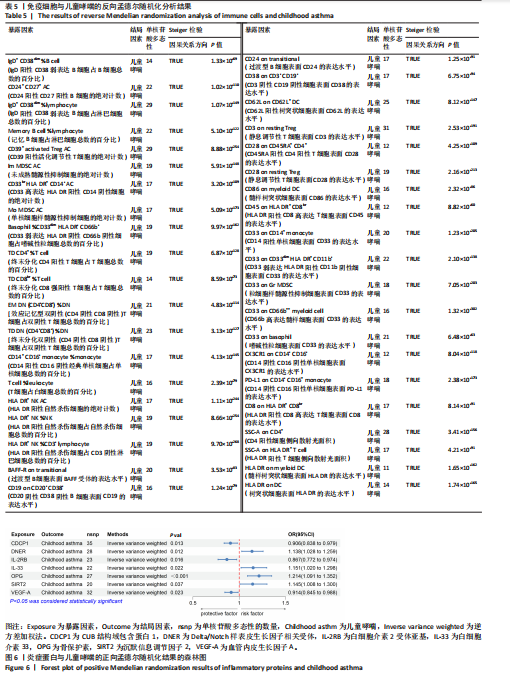
2.1 免疫细胞与儿童哮喘的孟德尔随机化分析结果 通过两样本孟德尔随机化方法对免疫细胞和儿童哮喘的关联进行分析,结果显示有40种免疫细胞与儿童哮喘存在因果关系。在已鉴定的免疫细胞中,CD24+ CD27+ AC (CD24阳性CD27阳性B细胞的绝对计数)、Memory B cell %lymphocyte(记忆B细胞占淋巴细胞总数的百分比)、Im MDSC AC (未成熟髓源性抑制细胞的绝对计数)、CD33br HLA DR+ CD14- AC (CD33高表达HLA DR阳性CD14阴性细胞的绝对计数)、Mo MDSC AC (单核细胞样髓源性抑制细胞的绝对计数)、Basophil %CD33dim HLA DR- CD66b- (CD33弱表达HLA DR阴性CD66b阴性细胞占嗜碱性粒细胞总数的百分比)、TD DN (CD4-CD8-) %DN [终末分化双阴性(CD4阴性CD8阴性)T细胞占双阴性T细胞总数的百分比]、CD14+ CD16- monocyte% monocyte (CD14阳性CD16阴性经典单核细胞占单核细胞总数的百分比)、T cell %leukocyte (T细胞占白细胞总数的百分比)、BAFF-R on transitional (过渡型B细胞表面BAFF受体的表达水平)、CD24 on transitional (过渡型B细胞表面CD24的表达水平)、CD38 on CD3- CD19- (CD3阴性CD19阴性细胞表面CD38的表达水平)、CD86 on myeloid DC (髓样树突状细胞表面CD86的表达水平)、CD33 on CD14+ monocyte (CD14阳性单核细胞表面CD33的表达水平)、CD33 on CD33dim HLA DR+ CD11b- (CD33弱表达HLA DR阳性CD11b阴性细胞表面CD33的表达水平)、CD33 on Gr MDSC (粒细胞样髓源性抑制细胞表面CD33的表达水平)、CD33 on CD66b++ myeloid cell (CD66b高表达髓样细胞表面CD33的表达水平)、CD33 on basophil (嗜碱性粒细胞表面CD33的表达水平)、CX3CR1 on CD14- CD16- (CD14阴性CD16阴性单核细胞表面CX3CR1的表达水平) 共19种免疫细胞与儿童哮喘风险增加有关。IgD+ CD38dim %B cell (IgD阳性CD38弱表达B细胞占B细胞总数的百分比)、IgD+ CD38dim %lymphocyte (IgD阳性CD38弱表达B细胞占淋巴细胞总数的百分比)、CD39+ activated Treg AC (CD39阳性活化调节性T细胞的绝对计数)、TD CD4+ %T cell (终末分化CD4阳性T细胞占T细胞总数的百分比)、TD CD8br %T cell (终末分化CD8强阳性T细胞占T细胞总数的百分比)、EM DN (CD4-CD8-) %DN[效应记忆型双阴性(CD4阴性CD8阴性)T细胞占双阴性T细胞总数的百分比]、HLA DR+ NK AC (HLA DR阳性自然杀伤细胞的绝对计数)、HLA DR+ NK %NK (HLA DR阳性自然杀伤细胞占自然杀伤细胞总数的百分比)、HLA DR+ NK %CD3- lymphocyte (HLA DR阳性自然杀伤细胞占CD3阴性淋巴细胞总数的百分比)、CD19 on CD20- CD38- (CD20阴性CD38阴性B细胞表面CD19的表达水平)、CD62L on CD62L+ DC (CD62L阳性树突状细胞表面CD62L的表达水平)、CD3 on resting Treg (静息调节性T细胞表面CD3的表达水平)、CD28 on CD45RA+ CD4+ (CD45RA阳性CD4阳性T细胞表面CD28的表达水平)、CD28 on resting Treg (静息调节性T细胞表面CD28的表达水平)、CD45 on HLA DR+ CD8br (HLA DR阳性CD8高表达T细胞表面CD45的表达水平)、PD-L1 on CD14- CD16+ monocyte (CD14阴性CD16阳性单核细胞表面PD-L1的表达水平)、CD8 on HLA DR+ CD8br (HLA DR阳性CD8高表达T细胞表面CD8的表达水平)、SSC-A on CD4+ (CD4阳性细胞侧向散射光面积)、SSC-A on HLA DR+ T cell (HLA DR阳性T细胞侧向散射光面积)、HLA DR on myeloid DC (髓样树突状细胞表面HLA DR的表达水平)、HLA DR on DC (树突状细胞表面HLA DR的表达水平) 共21种免疫细胞与儿童哮喘风险降低有关。结果见图2。"
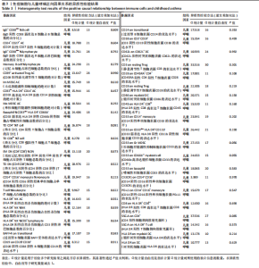
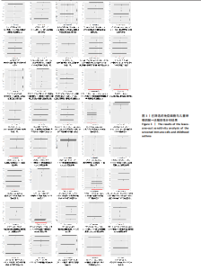
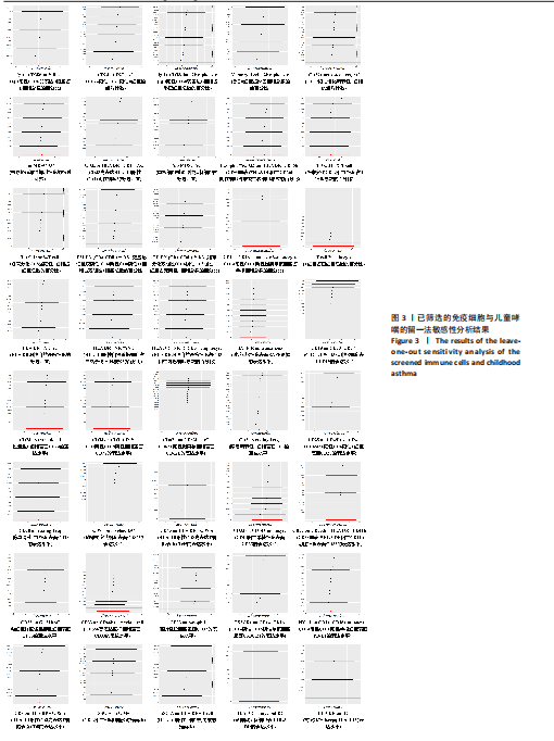
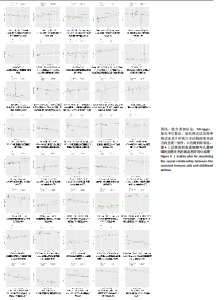
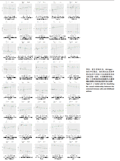
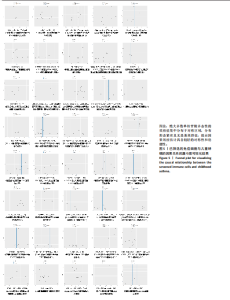
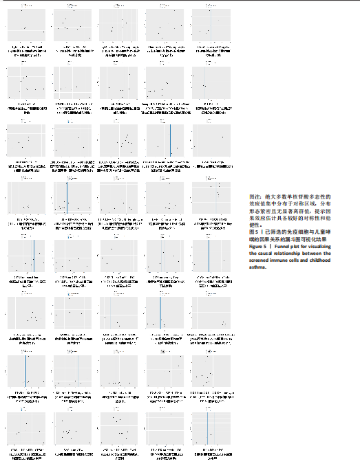
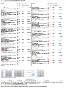
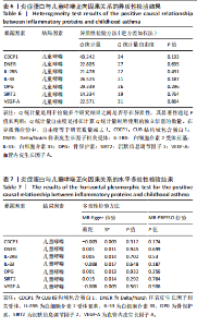
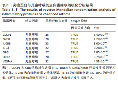
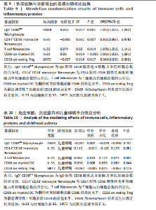
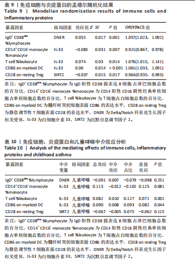
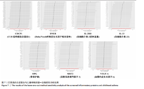
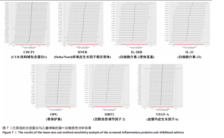
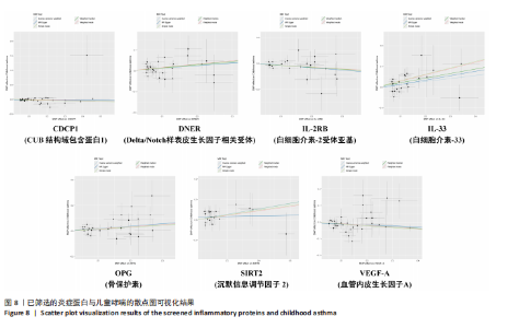
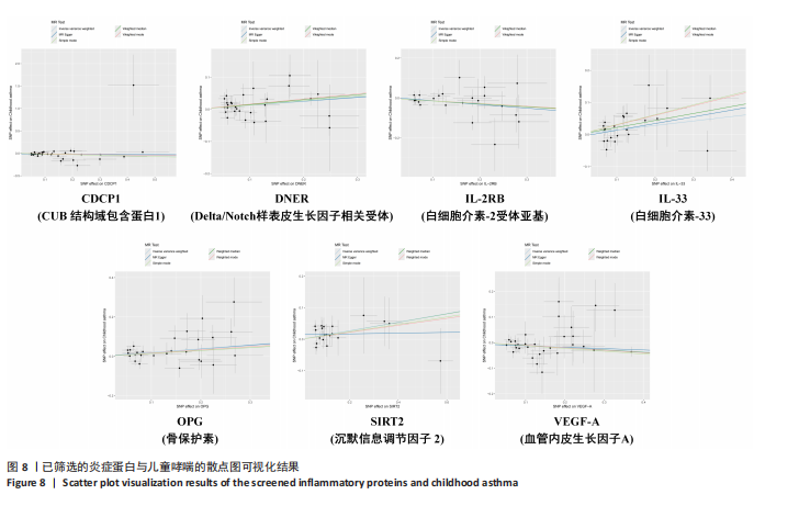
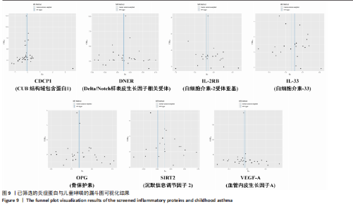
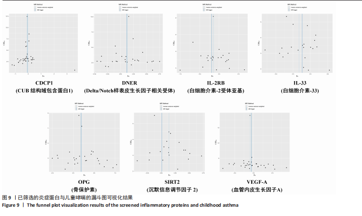
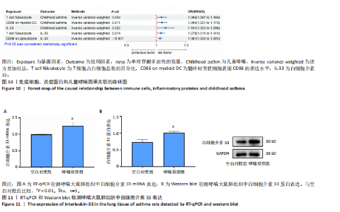
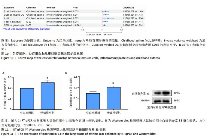
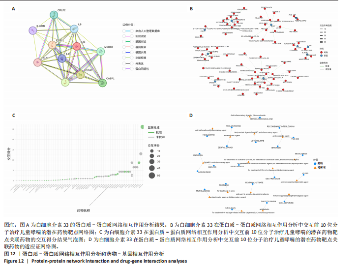
Cochran’Q 统计量检验表明已筛选的40种免疫细胞的单核苷酸多态性中未发现异质性的证据(P > 0.05)(表3),MR-Egger截距检验和MR-PRESSO检验两项测试均未显示潜在的水平多效性(P > 0.05)(表4)。留一法敏感性分析结果显示,逐一剔除各单核苷酸多态性后,总体效应估计值保持稳定,结果具有较好的稳健性(图3)。Steiger检验结果显示,40种免疫细胞的因果关系方向为TRUE,且P值均 < 0.05,表明该暴露因素对结局产生影响,确保了方向性(表5)。散点图分析显示,逆方差加权法、MR-Egger、加权中位数法、加权模式法及简单模式法共5种统计方法的因果效应方向呈现一致性,未检测到异常值(图4)。漏斗图检验结果表明,绝大多数单核苷酸多态性的效应值集中分布于对称区域,分布形态紧密且无显著离群值,提示因果效应估计具备较好的对称性和稳健性(图5)。 2.2 炎症蛋白与儿童哮喘的孟德尔随机化分析结果 将炎症蛋白作为暴露因素,儿童哮喘作为结局因素,以逆方差加权法为主要分析方法,经过一系列筛选后,发现7种炎症蛋白与儿童哮喘存在显著相关性。其中,Delta/Notch样表皮生长因子相关受体、白细胞介素33、骨保护素、沉默信息调节因子2是儿童哮喘的危险因素,CUB结构域包含蛋白1、白细胞介素2受体亚基、血管内皮生长因子A是儿童哮喘的保护因素(图6)。敏感性分析结果表明,已筛选的7种炎症蛋白与儿童哮喘均不存在异质性(PCochran’Q > 0.05)和水平多效性(PMR-Egger > 0.05和PMR-PRESSO > 0.05)(表6,7)。留一法敏感性检验表明,当逐一排除单个单核苷酸多态性时,因果效应的整体估计值未发生显著偏移(图7)。散点图和漏斗图未识别到异常值和工具变量的选择偏倚(图8,9)。Steiger检验结果未发现反向因果关系的证据(表8)。 2.3 中介孟德尔随机化分析结果 为了探索儿童哮喘发展和进展的潜在机制,利用中介分析以鉴定介导从免疫细胞到儿童哮喘致病途径的炎症蛋白。以逆方差加权法方法作为主要方法进行双样本孟德尔随机化分析,探究免疫细胞与炎症蛋白之间的因果关系。分析过程同上述过程一致,显著性水平为P < 0.05。结果确定了5种免疫细胞与炎症蛋白之间存在因果关系。具体而言,IgD+ CD38dim %lymphocyte (IgD阳性CD38弱表达B细胞占淋巴细胞总数的百分比)与Delta/Notch样表皮生长因子相关受体、T cell %leukocyte (T细胞占白细胞总数的百分比)与白细胞介素33、CD86 on myeloid DC (髓样树突状细胞表面CD86的表达水平)与白细胞介素33呈正相关,CD14+ CD16- monocyte %monocyte (CD14阳性CD16阴性经典单核细胞占单核细胞总数的百分比)与白细胞介素33、CD28 on resting Treg (静息调节性T细胞表面CD28的表达水平)与沉默信息调节因子2呈负相关(表9)。随后,中介效应模型显示,白细胞介素33在T cell %leukocyte (T细胞占白细胞总数的百分比)、CD86 on myeloid DC (髓样树突状细胞表面CD86的表达水平)与儿童哮喘的因果关系中起到了介导作用(图10)。其中,白细胞介素33在T cell %leukocyte (T细胞占白细胞总数的百分比)到儿童哮喘路径中发挥中介作用的比例为12.7%,中介效应为0.010;白细胞介素33在CD86 on myeloid DC(髓样树突状细胞表面CD86的表达水平)到儿童哮喘路径中发挥中介作用的比例为9.3%,中介效应为0.008(表10)。 2.4 哮喘小鼠肺组织中白细胞介素33基因和蛋白表达的实验验证结果 RT-qPCR检测结果显示,与空白对照组相比,哮喘模型组大鼠肺组织中白细胞介素33 mRNA表达水平显著升高(P < 0.01)(图11A)。Western blot检测结果显示,与空白对照组相比,哮喘模型组大鼠肺组织中白细胞介素33蛋白表达水平显著升高(P < 0.01)。综合以上结果表明,白细胞介素33可能在哮喘的发病机制中发挥实际作用,并且白细胞介素33表达与疾病状态相关(图11B)。 2.5 蛋白质-蛋白质网络相互作用分析和药物-基因相互作用分析结果 针对分子靶点炎症因子白细胞介素33构建的蛋白质-蛋白质相互作用网络分析显示,白细胞介素33作为核心节点与白细胞介素1受体样1 (IL1RL1)、白细胞介素1受体辅助蛋白(IL1RAP)、白细胞介素1受体1型(IL1R1)、半胱天冬酶1 (CASP1)、髓样分化初级反应蛋白88(MYD88)、细胞因子受体样因子2(CRLF2)、白细胞介素5 (IL5)、白细胞介素13 (IL13)、白细"
胞介素17受体B (IL17RB)、胸腺基质淋巴细胞生成素(TSLP)前10位交互分子形成复杂调控网络(图12A)。具体连接模式表明,白细胞介素33通过多类型相互作用与白细胞介素1受体样1、白细胞介素1受体辅助蛋白、髓样分化初级反应蛋白88等关键分子形成紧密关联,其中白细胞介素1受体样1作为白细胞介素33的高亲和力结合蛋白,构成信号转导的核心轴。网络中节点特征显示,白细胞介素33与白细胞介素5、白细胞介素13、白细胞介素17受体B、胸腺基质淋巴细胞生成素等2型免疫相关分子形成局部聚类,提示白细胞介素33在Th2型炎症中的枢纽地位;而与半胱天冬酶1、髓样分化初级反应蛋白88的连接则揭示白细胞介素33通过炎症小体及Toll样受体信号通路参与先天免疫调控。在此基础上,进一步借助药物-基因相互作用数据库,聚焦包含白细胞介素33及其10个核心交互分子,显示药物Astegolimab、Torudokimab、Etokimab和Itepekimab与白细胞介素33存在高交互得分(图12B)。此外,针对白细胞介素33在相互作用网络中的核心交互分子白细胞介素5和白细胞介素13,已有靶向药物开发并应用于哮喘治疗。靶向白细胞介素5的药物Benralizumab、Reslizumab、Mepolizumab以及靶向"

白细胞介素13的药物Tralokinumab正在作为平喘剂使用,且已获得监管机构批准上市(图12C,D)。"
"
"
"
"

"

"
"
"
"
"
"
| [1] FAN GZ, CHEN KY, LIU XM, et al. Mendelian randomization study of childhood asthma and chronic obstructive pulmonary disease in European and East Asian population. World Allergy Organ J. 2024;17(9):100960. [2] PRASAD B, NYENHUIS SM, IMAYAMA I, et al. Asthma and Obstructive Sleep Apnea Overlap: What Has the Evidence Taught Us? Am J Respir Crit Care Med. 2020;201(11):1345-1357. [3] ZANOBETTI A, RYAN PH, COULL B, et al. Childhood Asthma Incidence, Early and Persistent Wheeze, and Neighborhood Socioeconomic Factors in the ECHO/CREW Consortium. JAMA Pediatr. 2022;176(8):759-767. [4] ZHANG Y, HAI Y, SONG B, et al. Screening and Validation of Potential Biomarkers of Immune Cells in Childhood Asthma Patients via Mendelian Randomization and Machine Learning. J Inflamm Res. 2025;18:2583-2600. [5] QIU F, SHAO W, QIN X, et al. Causal association between cathepsins and asthma: a Mendelian randomization study. Sci Rep. 2025;15(1):23984. [6] EL-HUSSEINI ZW, GOSENS R, DEKKER F, et al. The genetics of asthma and the promise of genomics-guided drug target discovery. Lancet Respir Med. 2020;8(10):1045-1056. [7] PIJNENBURG MW, FLEMING L. Advances in understanding and reducing the burden of severe asthma in children. Lancet Respir Med. 2020;8(10):1032-1044. [8] HABIB N, PASHA MA, TANG DD. Current Understanding of Asthma Pathogenesis and Biomarkers. Cells. 2022;11(17):2764. [9] CHOI BS. Eosinophils and childhood asthma. Clin Exp Pediatr. 2021;64(2): 60-67. [10] SHAILESH H, NOOR S, HAYATI L, et al. Asthma and obesity increase inflammatory markers in children. Front Allergy. 2025;5:1536168. [11] 孟娜娜,孙璐,黄诚花.支气管哮喘患儿上呼吸道菌群和炎症因子水平变化与病情严重程度的关系[J].中国微生态学杂志,2024,36(2):191-195+200. [12] SKRIVANKOVA VW, RICHMOND RC, WOOLF BAR, et al. Strengthening the Reporting of Observational Studies in Epidemiology Using Mendelian Randomization: The STROBE-MR Statement. JAMA. 2021;326(16): 1614-1621. [13] ZHANG Z, WANG Y, LI J, et al. Exploring the causal link between childhood maltreatment and asthma: a Mendelian randomization study. Eur J Psychotraumatol. 2025;16(1):2383127. [14] ORRÙ V, STERI M, SIDORE C, et al. Complex genetic signatures in immune cells underlie autoimmunity and inform therapy. Nat Genet. 2020;52(10): 1036-1045. [15] ZHAO JH, STACEY D, ERIKSSON N, et al. Genetics of circulating inflammatory proteins identifies drivers of immune-mediated disease risk and therapeutic targets. Nat Immunol. 2023;24(9):1540-1551. [16] KURKI MI, KARJALAINEN J, PALTA P, et al. FinnGen provides genetic insights from a well-phenotyped isolated population. Nature. 2023;613(7944):508-518. [17] XU J, SI S, HAN Y, et al. Genetic insight into dissecting the immunophenotypes and inflammatory profiles in the pathogenesis of Sjogren syndrome. J Transl Med. 2025;23(1):56. [18] LIU QP, DU HC, XIE PJ, et al. Effect of the immune cells and plasma metabolites on rheumatoid arthritis: a mediated mendelian randomization study. Front Endocrinol (Lausanne). 2024;15:1438097. [19] MA S, HU W, BI Y, et al. Exploring the causal role of plasma metabolites in pediatric asthma: a Mendelian randomization study. J Asthma. 2025; 62(12):2070-2083. [20] HU Z, XU P, WU J, et al. Causal Relationships Between Immune Cell Traits, Plasma Metabolites, and Asthma: A Two-Step, Two-Sample Mendelian Randomization Study. Clin Respir J. 2025;19(6):e70097. [21] ZHANG S, ZHANG X, WEI C, et al. Causality Between 91 Circulating Inflammatory Proteins and Various Asthma Phenotypes: A Mendelian Randomization Study. Immunotargets Ther. 2024;13:617-629. [22] JIANG Y, WANG Y, GUO J, et al. Exploring potential therapeutic targets for asthma: a proteome-wide Mendelian randomization analysis. J Transl Med. 2024;22(1):978. [23] WANG C, FAN H, SUN Y, et al. Mendelian randomization analysis of the causal relationship between plasma proteins and childhood asthma. Medicine (Baltimore). 2025;104(14):e42050. [24] GAN Q, LIU Q, WU Y, et al. The Causal Association Between Obstructive Sleep Apnea and Child-Onset Asthma Come to Light: A Mendelian Randomization Study. Nat Sci Sleep. 2024;16:979-987. [25] TAN T, YANG F, WANG Z, et al. Mediated Mendelian randomization analysis to determine the role of immune cells in regulating the effects of plasma metabolites on childhood asthma. Medicine (Baltimore). 2024; 103(30):e38957. [26] YE CJ, LIU D, CHEN ML, et al. Mendelian randomization evidence for the causal effect of mental well-being on healthy aging. Nat Hum Behav. 2024; 8(9):1798-1809. [27] CAO S, GU Y, LU G, et al. Causal Correlations Between Plasma Metabolites, Inflammatory Proteins, and Chronic Obstructive Pulmonary Disease: A Mendelian Randomization and Bioinformatics-Based Investigation. J Inflamm Res. 2025;18:4057-4073. [28] 楼金成,苗镡允,苏嘉琪,等.过敏性哮喘大鼠模型的建立方法与评价[J].中国比较医学杂志,2023,33(1):130-137. [29] WOODROW JS, SHEATS MK, COOPER B, et al. Asthma: The Use of Animal Models and Their Translational Utility. Cells. 2023;12(7):1091. [30] 刘雀.捏脊法调节AMPK/PGC-1α通路防治未成年鼠哮喘的机制研究[D].南京:南京中医药大学,2025. [31] 梁雪杰.二甲双胍通过IL33/ST2信号通路缓解OVA诱导的小鼠过敏性气道炎症[D].兰州:兰州大学,2023. [32] SZKLARCZYK D, GABLE AL, LYON D, et al. STRING v11: protein-protein association networks with increased coverage, supporting functional discovery in genome-wide experimental datasets. Nucleic Acids Res. 2019; 47(D1):D607-D613. [33] DI PALMO E, CANTARELLI E, CATELLI A, et al. The Predictive Role of Biomarkers and Genetics in Childhood Asthma Exacerbations. Int J Mol Sci. 2021;22(9):4651. [34] WANG B, CHEN H, CHAN YL, et al. Why Do Intrauterine Exposure to Air Pollution and Cigarette Smoke Increase the Risk of Asthma? Front Cell Dev Biol. 2020;8:38. [35] MORALES E, DUFFY D. Genetics and Gene-Environment Interactions in Childhood and Adult Onset Asthma. Front Pediatr. 2019;7:499. [36] AKDIS M, AAB A, ALTUNBULAKLI C, et al. Interleukins (from IL-1 to IL-38), interferons, transforming growth factor β, and TNF-α: Receptors, functions, and roles in diseases. J Allergy Clin Immunol. 2016;138(4):984-1010. [37] DUVALL MG, BARNIG C, CERNADAS M, et al. National Heart, Lung, and Blood Institute’s Severe Asthma Research Program-3 Investigators. Natural killer cell-mediated inflammation resolution is disabled in severe asthma. Sci Immunol. 2017;2(9):eaam5446. [38] IZUMI G, NAKANO H, NAKANO K, et al. CD11b+ lung dendritic cells at different stages of maturation induce Th17 or Th2 differentiation. Nat Commun. 2021;12(1):5029. [39] 徐玉东,陈艳焦,汪丽婷,等.树突状细胞在哮喘慢性气道炎症中的作用[J].生命科学,2022,34(12):1465-1475. [40] THEODOROU J, NOWAK E, BÖCK A, et al. Mitogen-activated protein kinase signaling in childhood asthma development and environment-mediated protection. Pediatr Allergy Immunol. 2022;33(1):e13657. [41] HELLINGS PW, STEELANT B. Epithelial barriers in allergy and asthma. J Allergy Clin Immunol. 2020;145(6):1499-1509. [42] 盛红玲,柏翠,张秋业,等.支气管哮喘病儿外周血树突状细胞功能变化[J].现代生物医学进展,2012,12(8):1482-1485. [43] 吴剑卿,许伟,孙芸,等.树突状细胞表面共刺激分子在哮喘小鼠中的作用研究[J].南京医科大学学报(自然科学版),2007,27(1):31-35. [44] SAIKUMAR JAYALATHA AK, HESSE L, KETELAAR ME, et al. The central role of IL-33/IL-1RL1 pathway in asthma: From pathogenesis to intervention. Pharmacol Ther. 2021;225:107847. [45] GAURAV R, POOLE JA. Interleukin (IL)-33 immunobiology in asthma and airway inflammatory diseases. J Asthma. 2022;59(12):2530-2538. [46] GUO W, HONG E, MA H, et al. Effect of the gut microbiome, skin microbiome, plasma metabolome, white blood cells subtype, immune cells, inflammatory proteins, and inflammatory cytokines on asthma: a two-sample Mendelian randomized study and mediation analysis. Front Immunol. 2025;16:1436888. [47] WU M, ZHENG X, HUANG J, et al. Association of IL33, IL1RL1, IL1RAP Polymorphisms and Asthma in Chinese Han Children. Front Cell Dev Biol. 2021;9:759542. [48] LIU X, LI M, WU Y, et al. Anti-IL-33 antibody treatment inhibits airway inflammation in a murine model of allergic asthma. Biochem Biophys Res Commun. 2009;386(1):181-185. [49] BESNARD AG, TOGBE D, GUILLOU N, et al. IL-33-activated dendritic cells are critical for allergic airway inflammation. Eur J Immunol. 2011;41(6):1675-1686. [50] ANEAS I, DECKER DC, HOWARD CL, et al. Asthma-associated genetic variants induce IL33 differential expression through an enhancer-blocking regulatory region. Nat Commun. 2021;12(1):6115. [51] 陈芬,许春华,姚彤,等.儿童哮喘患者外周血Th17,Th2细胞及不同来源样本相关细胞因子表达与气道炎症诱导痰细胞学分类的相关性研究[J].现代检验医学杂志,2021,36(3):62-67. |
| [1] | Guo Ying, Tian Feng, Wang Chunfang. Potential drug targets for the treatment of rheumatoid arthritis: large sample analysis from European databases [J]. Chinese Journal of Tissue Engineering Research, 2026, 30(6): 1549-1557. |
| [2] | Wu Zhilin, , He Qin, Wang Pingxi, Shi Xian, Yuan Song, Zhang Jun, Wang Hao . DYRK2: a novel therapeutic target for rheumatoid arthritis combined with osteoporosis based on East Asian and European populations [J]. Chinese Journal of Tissue Engineering Research, 2026, 30(6): 1569-1579. |
| [3] | Liu Hongtao, Wu Xin, Jiang Xinyu, Sha Fei, An Qi, Li Gaobiao. Causal relationship between age-related macular degeneration and deep vein thrombosis: analysis based on genome-wide association study data [J]. Chinese Journal of Tissue Engineering Research, 2026, 30(6): 1602-1608. |
| [4] | Gao Zengjie, , Pu Xiang, Li Lailai, Chai Yihui, Huang Hua, Qin Yu. Increased risk of osteoporotic pathological fractures associated with sterol esters: evidence from IEU-GWAS and FinnGen databases [J]. Chinese Journal of Tissue Engineering Research, 2026, 30(5): 1302-1310. |
| [5] | Liu Fengzhi, Dong Yuna, Tian Wenyi, Wang Chunlei, Liang Xiaodong, Bao Lin. Gene-predicted associations between 731 immune cell phenotypes and rheumatoid arthritis [J]. Chinese Journal of Tissue Engineering Research, 2026, 30(5): 1311-1319. |
| [6] | Zhang Cuicui, Chen Huanyu, Yu Qiao, Huang Yuxuan, Yao Gengzhen, Zou Xu. Relationship between plasma proteins and pulmonary arterial hypertension and potential therapeutic targets [J]. Chinese Journal of Tissue Engineering Research, 2026, 30(5): 1331-1340. |
| [7] | Zeng Hao, Sun Pengcheng, Chai Yuan, Huang Yourong, Zhang Chi, Zhang Xiaoyun. Association between thyroid function and osteoporosis: genome-wide data analysis of European populations [J]. Chinese Journal of Tissue Engineering Research, 2026, 30(4): 1019-1027. |
| [8] | Rong Xiangbin, , Zheng Haibo, Mo Xueshen, Hou Kun, Zeng Ping, . Plasma metabolites, immune cells, and hip osteoarthritis: causal inference based on GWAS data from European populations [J]. Chinese Journal of Tissue Engineering Research, 2026, 30(4): 1028-1035. |
| [9] | He Qiwang, , , Chen Bo, Liang Fuchao, Kang Zewei, Zhou Yuan, Ji Anxu, Tang Xialin, . Relationship between Alzheimer’s disease and sarcopenia and body mass index: analysis of GWAS datasets for European populations [J]. Chinese Journal of Tissue Engineering Research, 2026, 30(4): 1036-1046. |
| [10] | Ding Yu, Chen Jingwen, Chen Xiuyan, Shi Huimin, Yang Yudie, Zhou Meiqi, Cui Shuai, . Circulating inflammatory proteins and myocardial hypertrophy: large sample analysis of European populations from GWAS Catalog and FinnGen databases [J]. Chinese Journal of Tissue Engineering Research, 2026, 30(4): 1047-1057. |
| [11] | Wang Xiaoxuan, Xiao Lu, Miao Changhong, Yuan Weijie, Guo Dengzhou, Yuan Ziwei. Causal association study between resting-state brain network functional and structural connectivity and autoimmune diseases [J]. Chinese Journal of Tissue Engineering Research, 2026, 30(36): 9640-9648. |
| [12] | Chen Cai, Hong Zhongyuan, Deng Huaidong, Zeng Qin, Chen Jiancong. Therapeutic targets for knee osteoarthritis: identification via a bioinformatics approach [J]. Chinese Journal of Tissue Engineering Research, 2026, 30(34): 8878-8888. |
| [13] | Duan Yudong, Zhao Piqian, Guo Qianping, Xie Jile. Regulatory role of mediating plasma protein alpha-2-HS glycoprotein in celiac disease and its predictive efficacy analysis for osteoporosis [J]. Chinese Journal of Tissue Engineering Research, 2026, 30(33): 8607-8617. |
| [14] | Mu Wenbo, Wang Yan, Cheng Yao. Association between peri-implantitis and tuberculosis: sample analysis based on GEO and GWAS databases [J]. Chinese Journal of Tissue Engineering Research, 2026, 30(33): 8822-8828. |
| [15] | Zhao Feifan, Cao Yujing. An artificial neural network model of ankylosing spondylitis and psoriasis shared genes and machine learning-based mining and validation [J]. Chinese Journal of Tissue Engineering Research, 2026, 30(3): 770-784. |
| Viewed | ||||||
|
Full text |
|
|||||
|
Abstract |
|
|||||