[1] DONNALLY CJ 3RD, PATEL PD, CANSECO JA, et al. Current Management of Cervical Spondylotic Myelopathy. Clin Spine Surg. 2022;35(1):E68-E76.
[2] MATSUNAGA S, KOMIYA S, TOYAMA Y. Risk factors for development of myelopathy in patients with cervical spondylotic cord compression. Eur Spine J. 2015;24 Suppl 2:142-149.
[3] KANG MS, LEE JW, ZHANG HY, et al. Diagnosis of Cervical OPLL in Lateral Radiograph and MRI: Is it Reliable? Korean J Spine. 2012;9(3):205-208.
[4] SUN Q, HU H, ZHANG Y, et al. Do intramedullary spinal cord changes in signal intensity on MRI affect surgical opportunity and approach for cervical myelopathy due to ossification of the posterior longitudinal ligament? Eur Spine J. 2011;20(9):1466-1473.
[5] MCCORMICK JR, SAMA AJ, SCHILLER NC, et al. Cervical Spondylotic Myelopathy: A Guide to Diagnosis and Management. J Am Board Fam Med. 2020;33(2):303-313.
[6] HARROP JS, NAROJI S, MALTENFORT M, et al. Cervical myelopathy: a clinical and radiographic evaluation and correlation to cervical spondylotic myelopathy. Spine (Phila Pa 1976). 2010;35(6):620-624.
[7] NOURI A, MARTIN AR, TETREAULT L, et al. MRI Analysis of the Combined Prospectively Collected AOSpine North America and International Data: The Prevalence and Spectrum of Pathologies in a Global Cohort of Patients With Degenerative Cervical Myelopathy. Spine (Phila Pa 1976). 2017;42(14): 1058-1067.
[8] BAO Y, ZHONG X, ZHU W, et al. Feasibility and Safety of Cervical Kinematic Magnetic Resonance Imaging in Patients with Cervical Spinal Cord Injury without Fracture and Dislocation. Orthop Surg. 2020;12(2):570-581.
[9] MAKHCHOUNE M, TRIFFAUX M, BOURAS T, et al. The value of dynamic MRI in cervical spondylotic myelopathy: About 24 cases. Ann Med Surg (Lond). 2022;83:104717.
[10] ENDO K, SUZUKI H, NISIMURA H, et al. Kinematic analysis of the cervical cord and cervical canal by dynamic neck motion. Asian Spine J. 2014;8(6): 747-752.
[11] ZHANG L, ZEITOUN D, RANGEL A, et al. Preoperative evaluation of the cervical spondylotic myelopathy with flexion-extension magnetic resonance imaging: about a prospective study of fifty patients. Spine (Phila Pa 1976). 2011;36(17):E1134-E1139.
[12] QI Q, HUANG S, LING Z, et al. A New Diagnostic Medium for Cervical Spondylotic Myelopathy: Dynamic Somatosensory Evoked Potentials. World Neurosurg. 2020;133:e225-e232.
[13] 李智斐,钟远鸣,罗清,等. 一种用于颈椎过伸和过屈位磁共振的装置[P]. 广西壮族自治区: CN215738941U,2022-02-08.
[14] PARK WT, MIN WK, SHIN JH, et al. High reliability and accuracy of dynamic magnetic resonance imaging in the diagnosis of cervical Spondylotic myelopathy: a multicenter study. BMC Musculoskelet Disord. 2022;23(1):1107.
[15] HARADA T, TSUJI Y, MIKAMI Y, et al. The clinical usefulness of preoperative dynamic MRI to select decompression levels for cervical spondylotic myelopathy. Magn Reson Imaging. 2010;28(6):820-825.
[16] 钟远鸣, 李志斐, 许建文, 等. 动态颈段MRI在脊髓型颈椎病早期诊断中的价值[J]. 中国组织工程研究与临床康复,2008,12(22):4275-4278.
[17] FACON D, OZANNE A, FILLARD P, et al. MR diffusion tensor imaging and fiber tracking in spinal cord compression. AJNR Am J Neuroradiol. 2005; 26(6):1587-1594.
[18] HUANG X, YE L, WW Z, et al. Biomechanical Effects of Lateral Bending Position on Performing Cervical Spinal Manipulation for Cervical Disc Herniation: A Three-Dimensional Finite Element Analysis. Evid Based Complement Alternat Med. 2018;2018:2798396.
[19] XUE F, CHEN Z, YANG H, et al. Effects of cervical rotatory manipulation on the cervical spinal cord: a finite element study. J Orthop Surg Res. 2021; 16(1):737.
[20] BROLIN K, HALLDIN P. Development of a finite element model of the upper cervical spine and a parameter study of ligament characteristics. Spine (Phila Pa 1976). 2004;29(4):376-385.
[21] PERSSON C, EVANS S, MARSH R, et al. Poisson’s ratio and strain rate dependency of the constitutive behavior of spinal dura mater. Ann Biomed Eng. 2010;38(3):975-983.
[22] ICHIHARA K, TAGUCHI T, SHIMADA Y, et al. Gray matter of the bovine cervical spinal cord is mechanically more rigid and fragile than the white matter. J Neurotrauma. 2001;18(3):361-367.
[23] MANICKAM PS, ROY S. The biomechanical study of cervical spine: A Finite Element Analysis. Int J Artif Organs. 2022;45(1):89-95.
[24] PANJABI MM. Cervical spine models for biomechanical research. Spine (Phila Pa 1976). 1998;23(24):2684-2700.
[25] ZHENG ZY, LI P, AO X, et al. Characterization of a Novel Model of Lumbar Ligamentum Flavum Hypertrophy in Bipedal Standing Mice. Orthop Surg. 2021;13(8):2457-2467.
[26] PENG YX, ZHENG ZY, WANG MD WG, et al. Relationship between the location of ligamentum flavum hypertrophy and its stress in finite element analysis. Orthop Surg. 2020;12(3):974-982.
[27] KHUYAGBAATAR B, KIM K, MAN PARK W, et al. Biomechanical Behaviors in Three Types of Spinal Cord Injury Mechanisms. J Biomech Eng. 2016;138(8). doi: 10.1115/1.4033794.
[28] ZHU R, CHEN YH, YU QQ, et al. Effects of contusion load on cervical spinal cord: A finite element study. Math Biosci Eng. 2020;17(3):2272-2283.
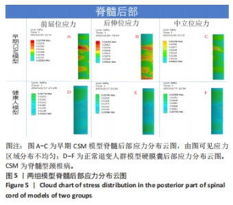
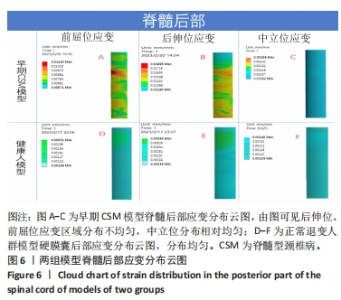
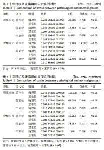
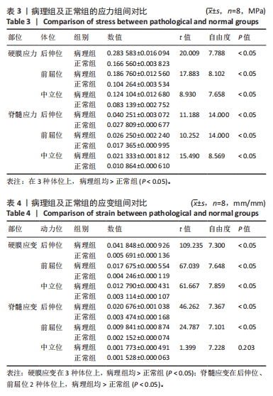
[29] 谭蓓, 李娜, 冯智超, 等. 脊髓型颈椎病患者三维有限元模型的构建与生物力学分析[J]. 中南大学学报(医学版),2019,44(5):507-514.
[30] XU ML, ZENG HZ, ZHENG LD, et al. Effect of degenerative factors on cervical spinal cord during flexion and extension: a dynamic finite element analysis. Biomech Model Mechanobiol. 2022;21(6):1743-1759.
[31] MUHLE C, WISKIRCHEN J, WEINERT D, et al. Biomechanical aspects of the subarachnoid space and cervical cord in healthy individuals examined with kinematic magnetic resonance imaging. Spine (Phila Pa 1976). 1998;23(5): 556-567.
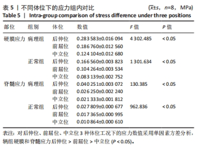
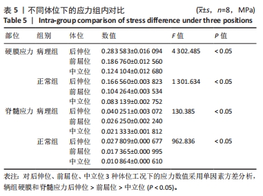
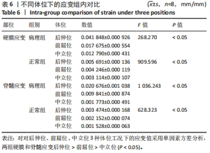
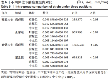
|