Chinese Journal of Tissue Engineering Research ›› 2023, Vol. 27 ›› Issue (36): 5760-5765.doi: 10.12307/2023.721
Previous Articles Next Articles
Finite element analysis of three internal fixation methods for distal tibial fractures
Wang Yichang1, Lin Wenjie2, Lin Tao1, Zhou Baian1, Huang Wenhua1, 3, Liang Zhenming1, Wei Jinsong1, Ouyang Hanbin1
- 1Orthopedic Center, Affiliated Hospital of Guangdong Medical University, Clinical Base of Guangdong Medical 3D Printing Application and Transformation Engineering Center, Zhanjiang 524002, Guangdong Province, China; 2Department of Extremities and Articular Orthopedics, Jiangmen Central Hospital, Jiangmen 529070, Guangdong Province, China; 3Department of Human Anatomy, School of Basic Medicine, Southern Medical University, Guangzhou 510515, Guangdong Province, China
-
Received:2022-09-21Accepted:2022-11-16Online:2023-12-28Published:2023-03-24 -
Contact:Wei Jinsong, MD, Chief physician, Orthopedic Center, Affiliated Hospital of Guangdong Medical University, Clinical Base of Guangdong Medical 3D Printing Application and Transformation Engineering Center, Zhanjiang 524002, Guangdong Province, China Ouyang Hanbin, MD, Associate chief physician, Orthopedic Center, Affiliated Hospital of Guangdong Medical University, Clinical Base of Guangdong Medical 3D Printing Application and Transformation Engineering Center, Zhanjiang 524002, Guangdong Province, China -
About author:Wang Yichang, Master candidate, Orthopedic Center, Affiliated Hospital of Guangdong Medical University, Clinical Base of Guangdong Medical 3D Printing Application and Transformation Engineering Center, Zhanjiang 524002, Guangdong Province, China -
Supported by:2018 Competitive Allocation Project of Special Science and Technology Development Fund of Zhanjiang, No. 2018A01036 (to OHB); Competitive Allocation Project of Special Fund for Science and Technology of Guangdong Province in 2021 (“Major Project+Task List”), No. 2021A05243 (to OHB)
CLC Number:
Cite this article
Wang Yichang, Lin Wenjie, Lin Tao, Zhou Baian, Huang Wenhua, Liang Zhenming, Wei Jinsong, Ouyang Hanbin. Finite element analysis of three internal fixation methods for distal tibial fractures[J]. Chinese Journal of Tissue Engineering Research, 2023, 27(36): 5760-5765.
share this article
Add to citation manager EndNote|Reference Manager|ProCite|BibTeX|RefWorks
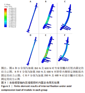
2.1 模型有效性验证 参照既往力学试验文献的加载条件,计算低轴向压缩工况中3组有限元模型的整体结构刚度,MDTP组为613.04 N/mm,RTN组为827.43 N/mm,ETN组为603.01 N/mm。与既往实体试验相比[16-17],ETN组为(888±249) N/mm,RTN 组为(609±149) N/mm,MDTP 组为(465±178) N/mm。总体上结果较为相近,证明了该有限元模型的有效性。 2.2 不同工况中3种内固定物应力分布及应力峰值 在轴向压缩工况中,3个模型组内固定物的应力分布云图,见图3。可以看出,分别施加350 N和600 N压缩载荷时,ETN组内固定高应力区主要位于E2主钉孔处,应力峰值分别为156.73 MPa和269.06 MPa;MDTP组内固定高应力区主要位于P3螺钉上,应力峰值为430.62 MPa和737.78 MPa;在RTN组内固定高应力区则位于R3螺钉上,应力峰值分别为145.81 MPa和242.67 MPa。"

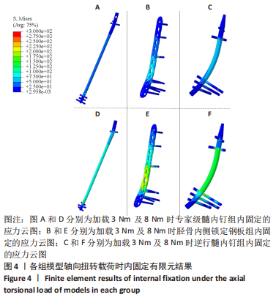
在轴向扭转工况中,3个模型组内固定物的应力分布云图,见图4。加载扭转3 Nm及8 Nm载荷时,ETN组内固定物的高应力区出现在骨折间隙间中未锁定的主钉孔内上,应力峰值分别为106.31 MPa及283.53 MPa;对于MDTP组,内固定的高应力区集中在P3螺钉上,应力峰值分别为450.27 MPa及913.07 MPa;RTN组在内固定的高应力区集中在R2螺钉的主钉孔上,应力峰值分别为163.54 MPa及435.42 MPa。"
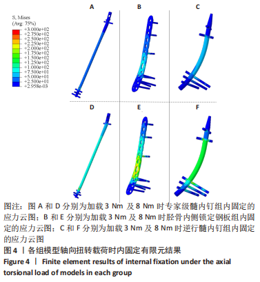
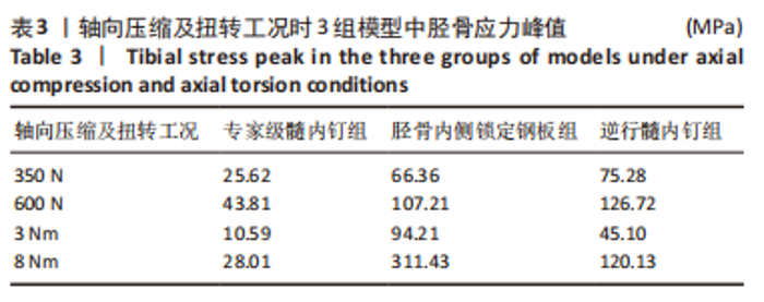
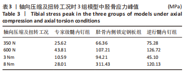
2.3 不同工况中3组模型胫骨应力分布及应力峰值 在轴向压缩工况中,3个模型组胫骨的应力峰值,见表3。在加载350 N及600 N载荷时,ETN组胫骨高应力区在E2内侧钉孔周围的皮质骨;MDTP组的胫骨高应力区在P4内侧钉孔周围的皮质骨;RTN组胫骨高应力区在R3内侧钉孔周围的皮质骨。在轴向扭转工况中,3个模型组胫骨的应力峰值,见表3。加载3 Nm及8 Nm扭转载荷,ETN组胫骨的高应力区也在E2钉孔外侧周围的皮质骨上;MDTP组中胫骨的高应力区为P3螺钉孔内侧周围的皮质骨;RTN组中胫骨高应力区在R3钉孔内侧周围皮质骨。"
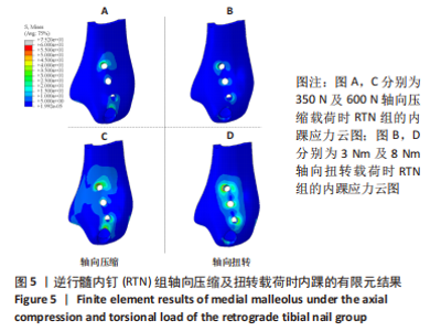
2.4 RTN内固定下内踝处骨骼的应力分布 RTN组内踝处的应力分布,见图5。在2种轴向压缩载荷时,内踝主钉孔周围皮质骨高应力区靠近骨折端,应力峰值分别为32.66 MPa及55.34 MPa。RTN组2种扭转载荷下主钉孔周围皮质骨高应力区靠近踝关节面,应力峰值为10.56 MPa及26.92 MPa。"

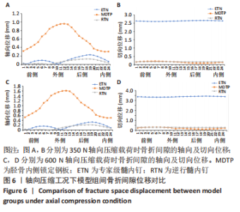
2.5 不同工况下3组模型组间的骨折间隙位移 在轴向压缩工况下,对比3组内固定模型的骨折间隙轴向位移结果,见图6。MDTP组所有区域均明显大于ETN及RTN组,MDTP组轴向位移最大值在外侧区,为1.62 mm;ETN组及RTN组则位于后侧区,为0.33 mm及0.22 mm。对比3组内固定模型的骨折间隙切向位移结果可以发现,ETN组所有区域均明显大于其他两组模型,MDTP及RTN骨折间隙切向位移基本一致,而MDTP组后内侧区位移要略低。"
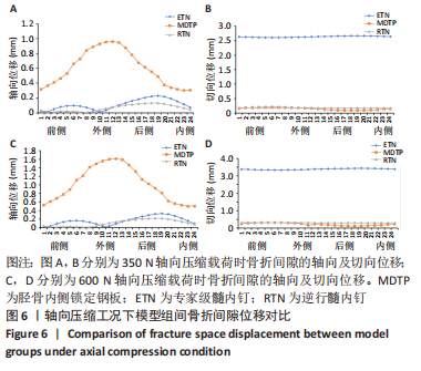
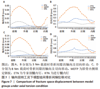
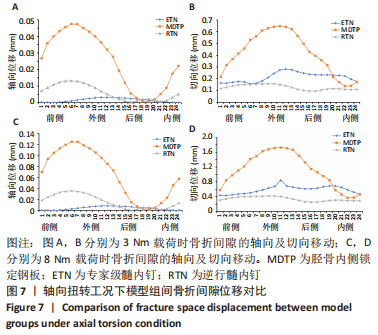
在轴向扭转工况下,对比3组内固定模型的骨折间隙轴向位移结果,见图7。MDTP组位移最大值位于前侧区,为0.13 mm;ETN组位于外侧区,为0.01 mm;RTN组则位于前侧区,为0.04 mm。对比3组内固定模型的骨折间隙切向位移结果,MDTP及ETN组在外侧区的位移值最大,分别为1.73 mm及0.84 mm;RTN组位移最大值则位于外侧区,为0.42 mm。"
| [1] 陈安富, 唐旭东, 黄凯. 髓内钉与钢板内固定治疗成人胫骨干远端骨折的疗效比较[J]. 中华创伤骨科杂志,2019,21(8):706-709. [2] SITNIK A, BELETSKY A, SCHELKUN S. Intra-articular fractures of the distal tibia: current concepts of management. Efort Open Rev. 2017;2(8): 352-361. [3] BLEEKER NJ, VAN DE WALL BJM, FFA IJ, et al. Plate vs. nail for extra-articular distal tibia fractures: How should we personalize surgical treatment? A meta-analysis of 1332 patients. Injury. 2021;52(3):345-357. [4] LIU XK, XU WN, XUE QY, et al. Intramedullary Nailing Versus Minimally Invasive Plate Osteosynthesis for Distal Tibial Fractures: A Systematic Review and Meta-Analysis. Orthop Surg. 2019;11(6):954-965. [5] HOFMANN GO, GONSCHOREK O, BÜHLER M, et al. Retrograde Nagelung der Tibia. Trauma Und Berufskrankheit. 2001;3(2 Supplement):S303-S308. [6] KUHN S, APPELMANN P, PAIRON P, et al. The Retrograde Tibial Nail: presentation and biomechanical evaluation of a new concept in the treatment of distal tibia fractures. Injury. 2014;45 Suppl 1:S81-S86. [7] KUHN S, APPELMANN P, MEHLER D, et al. Retrograde tibial nailing: a minimally invasive and biomechanically superior alternative to angle-stable plate osteosynthesis in distal tibia fractures. J Orthop Surg Res. 2014;9:35. [8] CHEN F, HUANG X, YA Y, et al. Finite element analysis of intramedullary nailing and double locking plate for treating extra-articular proximal tibial fractures. J Orthop Surg Res. 2018;13(1):12. [9] 杨志刚, 甘霖, 叶俊星. 胫骨远端骨折有限元模型的建立及稳定性分析[J]. 中国组织工程研究,2016,20(17):2540-2545. [10] SOWMIANARAYANAN S, CHANDRASEKARAN A, KUMAR RK. Finite element analysis of a subtrochanteric fractured femur with dynamic hip screw, dynamic condylar screw, and proximal femur nail implants-a comparative study. Proc Inst Mech Eng H. 2008;222(1):117-127. [11] 贾军锋, 唐承杰, 乐劲涛, 等. 胫骨远端骨折3种不同固定方式的有限元分析[J]. 中国组织工程研究,2019,23(32):5188-5194. [12] 张强, 巫宗德, 刘亮, 等. 胫骨内侧、外侧解剖锁定钢板固定胫骨中下段外旋型螺旋骨折的有限元分析[J]. 中国组织工程研究,2022,26(36):5750-5754. [13] GREENFIELD J, APPELMANN P, LAFON Y, et al. A comparative biomechanical study of the Distal Tibia Nail against compression plating for the osteosynthesis of supramalleolar corrective osteotomies. Sci Rep. 2021;11(1):1-12. [14] BEIRAMI S, NIKKHOO M, HASSANI K, et al. A comparative finite element simulation of locking compression plate materials for tibial fracture treatment. Comput Methods Biomech Biomed Engin. 2021;24(10): 1064-1072. [15] ALBAREDA J, IBARZ E, MATEO J, et al. Are the unreamed nails indicated in diaphyseal fractures of the lower extremity? A biomechanical study. Injury. 2021;52 Suppl 4: S61-s70. [16] KUHN S, APPELMANN P, PAIRON P, et al. A new angle stable nailing concept for the treatment of distal tibia fractures. Int Orthop. 2014;38(6):1255-1260. [17] KUHN S, GREENFIELD J, ARAND C, et al. Treatment of distal intraarticular tibial fractures: A biomechanical evaluation of intramedullary nailing vs. angle-stable plate osteosynthesis. Injury. 2015;46 Suppl 4:S99-S103. [18] BLEEKER NJ, REININGA IHF, VAN DE WALL BJM, et al. Difference in Pain, Complication Rates, and Clinical Outcomes After Suprapatellar Versus Infrapatellar Nailing for Tibia Fractures? A Systematic Review of 1447 Patients. J Orthop Trauma. 2021;35(8):391-400. [19] HU L, XIONG Y, MI B, et al. Comparison of intramedullary nailing and plate fixation in distal tibial fractures with metaphyseal damage: a meta-analysis of randomized controlled trials. J Orthop Surg Res. 2019; 14(1): 30. [20] SHEN J, XU J, TANG MJ, et al. Extra-articular distal tibia facture (AO-43A): A retrospective study comparing modified MIPPO with IMN. Injury. 2016;47(10): 2352-2359. [21] WANG B, ZHAO Y, WANG Q, et al. Minimally invasive percutaneous plate osteosynthesis versus intramedullary nail fixation for distal tibial fractures: a systematic review and meta-analysis. J Orthop Surg Res. 2019;14(1):456. [22] BAHAMONDE L, ZAMORANO A, ZECCHETTO P. Far proximal and far distal tibial fractures: management with intramedullary nails, Tibia Pathology and Fractures: Intech Open, 2020. [23] GIANNOUDIS PV, TZIOUPIS C, PAPE HC. Fat embolism: the reaming controversy. Injury. 2006;37 Suppl 4:S50-S58. [24] MORIZANE K, TAKEMOTO M, NEO M, et al. Occipital and external acoustic meatus to axis angle: a useful predictor of oropharyngeal space in rheumatoid arthritis patients with atlantoaxial subluxation. J Neurosurg Spine. 2019:1-8. doi: 10.3171/2019.3.SPINE181390. [25] 胡翰生, 王静成, 卢志华, 等. 髋关节三维有限元模型的构建及关节囊修复预后分析[J]. 南方医科大学学报,2020,40(12):1826-1830. [26] 林奕鹏, 岳凯, 徐盈舒, 等. 新型可吸收空心螺钉结合不可吸收缝线张力带固定治疗髌骨骨折:与传统改良张力带的对比[J]. 中国组织工程研究,2019,23(22):3506-3511. [27] SAMSAMI S, HERRMANN S, PÄTZOLD R, et al. Finite element analysis of Bi-condylar Tibial Plateau fractures to assess the effect of coronal splits. Med Eng Phys. 2020; 84:84-95. [28] DENG Y, OUYANG H, XIE P, et al. Biomechanical assessment of screw safety between far cortical locking and locked plating constructs. Comput Methods Biomech Biomed Engin. 2021;24(6):663-672. [29] GIANNOUDIS PV, GIANNOUDIS VP. Far cortical locking and active plating concepts: New revolutions of fracture fixation in the waiting? Injury. 2017;48(12):2615-2618. [30] AUGAT P, HOLLENSTEINER M, VON RÜDEN C. The role of mechanical stimulation in the enhancement of bone healing. Injury. 2021;52 Suppl 2:S78-S83. [31] NOURISA J, ROUHI G. Biomechanical evaluation of intramedullary nail and bone plate for the fixation of distal metaphyseal fractures. J Mech Behav Biomed Mater. 2016;56:34-44. [32] 史金友, 肖玉周, 吴敏, 等. 微动本质及骨折愈合生物力学分期的研究[J]. 中国修复重建外科杂志,2021,35(9):1205-1211. [33] EPARI DR, TAYLOR WR, HELLER MO, et al. Mechanical conditions in the initial phase of bone healing. Clin Biomech (Bristol, Avon). 2006;21(6):646-655. [34] SCHELL H, EPARI DR, KASSI JP, et al. The course of bone healing is influenced by the initial shear fixation stability. J Orthop Res. 2005;23(5):1022-1028. [35] 李刚, 杜新辉. 专家型胫骨髓内钉和锁定加压钢板内固定术在胫骨远端骨折治疗中的临床疗效对比观察[J]. 创伤外科杂志,2017,19(8):590-594. [36] 何敏, 李正茂, 谭文甫, 等. 新型逆行胫骨髓内钉治疗胫骨远端骨折的初步疗效分析[J]. 中华创伤骨科杂志,2022,24(4):334-338. [37] BASERI A, BAGHERI MA, ROUHI G, et al. Fixation of distal tibia fracture through plating, nailing, and nailing with Poller screws: A comparative biomechanical-based experimental and numerical investigation. Proc Inst Mech Eng H. 2020; 234(10):1129-1138. [38] PENG B, WAN T, TAN W, et al. Novel Retrograde Tibial Intramedullary Nailing for Distal Tibial Fractures. Front Surg. 2022;9:899483. [39] GARDNER MP, CHONG AC, POLLOCK AG, et al. Mechanical evaluation of large-size fourth-generation composite femur and tibia models. Ann Biomed Eng. 2010; 38(3):613-620. [40] 张雪玲, 任武, 王淑婷, 等. 人体下肢生物力学仿真和实验的研究现状与发展趋势[J]. 生物医学工程研究,2021,40(4):448-454. [41] BUZZATTI L, KEELSON B, VANLAUWE J, et al. Evaluating lower limb kinematics and pathology with dynamic CT. Bone Joint J. 2021;103-b (5):822-827. |
| [1] | Zhong Yizheng, Huang Peizhen, Cai Qunbin, Zheng Liqin, He Xingpeng, Dong Hang. Microstructural indexes that determine the trabecular bone maximum stress of micro-finite element models [J]. Chinese Journal of Tissue Engineering Research, 2023, 27(9): 1313-1318. |
| [2] | Wu Taoguang, Nie Shaobo, Chen Hua, Zhu Zhengguo, Qi Lin, Tang Peifu. Biomechanical characteristics of a new multi-dimensional cross locking plate in the treatment of subtrochanteric nonunion [J]. Chinese Journal of Tissue Engineering Research, 2023, 27(9): 1330-1334. |
| [3] | Peng Zhixin, Yan Wengang, Wang Kun, Zhang Zhenjiang. Finite element analysis and structural optimization design of 3D printed forearm braces [J]. Chinese Journal of Tissue Engineering Research, 2023, 27(9): 1340-1345. |
| [4] | Wu Tianliang, Tao Xiuxia, Xu Hongguang. Influence of different bone mineral densities on cage subsidence after stand-alone oblique lateral interbody fusion: three-dimensional finite element analysis [J]. Chinese Journal of Tissue Engineering Research, 2023, 27(9): 1352-1358. |
| [5] | Liu Jinyu, Zhang Hanshuo, Cui Hongpeng, Pan Lingzhi, Zhao Boran, Li Fei, Ding Yu. Finite element biomechanical analysis of minimally invasive treatment of cervical spondylotic myelopathy and accurate exercise rehabilitation [J]. Chinese Journal of Tissue Engineering Research, 2023, 27(9): 1359-1364. |
| [6] | He Yujie, Kang Zhijie, Xue Mingming, Jin Feng, Li Zhijun, Wang Xing, Xu Yangyang, Gao Mingjie, Li Jiawei, Li Xiaohe, Wang Haiyan. Finite element analysis of transarticular screw fixation of adolescent thoracic vertebra [J]. Chinese Journal of Tissue Engineering Research, 2023, 27(9): 1365-1370. |
| [7] | Wen Xinghua, Ding Huanwen, Cheng Kai, Yan Xiaonan, Peng Yuanhao, Wang Yuning, Liu Kang, Zhang Huiwu. Three-dimensional finite element model analysis of intramedullary nailing fixation design for large femoral defects in Beagle dogs [J]. Chinese Journal of Tissue Engineering Research, 2023, 27(9): 1371-1376. |
| [8] | Zheng Bo, Zhang Xiuli, Zhou Hao, He Zebi, Zhou Jin, Zhou Weiyun, Li Peng. Arthroscopy-assisted locking hollow screw fixation and open reduction plate internal fixation in the treatment of Schatzker II-III tibial plateau fractures: early CT evaluation [J]. Chinese Journal of Tissue Engineering Research, 2023, 27(9): 1410-1416. |
| [9] | Liu Jiaxin, Jia Peng, Men Yutao, Liu Lu, Wang Yeming, Ye Jinduo. Design and optimization of bone trabecular structure with triply periodic minimal surfaces [J]. Chinese Journal of Tissue Engineering Research, 2023, 27(7): 992-997. |
| [10] | Guo Tingting, Xie Hong, Xu Guanghua. Finite element analysis of elastic ankle brace performance [J]. Chinese Journal of Tissue Engineering Research, 2023, 27(7): 1031-1037. |
| [11] | Zhu Lin, Gu Weiping, Wang Can, Chen Gang. Biomechanical analysis of All-on-Four and pterygomaxillary implants under different maxillary bone conditions [J]. Chinese Journal of Tissue Engineering Research, 2023, 27(7): 985-991. |
| [12] | Sun Jiangwei, Wang Junxiang, Baibujiafu·Yellisi, Dai Huijuan, Nijati·Turson. Three-dimensional finite element analysis of stress distribution in different smooth collar implants [J]. Chinese Journal of Tissue Engineering Research, 2023, 27(7): 1004-1011. |
| [13] | Jiang Yifang, Cai Qimin, Chu Zhengyi, Qin Min, Shen Yurong, Gu Yuanping. Simulation analysis of stress distribution of NRT FILES in curved root canals [J]. Chinese Journal of Tissue Engineering Research, 2023, 27(7): 1038-1042. |
| [14] | Li Huiqin, Wang Chunjuan, Wang Yu, Wang Weifeng, Chen Dinggen, Li Na. Clear aligner orthodontic therapy of rotated mandibular teeth with different shapes: a three-dimensional finite element analysis [J]. Chinese Journal of Tissue Engineering Research, 2023, 27(7): 1050-1054. |
| [15] | Liu Qinghua, Cai Yongqiang, Jin Feng, Yu Jinghong, Wang Haiyan, Zhang Yunfeng, Wang Lidong, Li Jiawei, Wang Xing, He Yujie, Dai Lina, Wang Jianzhong, Wu Chao, Tong Ling, Kang Zhijie, Li Zhijun, Li Xiaohe. Finite element model of the 12-year-old child whole cervical spine: establishment and validity verification based on CT data [J]. Chinese Journal of Tissue Engineering Research, 2023, 27(4): 500-504. |
| Viewed | ||||||
|
Full text |
|
|||||
|
Abstract |
|
|||||