Chinese Journal of Tissue Engineering Research ›› 2023, Vol. 27 ›› Issue (29): 4658-4663.doi: 10.12307/2023.651
Previous Articles Next Articles
Effect of iron overload on cartilaginous tissue in a mouse model of knee osteoarthritis evaluated by micro-CT
Li Shaocong1, He Qi1, Pan Zhaofeng1, Yang Junzheng1, Chen Bohao1, Wang Haibin2, Zhou Chi2
- 1Guangzhou University of Chinese Medicine, Guangzhou 510405, Guangdong Province, China; 2First Affiliated Hospital, Guangzhou University of Chinese Medicine, Guangzhou 510405, Guangdong Province, China
-
Received:2022-07-05Accepted:2022-08-24Online:2023-10-18Published:2022-12-02 -
Contact:Zhou Chi, MD, Associate professor, Associate chief physician, Master’s supervisor, First Affiliated Hospital, Guangzhou University of Chinese Medicine, Guangzhou 510405, Guangdong Province, China Wang Haibin, MD, Professor, Chief physician, Doctoral supervisor, First Affiliated Hospital, Guangzhou University of Chinese Medicine, Guangzhou 510405, Guangdong Province, China -
About author:Li Shaocong, Master candidate, Guangzhou University of Chinese Medicine, Guangzhou 510405, Guangdong Province, China -
Supported by:National Natural Science Foundation of China, No. 82074462 (to WHB); Guangzhou University of Traditional Chinese Medicine "Double First-Class" and High-Level University Discipline Collaborative Innovation Team Project, No. 2021xk53 (to WHB)
CLC Number:
Cite this article
Li Shaocong, He Qi, Pan Zhaofeng, Yang Junzheng, Chen Bohao, Wang Haibin, Zhou Chi. Effect of iron overload on cartilaginous tissue in a mouse model of knee osteoarthritis evaluated by micro-CT[J]. Chinese Journal of Tissue Engineering Research, 2023, 27(29): 4658-4663.
share this article
Add to citation manager EndNote|Reference Manager|ProCite|BibTeX|RefWorks
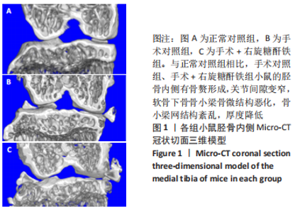
2.1 实验动物数量分析 手术+右旋糖酐铁组有1只小鼠于手术过程中死亡,系实验人员操作失误所致,给予补充造模,最终30只小鼠全部进入结果分析。 2.2 各组小鼠胫骨标本Micro-CT检测结果 三维成像显示,与正常对照组相比,手术对照组、手术+右旋糖酐铁组小鼠的胫骨内侧有骨赘形成,关节间隙变窄,软骨下骨骨小梁骨微结构恶化,骨小梁网结构紊乱,厚度降低,见图1。"
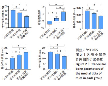
据各组小鼠胫骨内侧软骨下骨的骨小梁参数检测结果,与正常对照组相比,手术对照组、手术+右旋糖酐铁组骨小梁相对体积、骨小梁厚度、骨小梁数量明显减小(P < 0.05),骨小梁分离度、结构模型指数增加(P < 0.05);与手术+右旋糖酐铁组比较,手术对照组骨小梁相对体积、骨小梁厚度、骨小梁数量增加(P < 0.05),骨小梁分离度、结构模型指数相减小(P < 0.05),见图2。"

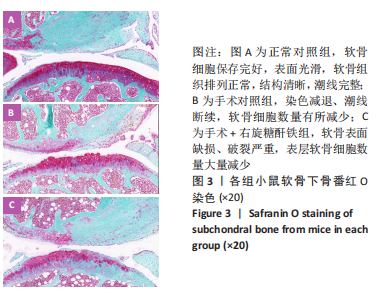
2.3 各组小鼠胫骨标本番红O染色结果 见图3。"


正常对照组软骨细胞保存完好,表面光滑,软骨组织排列正常,结构清晰,潮线完整;手术对照组染色减退、潮线断续,软骨细胞数量有所减少,OARSI评分有一定升高;软骨下骨与正常对照组、手术对照组相比,手术+右旋糖酐铁组小鼠软骨表面缺损、破裂严重,表层软骨细胞数量大量减少,OARSI评分更高,这些参数与图1三维成像相符合,表明手术+右旋糖酐铁组小鼠关节炎相比正常对照组更加严重。 正常对照组、手术对照组、手术+右旋糖酐铁组OARSI评分分别为0,2.30±0.52,7.40±1.20,组间两两比较差异均有显著性意义(P < 0.05)。 2.4 各组小鼠胫骨标本普鲁士蓝染色结果 见图4。"
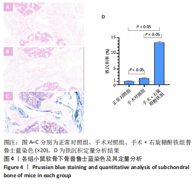

与正常对照组、手术对照组相比,手术+右旋糖酐铁组小鼠滑膜细胞质中可见大量被染成深蓝色的铁沉积颗粒,正常对照组、手术对照组小鼠滑膜细胞质中无明显铁沉积颗粒,提示铁超载造模成功。"

| [1] SAFIRI S, KOLAHI AA, SMITH E, et al. Global, regional and national burden of osteoarthritis 1990-2017: a systematic analysis of the Global Burden of Disease Study 2017. Ann Rheum Dis. 2020;79(6):819-828. [2] 窦晓丽,段晓琴,夏玲,等.骨关节炎:关节软骨退变的相关研究与进展[J].中国组织工程研究与临床康复,2011,15(20):3763-3766. [3] HUNTER DJ, SCHOFIELD D, CALLANDER E. The individual and socioeconomic impact of osteoarthritis. Nat Rev Rheumatol. 2014; 10(7):437-441. [4] HULSHOF CTJ, COLOSIO C, DAAMS JG, et al. WHO/ILO work-related burden of disease and injury: Protocol for systematic reviews of exposure to occupational ergonomic risk factors and of the effect of exposure to occupational ergonomic risk factors on osteoarthritis of hip or knee and selected other musculoskeletal diseases. Environ Int. 2019;125:554-566. [5] FELSON DT, ZHANG YQ. An update on the epidemiology of knee and hip osteoarthritis with a view to prevention. Arthritis Rheum. 1998;41(8): 1343-1355. [6] GKRETSI V, Simopoulou T, Tsezou A. Lipid metabolism and osteoarthritis: Lessons from atherosclerosis. Prog Lipid Res. 2011;50(2): 133-140. [7] BURR DB, GALLANT MA. Bone remodelling in osteoarthritis. Nat Rev Rheumatol. 2012;8(11):665-673. [8] BERENBAUM F, EYMARD F, HOUARD X. Osteoarthritis, inflammation and obesity. Curr Opin Rheumatol. 2013;25(1):114-118. [9] AZAMAR-LLAMAS D, HERNANDEZ-MOLINA G, RAMOS-AVALOS B, et al. Adipokine Contribution to the Pathogenesis of Osteoarthritis. Mediators Inflamm. 2017;2017:5468023. [10] DIXON SJ. Ferroptosis: bug or feature? Immunol Rev. 2017;277(1):150-157. [11] CALIŞKAN U, TONGUÇ MO, CIRIŞ M, et al. The Investigation of Gingival Iron Accumulation in Thalassemia Major Patients. J Pediatr Hematol Oncol. 2011;33(2):98-102. [12] 贾鹏,张增利,徐又佳,等.铁超载对小鼠单核细胞RAW264.7向破骨细胞分化的影响[J].中华骨质疏松和骨矿盐疾病杂志,2011,4(1): 44-49. [13] YU YY, JIANG L, WANG H, et al. Hepatic transferrin plays a role in systemic iron homeostasis and liver ferroptosis. Blood. 2020;136(6): 726-739. [14] LI J, CAO F, YIN HL, et al. Ferroptosis: past, present and future. Cell Death Dis. 2020;11(2):88. [15] YAO XD, SUN K, YU SN, et al. Chondrocyte ferroptosis contribute to the progression of osteoarthritis. J Orthop Translat. 2021;27:33-43. [16] MELCHIORRE D, MANETTI M, MATUCCI-CERINIC M. Pathophysiology of Hemophilic Arthropathy. J Clin Med. 2017;6(7):63. [17] WALDSTEIN W, PERINO G, GILBERT SL, et al. OARSI Osteoarthritis Cartilage Histopathology Assessment System: A Biomechanical Evaluation in the Human Knee. J Orthop Res. 2016;34(1):135-140. [18] MCILWRAITH CW, FRISBIE DD, KAWCAK CE, et al. The OARSI histopathology initiative - recommendations for histological assessments of osteoarthritis in the horse. Osteoarthritis Cartilage. 2010;18:S93-S105. [19] VAN LINTHOUDT D. Evaluation of the Adhesion To the Eular and Oarsi Recommendations for the Treatment of Knee and Hip Osteoarthritis in Current Practice. Osteoarthritis Cartilage. 2010;18:S245-S246. [20] 娄思权.骨关节炎的病理与发病因素[J].中华骨科杂志,1996,16(1): 57-60. [21] 邱贵兴.骨关节炎流行病学和病因学新进展[J].继续医学教育,2005, 19(7): 68-69. [22] 王斌,邢丹,董圣杰,等.中国膝骨关节炎流行病学和疾病负担的系统评价[J].中国循证医学杂志,2018,18(2):134-142. [23] LAWRENCE RC, FELSON DT, HELMICK CG, et al. Estimates of the prevalence of arthritis and other rheumatic conditions in the United States. Part II. Arthritis Rheum. 2008;58(1):26-35. [24] KRADER CG. Guidance on non-surgical management of knee osteoarthritis. Med Econ. 2014;91(9):12. [25] WANG X, CHEN B, SUN J, et al. Iron-induced oxidative stress stimulates osteoclast differentiation via NF-kappaB signaling pathway in mouse model. Metabolism. 2018;83:167-176. [26] YANG J, ZHANG J, DING C, et al. Regulation of Osteoblast Differentiation and Iron Content in MC3T3-E1 Cells by Static Magnetic Field with Different Intensities. Biol Trace Elem Res. 2018;184(1):214-225. [27] ISHII KA, FUMOTO T, IWAI K, et al. Coordination of PGC-1beta and iron uptake in mitochondrial biogenesis and osteoclast activation. Nat Med. 2009;15(3):259-266. [28] JENEY V. Clinical Impact and Cellular Mechanisms of Iron Overload-Associated Bone Loss. Front Pharmacol. 20171;8:77. [29] JING XZ, DU T, LI T, et al. The detrimental effect of iron on OA chondrocytes: Importance of pro-inflammatory cytokines induced iron influx and oxidative stres. J Cell Mol Med. 2021;25(12):5671-5680. [30] BLANCO FJ, REGO I, RUIZ-ROMERO C. The role of mitochondria in osteoarthritis. Nat Rev Rheumatol. 2011;7(3): 161-169. [31] CEN WJ, FENG Y, LI SS, et al. Iron overload induces G1 phase arrest and autophagy in murine preosteoblast cells. J Cell Physiol. 2018;233(9): 6779-6789. [32] AXFORD JS, BOMFORD A, REVELL P, et al. Hip arthropathy in genetic hemochromatosis. Radiographic and histologic features. Arthritis Rheum. 1991;34(3):357-361. [33] HEILAND GR, AIGNER E, DALLOS T, et al. Synovial immunopathology in haemochromatosis arthropathy. Ann Rheum Dis. 2010;69(6):1214-1219. [34] CHENG Q, ZHANG X, JIANG J, et al. Postmenopausal Iron Overload Exacerbated Bone Loss by Promoting the Degradation of Type I Collage. Biomed Res Int. 2017;2017:1345193. [35] WANG L, JIA P, SHAN Y, et al. Synergistic protection of bone vasculature and bone mass by desferrioxamine in osteoporotic mice. Mol Med Rep. 2017;16(5):6642-6649. [36] BURTON LH, RADAKOVICH LB, MAROLF AJ, et al. Systemic iron overload exacerbates osteoarthritis in the strain 13 guinea pig. Osteoarthritis Cartilage. 2020;28(9):1265-1275. [37] FLYNN C, HURTIG M, LINDEN AZ. Anionic Contrast-Enhanced MicroCT Imaging Correlates with Biochemical and Histological Evaluations of Osteoarthritic Articular Cartilage. Cartilage. 2021;13(2_suppl): 1388S-1397S. [38] 许宋锋,王臻.Micro-CT在骨科的应用和进展[J].中国骨肿瘤骨病, 2004,3(4):236-241+249. [39] 李冠武,汤光宇.Micro-CT及~1H-MRS在骨质疏松骨质量研究中的应用[J].国际医学放射学杂志,2010,33(6):525-528. [40] 魏占英,章振林. Micro-CT在骨代谢研究中骨微结构指标的解读及应用价值[J].中华骨质疏松和骨矿盐疾病杂志,2018,11(2):200-205. [41] BOUXSEIN ML, BOYD SK, CHRISTIANSEN BA, et al. Guidelines for assessment of bone microstructure in rodents using micro-computed tomography. J Bone Miner Res. 2010;25(7):1468-1486. [42] 杨真,罗海吉.缺铁性贫血及补铁剂研究进展[J].国外医学(卫生学分册),2006,33(2):90-93. [43] 王方海,赵维,陈建芳,等.补铁剂研究进展[J].药学进展,2016,40(9): 680-688. [44] JING X, LIN J, DU T, et al. Iron Overload Is Associated With Accelerated Progression of Osteoarthritis: The Role of DMT1 Mediated Iron Homeostasis. Front Cell Dev Biol. 2020;8:594509. [45] 刘卫华,唐曦,王智,等.Micro CT三维重建手舟骨相关显微影像解剖学数据测量[J].中国组织工程研究,2014,18(22):3537-3541. [46] 王德志,陈世昌,梁正洋,等. Micro CT定量研究去卵巢山羊胫骨平台松质骨微结构特点[J].中国组织工程研究,2014,18(24):3773-3778. [47] 彭云海,王琳,刘太运.退行性膝骨关节炎的影像学对比分析[J].中国现代医生,2012,50(27):97-98+101. [48] 陈连旭,余家阔,于长隆,等.大鼠、兔和人关节软骨组织形态学的比较[J].中国组织工程研究与临床康复,2007,11(41):8230-8233. [49] RAO VA. Iron chelators with topoisomerase-inhibitory activity and their anticancer applications. Antioxid Redox Signal. 2013;18(8):930-955. [50] JANSOVA H, SIMUNEK T. Cardioprotective Potential of Iron Chelators and Prochelators. Curr Med Chem. 2019;26(2):288-301. |
| [1] | Li Xiaomin, Tian Xiangdong, Tan Yetong, Zhu Guangyu, Wang Rongtian, Wang Jian, Xue Zhipeng, Ma Sheng, Hu Yuanyi, Huang Ye, Ding Tiansong. Changes of lower limb force line and knee function after high tibial osteotomy in osteoporotic medial ventricular knee osteoarthritis [J]. Chinese Journal of Tissue Engineering Research, 2023, 27(9): 1325-1329. |
| [2] | Zhang Lichuang, Gao Huali, Wang Jingchao, Lin Huijun, Wu Chonggui, Ma Yinghui, Huang Yunfei, Fang Xue, Zhai Weitao. Effect of tendon manipulation with equal emphasis on muscles and bones on accelerating the functional rehabilitation of quadriceps femoris after total knee arthroplasty [J]. Chinese Journal of Tissue Engineering Research, 2023, 27(9): 1383-1389. |
| [3] | Bi Gengchao, Zhang Yanlong, Li Qiuyue, Hu Longwei, Zhang Yu. Knee joint mechanics and activation characteristics of surrounding muscles during deep jumps at different heights and distances [J]. Chinese Journal of Tissue Engineering Research, 2023, 27(8): 1211-1218. |
| [4] | Xiong Bohan, Yu Yang, Lu Xiaojun, Wang Xu, Yang Tengyun, Zhang Yaozhang, Liao Xinyu, Zhou Xiaoxiang, He Lu, Li Yanlin. Research progress in promoting tendon to bone healing during anterior cruciate ligament reconstruction [J]. Chinese Journal of Tissue Engineering Research, 2023, 27(5): 779-786. |
| [5] | Li Yaping, Liu Hong, Gao Zhen, Chen Xiaolin, Huang Wujie, Jiang Zheng. Three-dimensional motion analysis of lower limb biomechanical performance in Tai Chi practitioners accompanied by knee joint pain [J]. Chinese Journal of Tissue Engineering Research, 2023, 27(4): 520-526. |
| [6] | Wei Bo, Yao Qingqiang, Tang Cheng, Li Xuxiang, Xu Yan, Wang Liming. Advantage of medial pivot prosthesis in total knee arthroplasty via medial subvastus approach [J]. Chinese Journal of Tissue Engineering Research, 2023, 27(4): 552-557. |
| [7] | Wan Guoli, Shi Chenhui, Wang Weishan, Li Ang, Shi Xunda, Cai Yi. Retrospective analysis of the influencing factors of chronic pain after total knee arthroplasty [J]. Chinese Journal of Tissue Engineering Research, 2023, 27(4): 558-564. |
| [8] | Gu Mingxi, Wang Bo, Tian Fengde, An Ning, Hao Ruihu, Wang Changcheng, Guo Lin. Comparison of early efficacy and safety of simultaneous and staged bilateral total knee arthroplasty [J]. Chinese Journal of Tissue Engineering Research, 2023, 27(4): 565-571. |
| [9] | Zhang Jinbiao, Li Xiaoming, Xing Wanlin, Ma Fei, Yu Qiaoya, Dai Rongqin. Early warning efficacy of thrombus molecular markers after total knee arthroplasty in patients complicated with venous thromboembolism [J]. Chinese Journal of Tissue Engineering Research, 2023, 27(4): 572-577. |
| [10] | Guo Yingqi, Gong Xianxu, Zhang Yan, Xiao Han, Wang Ye, Gu Wenguang. Meniscus extrusion and patellofemoral joint cartilage injury and bone marrow lesions: MRI semi-quantitative score [J]. Chinese Journal of Tissue Engineering Research, 2023, 27(4): 600-605. |
| [11] | Xu Xiangjun, Wang Chao, Song Qunshan, Li Bingyan, Zhang Jichao, Wang Guodong, Dong Yuefu. Optimal angle for prosthesis implantation in total knee arthroplasty [J]. Chinese Journal of Tissue Engineering Research, 2023, 27(4): 612-618. |
| [12] | Li Shihao, Li Qi, Li Zhen, Zhang Yuanyuan, Liu Miaomiao, Ouyang Yi, Xu Weiguo. Plantar pressure and gait analysis in patients with anterior cruciate ligament injury and reconstruction [J]. Chinese Journal of Tissue Engineering Research, 2023, 27(4): 626-631. |
| [13] | Yu Jiaan, Liu Xinwei, Lian Hongyu, Liu Kexin, Li Zitao. Medial open-wedge tibial osteotomy versus lateral closed-wedge tibial osteotomy for unicompartmental knee osteoarthritis: a meta-analysis [J]. Chinese Journal of Tissue Engineering Research, 2023, 27(4): 632-639. |
| [14] | Xu Yangyang, He Peiliang, Meng Qingqi, Li Siming. A meta-analysis of the effects of continuous adductor canal block and continuous femoral nerve block on early activity after knee arthroplasty [J]. Chinese Journal of Tissue Engineering Research, 2023, 27(4): 640-645. |
| [15] | Chai Hao, Yang Deyong, Zhang Lei, Shu Li. 3D printing personalized osteotomy guide technology versus conventional total knee arthroplasty on the accuracy of lower limb force alignment: a meta-analysis [J]. Chinese Journal of Tissue Engineering Research, 2023, 27(4): 646-654. |
| Viewed | ||||||
|
Full text |
|
|||||
|
Abstract |
|
|||||