Chinese Journal of Tissue Engineering Research ›› 2023, Vol. 27 ›› Issue (27): 4385-4392.doi: 10.12307/2023.613
Previous Articles Next Articles
Three-dimensional finite element method to analyze biomechanical characteristics of spinal manipulation
Liu Baijie, Zhou Honghai, He Xinyu, Qin Hongtu, Chen Longhao, Tian Junming, Lu Qingwang
- School of Orthopedics and Traumatology, Guangxi University of Chinese Medicine, Nanning 530001, Guangxi Zhuang Autonomous Region, China
-
Received:2022-05-24Accepted:2022-08-05Online:2023-09-28Published:2022-11-08 -
Contact:Zhou Honghai, MD, Professor, Chief physician, School of Orthopedics and Traumatology, Guangxi University of Chinese Medicine, Nanning 530001, Guangxi Zhuang Autonomous Region, China -
About author:Liu Baijie, Master candidate, School of Orthopedics and Traumatology, Guangxi University of Chinese Medicine, Nanning 530001, Guangxi Zhuang Autonomous Region, China -
Supported by:the National Natural Science Foundation of China (Regional Project), No. 81360552, 81660800 (to ZHH); Key Subject of Guangxi First-Class Discipline Construction Project, No. 2018XK057 (to ZHH); the Project of Improving the Basic Scientific Research Ability of Young and Middle-Aged Teachers in Guangxi Universities, No. 2022KY0272 (to CLH); Guangxi Postgraduate Education Innovation Project, No. YCSZ2022010 (to LBJ)
CLC Number:
Cite this article
Liu Baijie, Zhou Honghai, He Xinyu, Qin Hongtu, Chen Longhao, Tian Junming, Lu Qingwang. Three-dimensional finite element method to analyze biomechanical characteristics of spinal manipulation[J]. Chinese Journal of Tissue Engineering Research, 2023, 27(27): 4385-4392.
share this article
Add to citation manager EndNote|Reference Manager|ProCite|BibTeX|RefWorks
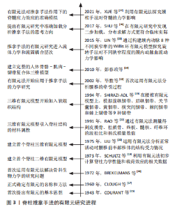
2.1 有限元法的发展历程 有限元法是一种求解微分方程的数值方法,其基本原理是将一个物体离散为有限的体单元,体单元之间通过有限个节点相连接,力则通过结点传递,使得每个体单元的变形、任意体单元或节点的应力分布可通过多种简单的方程式来求解[14]。自1943年提出有限元基本思想以来[15],有限元法的理论及应用发展迅速,在固体力学、流体力学、电磁学、温度场及生物力学等领域有着广泛的应用[16-17],20世纪70年代医学研究领域引入有限元法,1972年BREKELMANS等[18]首次运用有限元法解决骨生物力学的研究问题,此后的脊柱医学领域有了新的研究途径。 脊柱有限元分析研究的发展历程主要经历以下几个阶段:①二维有限元模型的建立。1973年,SCHULTZ等[19]最早建立椎间盘二维有限元模型探究脊柱的生物力学特性,提出韧带和小关节在脊柱承载载荷中的重要作用,初步计算脊柱力学性能和载荷效应的相关数据。②三维有限元模型的建立。1975年,LIU等[20]首次构建腰椎三维有限元模型以分析正常活动时腰椎前半部椎体的结构受力情况,但未完成椎体附件、椎旁软组织等层次的模拟和分析。③脊柱结构材料属性的引入。SIMON等[21]考虑到椎间盘的固液二相性,建立了腰椎间盘的多孔弹性有限元模型,RAO等[22]通过构建不同材料属性的椎体模型进行压力与位移实验测量,分别得到皮质骨、松质骨、终板、髓核、纤维环的波松比和杨氏模量数值。④椎旁软组织的加入。1994年,SHIRAZI-ADL等[23]在腰椎三维有限元模型的基础上,模拟前纵韧带、后纵韧带、关节囊韧带、黄韧带、横突间韧带、棘间韧带和棘上韧带等9种韧带,并证明腰椎小关节具有承受和传递应力的作用。2010年,彭春政等[24]建立包含人体骨骼、肌肉以及韧带的三维有限元模型,但肌肉的设定只留下被动响应的作用,未考虑其主动收缩的能力。脊柱推拿手法的有限元研究主要进程时间轴见图3[13,15,17-27]。"
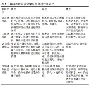
2.2 脊柱推拿手法生物力学的有限元分析流程 利用三维有限元法探究脊柱推拿手法的力学作用,首先是根据研究目的不同,构建正常或病变的脊柱三维有限元模型,而DICOM数据建模法是目前学者最常用的建模方法,见表2。"
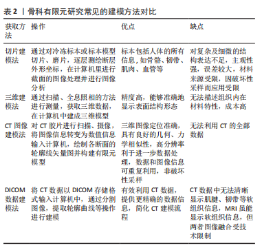
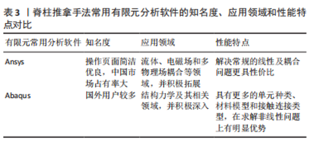
通过CT等影像学技术收集志愿者脊柱的相关扫描资料[28],运用Mimics,Geomagic和Solidworks等三维建模软件对所得的资料进行处理,“在软件中选取合适的阈值范围将所需的脊柱椎体结构提取出来,划分清椎体之间的边界,去除无用的部分,填充椎体内部的空洞,并通过桥梁工具、偏移工具及布尔运算等创建与上下椎体紧密贴合的椎间盘模型”等内容常常在mimics软件中完成,得到点云模型(是三维但非实体)[29]。其次“光滑、去除特征、闭合曲面”等操作常在Geomagic中完成,使点云模型变成光滑的闭合曲面模型[30]。而Solidworks软件常常用于建立在CT片上灰度值显影不佳的软组织部分,并且将之前的曲面模型实体化,以得到三维实体模型。然后将三维实体模型导入Ansys或Abaqus有限元分析软件中进行面网格和体网格划分,以及材料属性赋值等操作,完成脊柱椎体三维有限元模型的建立[31],并将模型各方向的活动度数据与相关的文献、尸体标本或临床观察的研究数据进行对比,验证其可靠性,见表3。"
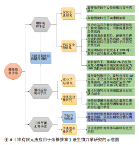
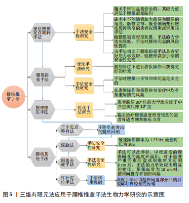
同时,为模拟脊柱推拿手法的具体操作,研究者需要了解手法操作的力学与运动学数据。脊柱推拿手法的力学数据包括手法操作各步骤作用力、最大作用力等参数,目前主要通过各种压力传感器、放大线路及数据采集分析软件等配套设备所组成的在体测力系统进行测量,而运动学数据包括手法操作引起脊柱各方位运动的角度、加速度、时间等参数,目前主要运用红外线摄像机、各种体表标志点及运动图像采集软件等配套仪器所组成的三维运动捕捉系统进行测量[3,32]。完成相应专家手法数据的收集工作后,在脊柱模型的上表面模拟其以上人体部分的质量载荷,在脊柱模型的下表面进行固定和约束处理,根据推拿手法的力学和运动学数据逐步进行模拟加载,在有限元分析软件中计算处理后得到三维有限元模型在推拿手法作用下的应变位移、等效应力分布云图等直观的实验结果[33]。 2.3 颈椎推拿手法生物力学的有限元研究 颈椎结构复杂,椎旁的重要神经和血管较多,具有活动度大、稳定性差的特点,是脊柱中最容易受损的节段,也是脊柱推拿手法治疗中不良反应报道最多的节段[34]。因此,研究者对颈椎推拿手法操作的安全性和技术优化尤为重视,并积极运用三维有限元法解答颈椎推拿手法的力学问题,研究的手法种类集中在颈椎旋转类和拔伸类手法,主要的应用成果见图4。"
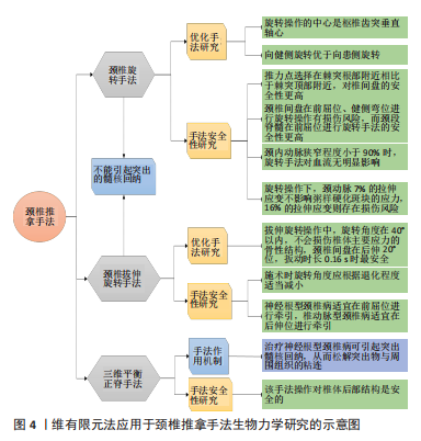
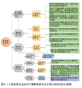
2.3.1 颈椎旋转手法 颈椎旋转手法是临床治疗颈部疾病的常用方法,流派众多、操作各异,但主要的操作步骤皆通过旋转使颈椎达到最大的生理活动限度,进行扳动,通常以引起颈椎的“咔嗒”声作为施术成功的标志[35]。有研究揭示了颈椎旋转手法的作用机制在于引起机体旋转恢复颈椎解剖位置,进而稳定生理旋转轴心[36-37]。其中手法的旋转操作中心是枢椎齿突垂直轴心,并非施术部位的枢椎棘突顶点,而旋转手法推力点选择在棘突根部附近相比于棘突顶部附近,对椎间盘的安全性更高。为探讨颈椎旋转手法合理的旋转方向和体位选择,黄学成等学者[38-40]构建前屈5°位、中立位、后屈5°位、健侧弯位、患侧弯位5种不同体位的颈椎有限元模型,研究发现模拟旋转手法操作时,从力学效应角度来说,医者向健侧旋转优于向患侧旋转;从安全性角度来看,患者在中立位和患侧弯位下受术要分别优于前后屈5°位和健侧弯位。而刘建辉等[30-31]的有限元实验提出后伸30°位的颈椎间盘髓内压低于中立位和前屈30°位,在后伸体位下施术安全性更高,因此,相关结论仍需进一步验证。此外,两者实验均发现在前屈位和健侧弯位下进行旋转操作有损伤颈椎间盘的风险,同时也有研究提出颈段脊髓在前屈位下进行旋转手法的安全性更高[27],而临床医师在实际操作中发现选择前屈位和健侧弯位可以达到最佳疗效,所以如何选择合适的受术体位以平衡手法疗效和安全性之间的关系以及手法操作的适应和禁忌条件等问题有待深入地探讨。同时,颈椎旋转手法仍可以根据流派和操作的不同进行细化分类,来相对应地开展有限元分析以补充现有研究的不足。 随着三维有限元模型椎旁结构设定的不断完善,目前已有学者完成模型的流固耦合,推拿手法的有限元研究应用已由固体力学发展至流体力学,为探究旋转手法对周围血管内血流状态的影响提供新途径,进而丰富推拿手法安全性研究的内涵。LIN等[25]构建颈内动脉8种不同狭窄率(狭窄比率包括0,15%,30%,45%,60%,70%,80%,90%)的Willis环流固耦合有限元模型,分析旋转手法对不同狭窄程度的颈内动脉血流动力学影响,结果显示当狭窄程度小于90%时,旋转手法对模型血流无明显影响。尽管该模型是基于奶牛的MRI数据建立的,但其对后续脑血管血流动力学的研究有着很强的借鉴意义。随后,陈奕历等[41]通过颈动脉粥样硬化患者的MRI资料构建相应的流体力学有限元模型,对比旋转操作下颈动脉不同程度拉伸应变(包含无拉伸组、7%拉伸组和16%拉伸组)的血流动力学效应,结果显示颈动脉7%的拉伸应变对斑块应力无明显影响,16%的拉伸应变因斑块剪切力增加而存在损伤风险。因此,颈椎旋转手法被证明是一种安全的临床应用技术,但颈动脉粥样硬化伴斑块甚至导致重度狭窄的患者应慎用该手法。 2.3.2 颈椎拔伸旋转手法 颈椎拔伸旋转手法是传统推拿技术治疗颈部疾病的有效手段之一,手法通过拔伸、旋转和扳动达到治疗效果,三维有限元法为研究颈椎拔伸旋转手法的作用机制和适宜的牵引体位、扳动时长提供一种新方法。有研究分析其作用机制可能在于调整钩椎关节的位置关系,降低椎体、椎间盘、关节突关节和韧带的应力,改变椎间孔大小并恢复颈椎生理曲度[35]。为探寻合理的牵引角度,李勇等[42]通过对比颈椎前屈10°和后伸10°两种牵引体位下的有限元模型力学效应,发现在前屈10°位牵引,椎体和椎间盘的应力主要集中在椎体两侧后外方神经根出口处,椎体后缘产生位移;在后伸10°位牵引,椎体和椎间盘的应力主要集中在椎体两侧钩锥关节处,椎体前缘产生位移。因此提出神经根型颈椎病适宜在前屈位进行牵引以缓解神经根型颈椎病的症状,椎动脉型颈椎病适宜在后伸位进行牵引以改善两侧椎动脉的受压情况。王宇等[43]研究发现随着退化程度增加,椎间盘应力效应减小,纤维环和关节的受力增大,提示临床医师在实施旋转操作时应根据颈椎间盘退化程度适当减小旋转的角度。为优化手法参数,有研究运用三维有限元法分析颈椎间盘在5种体位(前屈20°、前屈10°、中立、后伸10°、后伸20°)和拔伸旋转手法3种扳动时长(0.06 s,0.11 s, 0.16 s)作用下的应力变化[44-45],结果显示拔伸旋转操作中,向健侧旋转比向患侧旋转对颈椎间盘更为安全合理,手法旋转角度在40°以内,不会损伤椎体主要应力的骨性结构,颈椎间盘在后伸20°位,扳动时长0.16 s时最为安全。 2.3.3 三维平衡正脊手法 此手法是在脊柱力学三维空间理论的基础上融合传统拔伸、斜扳、旋转及推压等手法所形成的,在脊柱相关疾病的保守治疗中应用广泛[46]。CAO等[47-48]运用三维有限元法证明三维平衡正脊手法治疗神经根型颈椎病的过程中,使颈椎椎间隙和后纵韧带张力增宽,利于髓核回纳,从而松解突出物与周围组织的粘连以达到治疗效果,同时也验证该手法操作对椎体后部结构的安全性。虽然临床上该手法适用于脊柱各关节病变的治疗,但其有限元研究目前集中在颈椎方面,后续也将期待该手法作用于脊柱其他节段的文献报道。 2.4 胸腰椎推拿手法生物力学的有限元研究 胸腰段脊柱是椎体骨折的好发部位[49-50],过伸复位手法治疗椎体骨折具有疗效好、损伤轻的应用优势[51]。在模拟胸腰椎压缩性骨折有限元模型的过伸复位手法操作中,李孝林等[51-53]发现该手法的作用机制是通过前纵韧带和伤椎上下相邻椎间盘对受损椎体的牵拉作用而实现的,过伸复位操作的最佳支点在伤椎棘突顶点。同时,该团队提出关节突关节在该手法操作中承受较大应力,其受损概率随过伸复位程度的增加而增加,而复位疗效和手法安全性的权衡问题有待进一步研究。总体而言,胸椎推拿手法的有限元研究要明显少于颈椎和腰椎推拿手法,胸椎部分仍有许多手法没有得到有限元学者的挖掘与研究,主要原因是该部分手法的力学分析需要考虑肋骨整体的影响,在建模方面具有一定的挑战性,而相关研究的空白有待学者们的探究与补充。 2.5 腰椎推拿手法生物力学的有限元研究 腰椎是人体脊柱活动量和承重量最大的部位,具有维持脊柱稳定的重要作用,一旦其力学结构失衡,将极易引发腰椎疾病。因此腰椎推拿手法更需要注意操作的规范化和优化研究,在治疗中更好地恢复腰椎力学结构的平衡。而目前研究者通过三维有限元法不断挖掘腰椎各派系的特色手法以完善相应的基础研究,总之,研究的手法类型仍集中在腰椎旋转、斜板和拔伸类手法,主要的应用进展示意图见图5。"

2.5.1 坐位腰椎定点旋转手法 坐位腰椎定点旋转手法是冯天有教授在传统推拿手法的基础上改良后形成,大量临床实验证实其疗效显著[54],利用三维有限元法可以对其手法的作用机制、旋转方向及受术体位等进行研究及评价。张慧[55]通过有限元分析发现坐位腰椎旋转手法操作过程中,腰椎后部结构的应力值远高于椎间盘,关节突关节的软骨在手法扭矩作用下会发生相对滑移,从而推导该手法对腰椎间盘是安全的,其作用机制在于改善关节突关节活动度。为优化手法操作,有学者在腰椎有限元模型上进行坐位腰椎定点旋转手法的模拟,发现该手法操作中不能随意加大前屈和侧屈的角度,向健侧进行旋转操作更为安全,并提示临床面对骶髂关节、股骨颈病变和腰椎椎管狭窄的患者应慎用或禁用该手法[56-59]。有研究通过构建正常、轻度退变与中度退变3种腰椎有限元模型[26,60-62],比较不同退变程度的椎间盘在坐位腰椎定点旋转手法和斜扳手法作用下的力学效应,结果显示腰椎定点旋转手法具有更好的生物力学效应,而斜扳手法的安全性更高。同时,随着模型退变程度加重,两种手法的力学效应降低,手法操作对腰椎间盘的风险性越高,提示腰椎间盘退变程度越严重,手法操作越需谨慎。而多运用三维有限元法对比不同脊柱推拿手法的力学效应,将更有利于临床医师在手法治疗中做出更优的选择。 2.5.2 腰椎斜扳手法 此手法广泛应用于腰椎疾病的保守治疗中[63-64],三维有限元法可以在其手法成效性和安全性研究中提供相应依据。毕胜等[65]最早应用有限元技术证实腰椎斜扳手法的作用机制在于改变椎间盘与神经根的位置关系,解除突出物对神经根的压迫,实验中关节软骨的最大应力值小于开始骨折的临界值,证明斜扳手法对腰椎小关节是安全的。徐海涛等[66]在分析腰椎斜扳手法对椎间盘的力学效应中发现,椎间盘应力值较低,安全性高,但旋转侧后方存在位移趋势,因此在患侧操作和椎管狭窄治疗中均有加重病情的风险。卢钰等[67]构建前屈30°、水平0°和后伸10°共3种体位的腰椎有限元模型,对比不同体位的腰椎间盘在斜扳手法作用下的力学效应,结果显示在前屈位进行斜扳操作更利于改善椎间盘和神经根的位置关系。 2.5.3 拔伸按压手法 此手法是宋贵杰教授提出的“三步三位九法”的重要操作之一,在腰椎疾病的临床治疗中有着广泛的应用[68],三维有限元法适用于该手法的作用机制和优化研究。张晓刚等[69-71]运用有限元技术分析拔伸按压手法治疗腰椎间盘突出症的最佳体位,证明前屈30°位的力学效应优于中立位和后伸10°位,而拔伸按压手法的作用机制符合“松解粘连”学说。尚永等[72]通过构建轻度退变和中度退变2种腰椎有限元模型探讨腰椎退变程度对拔伸按压手法力学效应的影响,结果显示腰椎退变不影响小关节的应力情况,但不利于手法的力学效应,提示临床工作者在面对腰椎退变程度较重的患者可适当增加按压力度以达到预期疗效。 2.5.4 腰椎其他手法 杠杆定位手法、三小定点整脊法、踩跷法以及倒盖金被手法是保守治疗腰椎疾病的特色手法,研究者相继运用有限元技术探讨这些手法的力学效应。吕立江等[73]在研究杠杆定位手法的作用机制中发现手法可通过椎间盘部分回纳作用以缓解对神经根的压迫。张仁倩等[74]通过有限元研究发现三小定点整脊法的作用机制在于调整椎体与神经根的位置关系,进而解除粘连达到治疗效果。罗建等[75]构建正常与病变的腰椎模型,通过对比6种不同频率和6种不同时长的工况,发现踩跷法最佳操作频率为1.25 Hz,最佳时长为30 s,完善该手法的参数研究。还有学者在腰椎有限元模型上进行倒盖金被手法模拟,发现该手法可以改善轻、中度退变的腰椎间孔的高度和面积,但对于退变严重的椎间盘或顶推高度达到8 cm后,腰椎间孔的高度和面积没有明显的变化,而当顶推高度为10 cm时,腰椎间盘因应力集中存在损伤风险,因此提示手法操作时应选择适宜的顶推高度[76-77]。"

| [1] 孔博,薛彬,贾友冀,等.传承中不断发展的中医正骨流派现状简析[J].中国中医骨伤科杂志,2016,24(11):70-73. [2] CASSIDY JD, BRONFORT G, HARTVIGSEN J. Should we abandon cervical spine manipulation for mechanical neck pain? BMJ. 2012;344:e3680. [3] 梁龙,朱立国,于杰,等.中医手法量化及生物力学研究探析[J].世界中西医结合杂志,2020,15(6):1165-1168. [4] 郭建鹏,马迅,梁凯恒,等.脊柱颈胸结合部有限元模型的建立[J].临床医药实践,2009,18(1):7-9. [5] 徐浩翔,文王强,张泽佩,等.腰椎间盘生物力学体内外研究的新进展[J].中国组织工程研究,2020,24(15):2425-2432. [6] 林松青,王彬,张磊,等.有限元分析在骨科中的应用及研究进展[J].中国中医骨伤科杂志,2013,21(4):69-73. [7] 宫玉锁,周君,李盛华,等.骨科三维有限元研究进展[J].世界最新医学信息文摘,2019,19(93):40-41, 43. [8] GALBUSERA F, FANTIGROSSI A, RAIMONDI MT, et al. Biomechanics of the C5-C6 spinal unit before and after placement of a disc prosthesis. Biomechan Model Mechanobiol. 2006;5(4):253-261. [9] 李斌,赵文志,陈秉智.有限元分析:椎间盘退变对颈椎生物力学的影响[J].中国组织工程研究,2017,21(11):1748-1752. [10] 秦大平,张晓刚,宋敏.有限元分析在中医正骨手法治疗腰椎疾病作用机制中的应用[J].中国组织工程研究,2012,16(26):4913-4917. [11] STRANGE DG, FISHER ST, BOUGHTON PC, et al. Restoration of compressive loading properties of lumbar discs with a nucleus implant-a finite element analysis study. Spine J. 2010;10(7): 602-609. [12] 毕胜,张德文,张明,等.模拟腰部推拿手法三维有限元模型分析[J].军医进修学院学报,2002,23(1):67-69. [13] 毕胜,张德文,张明,等.腰椎牵引三维有限元模型分析[J].中国康复医学杂志,2002,17(2):20-22. [14] LIANG L, LIU M, MARTIN C, et al. A deep learning approach to estimate stress distribution: a fast and accurate surrogate of finite-element analysis. J R Soc Interface. 2018;15(138):20170844. [15] COURANT R. Variational methods for the solution of problems of equilibrium and vibrations. Bull. 1943;49(1):1-24. [16] WELCH-PHILLIPS A, GIBBONS D, AHERN DP, et al. What is finite element analysis? Clin Spine Surg. 2020;33(8):323-324. [17] CLOUGH RW. The finite element in plane stress analysis. Proc Asce Conferon Electric Computation, 1960. [18] BREKELMANS WA, POORT HW, SLOOFF TJ. A new method to analyse the mechanical behaviour of skeletal parts. Acta Orthop Scand. 1972; 43(5):301-317. [19] SCHULTZ AB, BELYTSCHKO TB, ANDRIACCHI TP, et al. Analog studies of forces in the human spine: mechanical properties and motion segment behavior. J Biomech. 1973;6(4):373-383. [20] LIU YK, RAY G, HIRSCH C. The resistance of the lumbar spine to direct shear. Orthop Clin North Am. 1975;6(1):33-49. [21] SIMON BR, WU JS, CARLTON MW, et al. Structural models for human spinal motion segments based on a poroelastic view of the intervertebral disk. J Biomechan Eng. 1985;107(4):327-335. [22] RAO AA, DUMAS GA. Influence of material properties on the mechanical behaviour of the L5-S1 intervertebral disc in compression: a nonlinear finite element study. J Biomechan Eng. 1991;13(2):139-151. [23] SHIRAZI-ADL A. Nonlinear stress analysis of the whole lumbar spine in torsion--mechanics of facet articulation. J Biomechan. 1994;27(3): 289-299. [24] 彭春政,张胜年,陆爱云.人体躯干骨骼-肌肉-韧带结构三维有限元模型的建立和验证[J].中国运动医学杂志,2010,29(6):702-705. [25] LIN W, MA X, DENG D, et al. Hemodynamics in the circle of willis with internal carotid artery stenosis under cervical rotatory manipulation: a finite element analysis. Med Sci Monit. 2015;21:1820-1826. [26] SHU XN, MU WZ, CHEN JF, et al. Comparison of biomechanical effect between oblique Ban-pulling manipulation and lumbar erection-rotation manipulation in sitting position for lumbar intervertebral disc herniation. Zhenjiu Tuita Yixue (Yingwenban). 2017;15(5):317-321. [27] XUE F, CHEN Z, YANG H, et al. Effects of cervical rotatory manipulation on the cervical spinal cord: a finite element study. J Orthop Surg Res. 2021;16(1):737. [28] 曾雪玲,张帆,王芳莉.有限元分析技术在腰椎推拿手法生物力学中的应用现状[J].西部中医药,2022,35(5):157-161. [29] 文毅,苏峰,刘肃,等.L4-5椎体有限元模型建立及退变椎间盘力学分析[J].中国组织工程研究,2019,23(8):1222-1227. [30] 刘建辉,朱建忠,潘福勤,等.不同体位下推拿旋转手法对颈椎间盘有限元模型应力分布的影响[J].世界中西医结合杂志,2020, 15(7):1304-1307. [31] 刘建辉, 朱建忠,潘福勤,等.颈椎间盘有限元模型分析旋转手法髓核受力与体位的关系[J].内蒙古医科大学学报,2020,42(2):173-175,204. [32] 周鑫,朱清广,孔令军,等.推拿手法生物力学研究方法的基本方向[J].中华中医药杂志,2019,34(3):1120-1123. [33] 王辉昊,范鑫,邓真,等.中医整骨手法对颈椎间孔减压术(PECD)后模型的有限元分析[J].医用生物力学, 2021, 36(S1):225. [34] 王辉昊,詹红生,张明才,等.手法治疗颈椎病意外事件分析与预防策略思考[J].中国骨伤,2012,25(9):730-736. [35] 邓真,牛文鑫,王辉昊,等.生物力学在中医骨伤手法治疗颈椎病中的应用[J].医用生物力学,2015,30(6):569-573. [36] 万磊,陈静,李义凯.颈椎定点旋转手法“点”的三维空间解剖位置的研究[J].南方医科大学学报,2008,28(4):548-550, 554. [37] DENG Z, WANG K, WANG H, et al. A finite element study of traditional Chinese cervical manipulation. Eur Spine J. 2017;26(9):2308-2317. [38] 黄学成,叶林强,梁德,等.三维有限元模型分析旋转手法中旋转方向对颈椎间盘位移和椎间孔容积的影响[J].中国组织工程研究, 2018,22(3):404-408. [39] HUANG X, YE L, WU Z, et al. Biomechanical effects of lateral bending position on performing cervical spinal manipulation for cervical disc herniation: a three-dimensional finite element analysis. Evid Based Complement Alternat Med. 2018;2018:2798396. [40] 黄学成,叶林强,江晓兵,等.不同体位下颈椎旋转手法对颈椎间盘位移和内在应力的影响[J].中国康复理论与实践,2017,23(12): 1470-1475. [41] 陈奕历, 劳永华, 张少群,等.颈动脉粥样硬化的流体力学模型:旋转手法下颈动脉粥样硬化斑块的血流动力学变化[J].中国组织工程研究,2019,23(15):2403-2408. [42] 李勇,张泽胜,王伶俐,等.不同牵引角度治疗颈椎病的三维有限元分析研究[J].新中医,2008,40(9):63-64. [43] 王宇,雷建银,辛浩,等.椎间盘退变颈椎(C2-C7)在正常承载与推拿下的有限元分析[J].中国组织工程研究,2020,24(27):4278-4284. [44] 邬黎平.颈椎推拿的作用机理及优化研究[D].广州:南方医科大学, 2010. [45] WU LP, HUANG YQ, MANAS D, et al. Real-time monitoring of stresses and displacements in cervical nuclei pulposi during cervical spine manipulation: a finite element model analysis. J Manipulative Physiol Ther. 2014;37(8):561-568. [46] 孙国栋,师彬,王丹丹,等.手势动作捕捉技术及有限元分析在神经根型颈椎病三维正脊手法机制研究中的应用分析[J].四川医学, 2018,39(2):223-225. [47] CAO SN, WANG DD, WANG CA, et al. Finite element analysis of the treatment of cervical spondylotic radiculopathy with three dimensional balanced manipulation. Zhongguo Gu Shang. 2020;33(9):867-872. [48] CAO S, CHEN Y, ZHANG F, et al. Clinical efficacy and safety of “three-dimensional balanced manipulation” in the treatment of cervical spondylotic radiculopathy by finite element analysis. BioMed Res Int. 2021;2021:5563296. [49] PEDRAM H, REZA ZM, REZA RM, et al. Spinal fractures resulting from traumatic injuries. Chin J Traumatol. 2010;13(1):3-9. [50] ZHANG D, WU H, WANG X. Comparative study of the clinical effect and safety of anterior surgical approach and posterior surgical approach in the treatment of thoracolumbar spinal fracture. Open Med. 2015; 10(1):410-415. [51] 刘迎军,李孝林.有限元分析过伸复位治疗胸腰椎压缩性骨折的椎间盘动态力学[J].中国组织工程研究与临床康复,2011,15(4):589-592. [52] 舒先涛,李孝林.胸腰椎压缩性骨折患者过伸复位过程中前纵韧带动态力学的有限元分析[J].中国组织工程研究与临床康复,2009, 13(48):9567-9956. [53] 任伯绪,李孝林.过伸复位治疗胸腰椎压缩性骨折关节突关节动态力学有限元分析[J].山东医药,2010,50(14):4-5. [54] 韩雪,韩磊,张军,等.冯氏坐位脊柱定点旋转法与中医传统侧卧不定点斜扳法治疗腰椎间盘突出症比较研究[J].北京中医药,2015, 34(8):598-602. [55] 张慧.腰椎坐位旋转手法治疗非特异性下腰痛的临床疗效观察及腰椎有限元初步探讨[D].北京:中国中医科学院,2015. [56] 徐海涛,徐达传,李云贵,等.坐位旋转手法时退变腰椎间盘内在应力和位移的有限元分析[J].中国康复医学杂志,2007,22(9):769-771,867. [57] 徐海涛, 张美超, 徐达传,等.三种前屈角度下坐位旋转手法对腰椎间盘作用的有限元分析[J].中国疗养医学,2008,17(2):65-67. [58] HU H, XIONG CY, HAN GW. Finite element analysis of lumbar pelvic and proximal femur model with simulate lumbar rotatory manipulation. Zhongguo Gu Shang. 2012;25(7):582-586. [59] 陈忻,于杰,冯敏山,等.坐位旋转手法治疗退行性腰椎滑脱的椎间盘力学分析[J].中华中医药杂志,2019,34(4):1395-1400. [60] 陈金凤,舒新农,唐树杰,等.腰椎退变对坐位腰椎定点旋转手法疗效影响的有限元分析(英文)[J]. 针灸推拿医学(英文版),2016, 14(4):295-299. [61] ZHANG R, MO Z, LI D, et al. Biomechanical comparison of lumbar fixed-point oblique pulling manipulation and traditional oblique pulling manipulation in treating lumbar intervertebral disk protrusion. J Manipulative Physiol Ther. 2020;43(5):446-456. [62] LI L, SHEN T, LI YK. A Finite element analysis of stress distribution and disk displacement in response to lumbar rotation manipulation in the sitting and side-lying positions. J Manipulative Physiol Ther. 2017;40(8):580-586. [63] 张人文,莫灼锚,常敏敏,等.坐位定点旋转手法与斜扳手法治疗腰椎间盘突出症的系统评价[J].中医正骨,2018,30(12):30-36. [64] MO Z, ZHANG R, CHEN J, et al. Comparison between oblique pulling spinal manipulation and other treatments for lumbar disc herniation: a systematic review and meta-analysis. J Manipulative Physiol Ther. 2018;41(9):771-779. [65] 毕胜,李义凯,赵卫东,等.腰部推拿手法生物力学和有限元比较研究[J].中华物理医学与康复杂志,2002,24(9):16-19. [66] 徐海涛,李松,刘澜,等.腰椎斜扳手法时椎间盘的有限元分析[J].中国组织工程研究与临床康复,2011,15(13):2335-2338. [67] 卢钰,郑太才,王琪,等.不同体位下斜扳手法治疗腰椎间盘突出的三维有限元分析[J].中国组织工程研究,2021,25(36):5872-5877. [68] 贾龙,张华,宋敏,等.“三步三位九法”治疗腰椎间盘突出症临床经验总结[J].世界最新医学信息文摘,2017,17(A2):185-187. [69] 李延红,张晓刚,李具宝,等.腰椎拔伸手法三维有限元模型分析[J].浙江中医杂志,2010,45(12):879-880. [70] 杨学锋,张晓刚,李具宝,等.模拟拔伸按压手法腰椎运动节段三维有限元模型分析[J].中国中医骨伤科杂志,2013,21(7):4-6. [71] 张晓刚,秦大平,宋敏,等.拔伸按压手法对退变腰椎节段应力分布影响的有限元分析[J].中华中医药杂志,2013,28(10):3108-3114. [72] 尚永,张人文,莫灼锚,等.腰椎退变影响拔伸按压手法生物力学作用的有限元研究[J].山东中医药大学学报,2019,43(2):151-154. [73] 吕立江,冯喆,廖胜辉,等.杠杆定位手法对腰椎间盘影响的有限元分析[J].中华中医药学刊,2014,32(5):971-973. [74] 张仁倩,赵志恒,王剑歌,等.三小定点整脊法对腰椎间盘突出症的有限元分析[J].湖南中医杂志,2014,30(8):90-92. [75] 罗建,金龙,徐侥,等.三维有限元模型下的踩跷法时效性研究[J].四川中医,2017,5(9):148-151. [76] 李雁婷,陈剑,刘梦兰,等.倒盖金被手法在腰椎间盘生物力学中的三维有限元分析[J].中国组织工程研究,2022,26(3):356-259. [77] DING H, LIAO L, YAN P, et al. Three-dimensional finite element analysis of l4-5 degenerative lumbar disc traction under different pushing heights. J Healthc Eng. 2021;2021:1322397. [78] 胡华,熊昌源,韩国武.模拟腰部推拿手法建立“腰-盆-髋”有限元模型[J].中国组织工程研究与临床康复,2011,15(48):8935-8938. [79] 汪芳俊,魏威,廖胜辉,等.前屈位不同角度牵引治疗颈椎病的有限元分析[J].中国骨伤,2014,27(7):592-596. [80] 张人文,莫灼锚,李冬,等.二步加载分步求解在手法治疗腰椎间盘突出症有限元分析中的应用[J].中国中医骨伤科杂志,2019, 27(1):1-5. |
| [1] | Nong Fuxiang, Jiang Zhixiong, Li Yinghao, Xu Wencong, Shi Zhilan, Luo Hui, Zhang Qinglang, Zhong Shuang, Tang Meiwen. Bone cement augmented proximal femoral nail antirotation for type A3.3 intertrochanteric femoral fracturalysis [J]. Chinese Journal of Tissue Engineering Research, 2023, 27(在线): 1-10. |
| [2] | Zhong Yizheng, Huang Peizhen, Cai Qunbin, Zheng Liqin, He Xingpeng, Dong Hang. Microstructural indexes that determine the trabecular bone maximum stress of micro-finite element models [J]. Chinese Journal of Tissue Engineering Research, 2023, 27(9): 1313-1318. |
| [3] | Wu Taoguang, Nie Shaobo, Chen Hua, Zhu Zhengguo, Qi Lin, Tang Peifu. Biomechanical characteristics of a new multi-dimensional cross locking plate in the treatment of subtrochanteric nonunion [J]. Chinese Journal of Tissue Engineering Research, 2023, 27(9): 1330-1334. |
| [4] | Peng Zhixin, Yan Wengang, Wang Kun, Zhang Zhenjiang. Finite element analysis and structural optimization design of 3D printed forearm braces [J]. Chinese Journal of Tissue Engineering Research, 2023, 27(9): 1340-1345. |
| [5] | Liu Guangwei, Feng Minshan, Zhu Liguo, Yin Xunlu, Chen Zhuoxian. Dynamic analysis of structural changes in the lower cervical intervertebral foramen during rotation-traction manipulation by virtual reality technology [J]. Chinese Journal of Tissue Engineering Research, 2023, 27(9): 1346-1351. |
| [6] | Wu Tianliang, Tao Xiuxia, Xu Hongguang. Influence of different bone mineral densities on cage subsidence after stand-alone oblique lateral interbody fusion: three-dimensional finite element analysis [J]. Chinese Journal of Tissue Engineering Research, 2023, 27(9): 1352-1358. |
| [7] | Liu Jinyu, Zhang Hanshuo, Cui Hongpeng, Pan Lingzhi, Zhao Boran, Li Fei, Ding Yu. Finite element biomechanical analysis of minimally invasive treatment of cervical spondylotic myelopathy and accurate exercise rehabilitation [J]. Chinese Journal of Tissue Engineering Research, 2023, 27(9): 1359-1364. |
| [8] | He Yujie, Kang Zhijie, Xue Mingming, Jin Feng, Li Zhijun, Wang Xing, Xu Yangyang, Gao Mingjie, Li Jiawei, Li Xiaohe, Wang Haiyan. Finite element analysis of transarticular screw fixation of adolescent thoracic vertebra [J]. Chinese Journal of Tissue Engineering Research, 2023, 27(9): 1365-1370. |
| [9] | Wen Xinghua, Ding Huanwen, Cheng Kai, Yan Xiaonan, Peng Yuanhao, Wang Yuning, Liu Kang, Zhang Huiwu. Three-dimensional finite element model analysis of intramedullary nailing fixation design for large femoral defects in Beagle dogs [J]. Chinese Journal of Tissue Engineering Research, 2023, 27(9): 1371-1376. |
| [10] | You Zhengqiu, Zhang Zhongzu, Wang Qunbo. Early symptomatic intervertebral disc pseudocysts after discectomy detected on MRI [J]. Chinese Journal of Tissue Engineering Research, 2023, 27(9): 1403-1409. |
| [11] | Pan Zhongjie, Qin Zhihong, Zheng Tiejun, Ding Xiaofei, Liao Shijie. Targeting of non-coding RNAs in the pathogenesis of the osteonecrosis of the femoral head [J]. Chinese Journal of Tissue Engineering Research, 2023, 27(9): 1441-1447. |
| [12] | Cai Zhihao, Xie Zhaoyong. Femoral neck anteversion measurement assessment: how to establish a unified method and standard [J]. Chinese Journal of Tissue Engineering Research, 2023, 27(9): 1448-1454. |
| [13] | Dang Yi, Du Chengyan, Yao Honglin, Yuan Nenghua, Cao Jin, Xiong Shan, Zhang Dingmei, Wang Xin. Hormonal osteonecrosis and oxidative stress [J]. Chinese Journal of Tissue Engineering Research, 2023, 27(9): 1469-1476. |
| [14] | Wang Ji, Zhang Min, Yang Zhongya, Zhang Long. A review of physical activity intervention in type 2 diabetes mellitus with sarcopenia [J]. Chinese Journal of Tissue Engineering Research, 2023, 27(8): 1272-1277. |
| [15] | Nie Chenchen, Su Kaiqi, Gao Jing, Fan Yongfu, Ruan Xiaodi, Yuan Jie, Duan Zhaoyuan, Feng Xiaodong. The regulatory role of circular RNAs in cerebral ischemia-reperfusion injury [J]. Chinese Journal of Tissue Engineering Research, 2023, 27(8): 1286-1291. |
| Viewed | ||||||
|
Full text |
|
|||||
|
Abstract |
|
|||||