[1] 孟和.中西医结合骨科外固定治疗学[M].北京: 人民卫生出版社, 2005.
[2] 孔博,彦威,李飞跃,等.中医小夹板作用机理及发展[J].世界中医药,2018,13(1):229-232.
[3] 孔博,贾友冀,薛彬,等.中医骨伤小夹板历史及现状初探[J].中国中医骨伤科杂志,2017,25(1):80-82.
[4] 潘娅岚,郭杨,钱超,等.小夹板用于骨折外固定研究现状[J].中国中医药信息杂志,2014,21(6):123-125.
[5] 李学臻,姜亚明,齐业雄,等.基于纺织复合材料的医用骨伤夹板的研究进展[J].纺织导报,2020(2):76-80.
[6] 王娟娟,秦雪梅,高晓霞,等.杜仲化学成分、药理活性和质量控制现状研究进展[j].中草药,2017,48(15):3228-3237.
[7] 国家药典委员会.中华人民共和国药典: 一部[M].北京: 中国医药科技出版社,2020.
[8] WU YF, YAO KC, NIE HR, et al. Confirmation on the compatibility between cis-1, 4-polyisoprene and trans- 1, 4-polyisoprene. Polymer. 2018;153:271-276.
[9] HAN X, ZHANG D, NIE H, et al. Isothermal crystallization of trans-1, 4-polyisoprene from solutions: Kinetic parameters and crystal modification. Polymer. 2018;139:36-43.
[10] HU JL, ZHU Y, HUANG HH, et al. Recent advances in shape-memory polymers: Structure, mechanism, functionality, modeling and applications. Prog Polym Sci. 2012;37(12):1720-1763.
[11] WALKE PM, SHARP JS, AKIMOV AV, et al. Coherent elastic waves in a one-dimensional polymer hypersonic crystal. Appl Phys Lett. 2010; 97(7):073106.
[12] 崔树阳.个性化杜仲胶运动护具防护性能研究[D].北京:北京化工大学,2020.
[13] BECK LS, DEGUZMAN L, LEE W, et al. TGF-beta I induces bone closure of skull defects. Bone Miner Res. 1991;6(11):1257-1265.
[14] 冯作国. “骨愈合剂”促进兔糖尿病患骨折愈合的实验研究[D].南京:南京中医药大学,2014.
[15] EINHORN TA. The science of fracture healing. J Orthop Trauma. 2005; 19:19.
[16] GREIFF J. A method for the production of an undisplaced reproducible tibial fracture in the ratury. Injury. 1978;9(4):278-281.
[17] BONNARENS F, EINHORN TA. Production of a standard closed fracture in laboratory animal bone. J Orthop Res. 1984;2(1):97-101.
[18] AERSSENS J, BOONEN S, LOWET G, et al. Interspecies differences in bone conposition,density,and quality:potential implications for in vivo bone research. Endocrinology. 1998;139(2):663-670.
[19] KUBO H, PILGE H, WEIMANN-STAHLSCHMIDT K, et al. Use of the Tübingen splint for the initial management of severely dysplastic and unstable hips in newborns with DDH:An alternative to Fettweis plaster and Pavlik harness. Arch Orthop Trauma Surg. 2018;138(2):149-153.
[20] 王希军,李学明.科雷氏骨折夹板固定新法[J].吉林中医药, 1992(4):24.
[21] MARTURANO JE. An improved murine femur fracture device for bone healing studies. J Biomech. 2008;41(6):1222-1228.
[22] 都广全,都芳涛,都芳娟.急救用可调试小夹板:中国,CN200320121357.2[P].2004-12-29.
[23] 程瑞香,马岩.石膏-微米厚度刨花腕部骨伤固定夹板的制造工艺[J].林业科学,2010,46(4):178-182.
[24] 汤黎明,戚仕涛,陈锐华,等.应用高分子化学材料研制可拆卸型的骨科固定夹板[J].中国组织工程研究与临床康复,2007,11(48): 9756-9758.
[25] 孔莉莉,成玲,万培培,等.苎麻纤维复合材料医用夹板的开发及智能化[J].材料导报,2018,32(7):1202-1208.
[26] BLAYA F, PEDRO PS, SILVA JL, et al. Design of an orthopedic product by using additive manufacturing technology:The arm splint. J Med Syst. 2018;42(3):54.
[27] 颜威,蒋涛,吴昌桂,等.桡骨远端骨折固定夹板外观、材质、固定带等研究的新进展[J].中国组织工程研究,2020,24(9):1430-1434.
[28] 许树柴,袁凯,刘军,等.中医骨科小夹板的现状及今后发展的思考[J].医学与哲学(临床决策论坛版),2011,32(6):47-49.
[29] 李明华.天然高分子材料杜仲胶的特性及研究进展[J].应用化工, 2018,47(5):1026-1029.
[30] 王韵然,刘莉,王琎,等.反式-1,4-聚异戊二烯性能及其并用胶的研宄进展[J].橡胶工业,2010,57(5):316-319.
[31] 刘争男,李旭东,黄宝探,等.反式-1,4-聚异戊二烯的氯化改性[J].弹性体,2006,16(1):8-12.
[32] 季美琴.氢化天然橡胶及硅氢化反式-1,4-聚异戊二烯的制备与结构性能研宄[D].北京:北京化工大学,2017.
[33] 王彦,董月,夏琳,等.环氧化杜仲胶改性三元乙丙橡胶/白炭黑复合材料的制备与性能[J].弹性体,2018,28(8):1-16.
[34] 杨凤,孟样睛,于欢.杜仲胶接枝甲基丙烯酸丁酯的合成与表征[J].高分子材料科学与工程,2017,33(4):19-24.
[35] 任庆海,童晓梅,李欢乐,等.合成杜仲胶与天然杜仲胶的改性及性能研宄[J].陕西科技大学学报,2014,32(5):73-77.
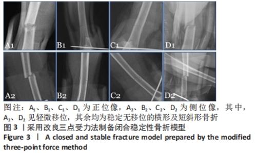
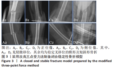
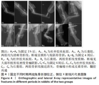
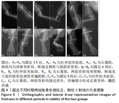
|