Chinese Journal of Tissue Engineering Research ›› 2022, Vol. 26 ›› Issue (14): 2172-2178.doi: 10.12307/2022.479
Previous Articles Next Articles
Parathyroid hormone promotes bone healing in rabbits after free reduction of mandibular condyle fractures
Huang Zhicai, Xie Liuqin, Wang Guangsu, Zhang Guoxing, Tang Zhenglong
- Guizhou Medical University School of Stomatology/Affiliated Stomatological Hospital, Guiyang 550004, Guizhou Province, China
-
Received:2021-02-22Revised:2021-02-25Accepted:2021-04-19Online:2022-05-18Published:2021-12-21 -
Contact:Tang Zhenglong, MD, Professor, Chief physician, Guizhou Medical University School of Stomatology/Affiliated Stomatological Hospital, Guiyang 550004, Guizhou Province, China -
About author:Huang Zhicai, Master candidate, Guizhou Medical University School of Stomatology/Affiliated Stomatological Hospital, Guiyang 550004, Guizhou Province, China -
Supported by:Special Field Project of Guizhou Provincial Department of Education, No. KY [2021]067 (to TZL); Guizhou Provincial Science and Technology Planning Project, No. [2017] 1151 (to TZL)
CLC Number:
Cite this article
Huang Zhicai, Xie Liuqin, Wang Guangsu, Zhang Guoxing, Tang Zhenglong. Parathyroid hormone promotes bone healing in rabbits after free reduction of mandibular condyle fractures[J]. Chinese Journal of Tissue Engineering Research, 2022, 26(14): 2172-2178.
share this article
Add to citation manager EndNote|Reference Manager|ProCite|BibTeX|RefWorks
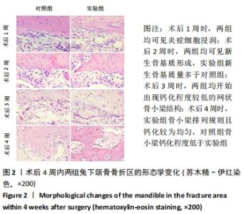
2.1 实验动物数量分析 48只新西兰大耳兔实验过程均未发生意外死亡,实验组及对照组各24只,全部进入结果分析。 2.2 大体标本愈合情况观察结果 术后第1周:所有标本骨折线均清晰可见,骨块活动度较大,未发生吸收或坏死,骨折线区域内可见大量纤维结缔组织。 术后第2周:两组标本均可见少量反应性骨组织增生,骨块活动度较术后1周时明显减弱。实验组骨折线处新生软骨样组织覆盖且量多于对照组;对照组见部分骨折线暴露。 术后第3周:两组标本骨折线处进一步融合、连接,游离骨块已与下颌骨融为一体,经细针穿刺发现所有标本骨折线区域新生骨组织已部分钙化。 术后第4周:实验组动物标本骨折线已消失,钙化程度较高;对照组仍隐约可见部分骨折线。 2.3 髁突骨折愈合组织形态学观察结果 见图2。"

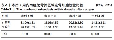
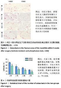
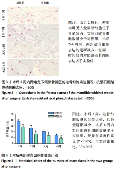
术后第1周:苏木精-伊红染色见两组标本骨折线两侧均可观察到大量密集的炎性细胞和成纤维细胞浸润骨折,肉芽组织形成,破骨细胞主要在骨折线外正常骨质内出现。实验组可见大量的软骨细胞在形成的纤维肉芽组织中,仅骨折线边缘可见少量成骨细胞聚集;对照组新生骨痂中纤维组织成分多,有散在少量软骨细胞及成骨细胞,对照组骨折线两侧有较多破骨细胞分布。 术后第2周:实验组新生骨组织增多并可见破骨细胞、炎性细胞浸润,成骨细胞数量增多,软骨性骨痂增多并向编织骨转化;对照组骨折线周围破骨细胞数量较多,骨基质生成现象不如实验组明显。 术后第3周:对照组仍可见少量炎细胞浸润,开始出现钙化程度较低的骨小梁,骨折线周围见破骨细胞散在分布;实验组断端已有编织骨形成,骨折断端间多为骨性骨痂,少量区域为软骨性骨痂,新生骨小梁呈网状,钙化程度较高且钙化较为均匀;实验组成骨细胞多于对照组。 术后第4周:实验组新生骨组织钙化区域继续增大,骨小梁排列规则且钙化较为均匀,破骨细胞数量较前明显减少;对照组骨小梁钙化区间隙较实验组大,钙化程度和骨小梁密集程度稍低于实验组。 2.4 破骨细胞计数结果分析 光学显微镜观察并统计骨折线周围区域破骨细胞,进行计数统计得出术后1,2,3,4周实验组及对照组中每个样本中破骨细胞个数的平均值,对两组破骨细胞数进行统计学分析,术后1周,两组破骨细胞数量均达到最大值,后数量逐渐减少,术后4周内对照组破骨细胞数量多于实验组,差异有显著性意义(P < 0.05),见表2及图3,4。"
"
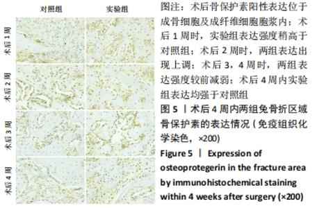
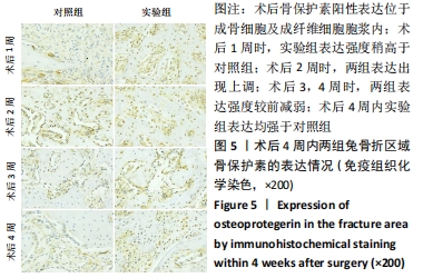
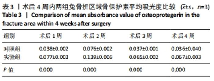
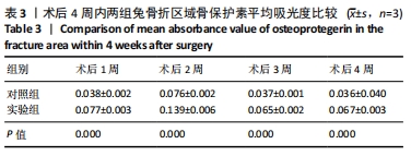
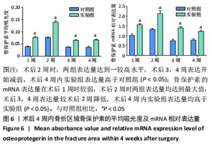
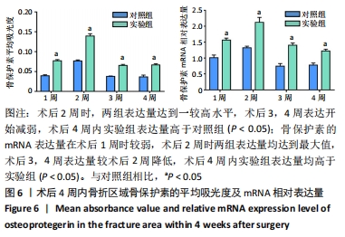
2.5 免疫组织化学及实施荧光定量PCR结果分析 2.5.1 骨保护素免疫组织化学分析 免疫组化染色骨保护素阳性表达主要存在于新生骨小梁边缘的成骨细胞及成纤维细胞胞浆中,在破骨细胞中也有表达,但成骨细胞中表达最强,见图5。术后2周内骨保护素表达逐渐增强,可观察到较多的阳性表达细胞,术后3周表达开始减弱。用平均吸光度进行统计比较,对照组于术后各个阶段骨保护素阳性表达均低于实验组且差异有显著性意义(P < 0.05),见表3。"
"
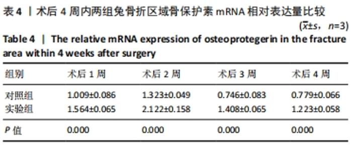
2.5.2 骨保护素的mRNA结果分析 骨保护素的mRNA的相对表达量在术后均出现上升,两组均于术后2周表达量达到较高水平后开始出现下降,术后4周内实验组表达量高于对照组,见表4及图6,且两组各时间点实验数据相比差异均有显著性意义(P < 0.05)。"
"
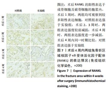
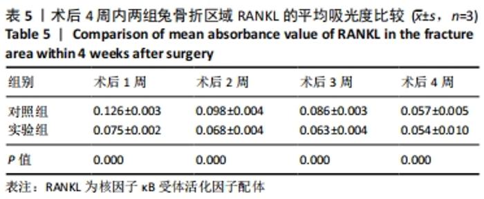
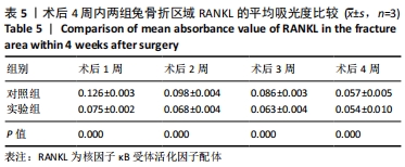
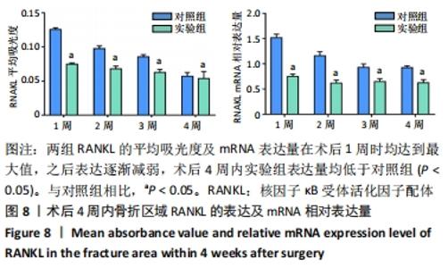
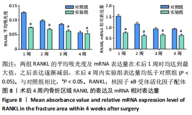
2.5.3 RANKL免疫组织化学分析 免疫组化显示RANKL的阳性表达主要存在于成骨细胞及破骨细胞的胞浆内,用平均吸光度值进行统计分析,见图7及表5,术后1周两组表达量均达到最大值,然后开始逐渐降低,实验组各阶段的表达量均低于对照组且差异有显著性意义(P < 0.05)。"
"

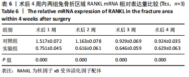
2.5.4 RANKL的mRNA结果分析 两组RANKL mRNA相对表达量均于术后1周时到达最高水平,然后逐渐降低;术后4周内实验组的RANKL mRNA相对表达量均低于对照组,差异有显著性意义(P < 0.05),见表6及图8。"
"
| [1] VINCENT AG, DUCIC Y,KELLMAN R. Fractures of the Mandibular Condyle. Facial Plast Surg. 2019;35(6):623-626. [2] KUMARAN A, SOH HJJOO, ORAL MSOJOTAAO, et al. Management of Nonunion and Malunion After Primary Mandibular Condylar Fracture Treatment: A Review and Recommendations. J Oral Maxillofac Surg. 2020;78(12):2267-2272. [3] KUNTAMUKKULA S, SINHA R, TIWARI P, et al. Dynamic Stability Assessment of the Temporomandibular Joint as a Sequela of Open Reduction and Internal Fixation of Unilateral Condylar Fracture. J Oral Maxillofac Surg. 2018;76(12):2598-2609. [4] MARJI FP, ANSTADT E, DAVIT A, et al. Pediatric Mandibular Condylar Fractures With Concomitant Cervical Spine Injury: A Treatment Protocol for Prevention of Temporomandibular Joint Ankylosis. J Craniofac Surg. 2020;31(3):248-250. [5] TAKANO H, TAKAHASHI T, NAKATA A, et al. Facilitation of bone resorption activities in synovial lavage fluid patients with mandibular condyle fractures. J Oral Rehabil. 2016;43(5):333-339. [6] XIA L, HE Y, AN J, et al. Condyle-preserved arthroplasty versus costochondral grafting in paediatric temporomandibular joint ankylosis: a retrospective investigation. Int J Oral Maxillofac Surg. 2019;48(4): 526-533. [7] ZHANG Y, HAN B, WEI Y, et al. Icariin Promotes Fracture Healing in Ovariectomized Rats. Med Sci Monit. 2020;26:e924554. [8] JULIA S, GEORG K, ANITA T, et al. The impact of nonosteogenic factors on the expression of osteoprotegerin and RANKL during human fracture healing. Bone Joint Res. 2019;8(7):349-356. [9] 谢冰洁,冯捷,韩向龙. 破骨细胞生物学特征的研究与进展[J].中国组织工程研究,2017,21(11):1770-1775. [10] TAKAHATA M, AWAD H, O’KEEFE R, et al. Endogenous tissue engineering: PTH therapy for skeletal repair. Cell Tissue Res. 2012;347(3):545-552. [11] GÜL KC, MERYEM K. Evaluation of single-dose applied teriparatide effect on bone healing with histomorphometric and micro-ct analysis. J Craniomaxillofac Surg. 2021;49(2):98-103. [12] ZHANG C, ZHU J, JIA J, et al. Once-weekly parathyroid hormone combined with ongoing long-term alendronate treatment promotes osteoporotic fracture healing in ovariectomized rats. J Orthop Res. 2020. doi: 10.1002/jor.24953. [13] 安宁, 李耀, 唐正龙, 等. 不同剂量甲状旁腺激素对下颌支截骨术后骨愈合的影响及其机制研究[J]. 中华口腔医学杂志,2018,53(6): 413-418. [14] 王冬香,王光素,唐正龙,等.下颌骨髁突高位骨折游离复位愈合过程中DKK-1/BMP-2的表达研究[J].口腔医学研究,2020,36(8): 748-752. [15] KOLK A, SCHEUNEMANN L, GRILL F, et al. Prognostic factors for long-term results after condylar head fractures: A comparative study of non-surgical treatment versus open reduction and osteosynthesis. J Craniomaxillofac Surg. 2020;48(12):1138-1145. [16] ALYAHYA A, BIN AHMED A, NUSAIR Y, et al. Mandibular condylar fracture: a systematic review of systematic reviews and a proposed algorithm for management. Br J Oral Maxillofac Surg. 2020;58(6):625-631. [17] YAMASHITA J, MCCAULEY LK. Effects of Intermittent Administration of Parathyroid Hormone and Parathyroid Hormone‐Related Protein on Fracture Healing: A Narrative Review of Animal and Human Studies. JBMR Plus. 2019;3(12):e10250. [18] SHIBAMOTO A, OGAWA T, DUYCK J, et al. Effect of high-frequency loading and parathyroid hormone administration on peri-implant bone healing and osseointegration. Int J Oral Sci. 2018;10(1): 6. [19] KOMRAKOVA M, STUERMER E, WERNER C, et al. Effect of human parathyroid hormone hPTH (1-34) applied at different regimes on fracture healing and muscle in ovariectomized and healthy rats. Bone. 2010;47(3):480-492. [20] KIM J, JANG H, KIM J, et al. Effects of pre-extraction intermittent PTH administration on extraction socket healing in bisphosphonate administered ovariectomized rats. Sci Rep. 2021;11(1):54. [21] STUTZ C, BATOOL F, PETIT C, et al. Influence of parathyroid hormone on periodontal healing in animal models: A systematic review. Arch Oral Biol. 2020;120:104932. [22] LIU Q, LIAO L, WU H, et al. PTH promotes rabbit tibial fracture healing via the Notch signaling pathway. Eur Rev Med Pharmacol Sci. 2020;24(4):1616-1623. [23] BI F, SHI Z, JIANG S, et al. Intermittently administered parathyroid hormone [1-34] promotes tendon-bone healing in a rat model. Int J Mol Sci. 2014;15(10):17366-17379. [24] KANEZAKI S, MIYAZAKI M, ISHIHARA T, et al. Enhancement of the effects of intermittent parathyroid hormone (1-34) by bone morphogenetic protein in a rat femoral open fracture model. J Orthop Surg Res. 2019; 14(1):299-304. [25] TANG Z, BAI S, ZHU P, et al. An Examination of Differences in the New Bone Formation Promoted by Different Doses of Recombinant Human Parathyroid Hormone during Mandibular Distraction Osteogenesis. Plastic Reconstr Surg. 2016;137(2):347e-354e. [26] PÉREZ-SAYÁNS M, SOMOZA-MARTÍN JM, BARROS-ANGUEIRA F, et al. RANK/RANKL/OPG role in distraction osteogenesis. Oral Surg Oral Med Oral Pathol Oral Radiol Endod. 2010;109(5):679-686. [27] LIU C, CHEN X, ZHI X, et al. Structure-based development of an osteoprotegerin-like glycopeptide that blocks RANKL/RANK interactions and reduces ovariectomy-induced bone loss in mice. Eur J Med Chem. 2018;145: 661-672. [28] 梁晨亮,赵振群,刘万林.OPG/RANKL/RANK信号通路在骨巨细胞瘤发病机制中的作用[J].中国组织工程研究,2020,24(23):3723-3729. [29] QUAX WJ, VAN ASSEN AHG, WANG YZ, et al. TNF-family member Receptor Activator of NF-B (RANK) and RANK-Ligand (RANKL) in bone remodelling. IOP Conf Series. 2018:185. [30] UDAGAWA N, KOIDE M, NAKAMURA M, et al. Osteoclast differentiation by RANKL and OPG signaling pathways. J Bone Miner Metab. 2021; 39(1):19-26. [31] HOU R, ZOU Z, ZHANG J, et al. Novel osteogenic growth peptide C-terminal pentapeptide grafted poly( d , l -lactic acid) improves the proliferation and differentiation of osteoblasts: The potential bone regenerative biomaterial. Int J Biol Macromol. 2018;119:874-881. [32] SPREAFICO A, FREDIANI B, CAPPERUCCI C, et al. Osteogenic growth peptide effects on primary human osteoblast cultures: potential relevance for the treatment of glucocorticoid-induced osteoporosis. J Cell Biochem. 2006;98(4):1007-1020. [33] SUN P, WANG M, YIN GJB, et al. Endogenous parathyroid hormone (PTH) signals through osteoblasts via RANKL during fracture healing to affect osteoclasts. Biochem Biophys Res Commun. 2020;525(4):850-856. [34] TAKEHITO O, MIKIHITO H, FUMIYUKI S, et al. RANKL biology: bone metabolism, the immune system, and beyond. Inflamm Regen. 2020; 40:582-590. |
| [1] | Wang Dong, Ding Hai, Chang Wenju, Wang Jinzi. Biological changes and mechanism of salvianolic acid B in ovariectomized osteoporosis rat models [J]. Chinese Journal of Tissue Engineering Research, 2022, 26(12): 1927-1930. |
| [2] | Li Ping, Lin Yu, Chen Xiang, Liu Zhentao, Xiao Lili, Lin Xueyi, Hua Peng . Characteristics of bone remodeling in female ovariectomized rat models of osteoporosis undergoing Erzhi Pill extract intervention [J]. Chinese Journal of Tissue Engineering Research, 2021, 25(2): 191-195. |
| [3] | Chen Jiayun, Li Anan, Lü Zhaohui, Wu Zixuan, Cai Minjie, Huang Xuyan . Effect of long-term use of proton pump inhibitors on bone mineral density and bone metabolism: a Meta-analysis [J]. Chinese Journal of Tissue Engineering Research, 2021, 25(17): 2775-2780. |
| [4] | Liu Hong, Wan Zhe, Zhang Zhen, Zhang Qin. Relationship between root resorption and intrusive force during maxillary molar intrusion in Beagle dogs [J]. Chinese Journal of Tissue Engineering Research, 2021, 25(14): 2172-2176. |
| [5] | Xie Liuqin, Huang Zhicai, Wang Guangsu, Zhang Guoxing, Li-Du Chenhui, Li Xiao, Tang Zhenglong. Effect of parathyroid hormone on cartilage healing in a rabbit model of mandibular condylar fracture [J]. Chinese Journal of Tissue Engineering Research, 2020, 24(32): 5114-5121. |
| [6] |
Dong Xiling, Zhang Xiaoming, Liu Tongbin.
Recombinant human parathyroid hormone (1-34): pro-osteogenic action and application in oral field [J]. Chinese Journal of Tissue Engineering Research, 2020, 24(26): 4231-4236. |
| [7] | Wang Hui . Optimization of culture and secretion of parathyroid cells via increasing the parathyroid hormone level in cell supernatant [J]. Chinese Journal of Tissue Engineering Research, 2019, 23(25): 4025-4030. |
| [8] |
Li Xianghe, Luo Wei, Hu Junxian, Yang Jing, Han Xinyun, Dong Shiwu, Yang Xianteng, Li Senlei, Yan Zhihui, Nie Yingjie, Tian Xiaobin, Sun Li.
Chrysin inhibits mouse osteoclastogenesis induced by receptor activator of nuclear factor kappa B ligand
[J]. Chinese Journal of Tissue Engineering Research, 2019, 23(25): 3978-3986.
|
| [9] | Huang Xiaolin, Liao Jian, Hong Wei, Li Fang, Cheng Yuting, Zhou Qian, Dong Qiang. Zoledronic acid in osteoclastogenesis: roles and mechanisms [J]. Chinese Journal of Tissue Engineering Research, 2019, 23(17): 2716-2721. |
| [10] | Zheng Rui, Xie Jing, Lu Shuai, Sun Yong. Beta-tricalcium phosphate combined with advanced platelet-rich fibrin contributes to bone regeneration: X-ray and immunohistochemical analysis [J]. Chinese Journal of Tissue Engineering Research, 2019, 23(10): 1514-1519. |
| [11] | Zhou Yi-fei, Zheng Qian, Mao Jie, Lv Jia-ling, Wu Xiao-ling, Xu Xiao-mei. Combined oral contraceptives facilitate periodontium remodeling during orthodontic tooth movement in rats [J]. Chinese Journal of Tissue Engineering Research, 2018, 22(4): 542-547. |
| [12] | Wang Ping-ting, Zhou Pan, Zheng Guo-ying, Liu Da-yong. Intervention with pyrrolidine dithioearbamate stimulates expression of RANKL and OPG in lipopolysaccharide-stimulated periodontal ligament stem cells [J]. Chinese Journal of Tissue Engineering Research, 2018, 22(21): 3365-3370. |
| [13] | Wan Lei1, Huang Hong-xing1, Huang Hong2, Wang Fan3, Chai Shuang4, Wang Ji-li4, Liu Shao-jin4, Huang Jia-chun4. Bushen Jianpi Huoxue decoction increases the proliferation of Sost-overexpressed adenovirus transfected osteoblasts and alkaline phosphatase activity [J]. Chinese Journal of Tissue Engineering Research, 2018, 22(16): 2461-2466. |
| [14] | Zhang Shai-lin, Cheng Xiang-yu, Shi Ji-xiang, Zhou Qiang, Shi Wen-jun, Liu Fu-ying, Ji Bin. Osteoclast activity of MG63 cells on silicon doped zirconia films prepared by cathodic arc deposition [J]. Chinese Journal of Tissue Engineering Research, 2018, 22(14): 2227-2232. |
| [15] | Tian Lu-ming1, Xin Zeng-xi1, Ma Yun-sheng2. Effect of concentrated growth factor fibrin membrane during distraction osteogenesis of the rabbit mandible [J]. Chinese Journal of Tissue Engineering Research, 2017, 21(8): 1172-1177. |
| Viewed | ||||||
|
Full text |
|
|||||
|
Abstract |
|
|||||