Chinese Journal of Tissue Engineering Research ›› 2021, Vol. 25 ›› Issue (26): 4162-4167.doi: 10.12307/2021.115
Previous Articles Next Articles
Effect of GOLM1 gene knockout on renal fibrosis in mice with unilateral ureteral obstruction
Qin Jianfang, Wang Huan, Wu Bingbing, Ma Xiaojing
- School of Life Science and Technology, Shanghai Jiaotong University, Shanghai 200240, China
-
Received:2020-04-21Revised:2020-04-25Accepted:2020-06-12Online:2021-09-18Published:2021-05-10 -
Contact:Ma Xiaojing, Professor, School of Life Science and Technology, Shanghai Jiaotong University, Shanghai 200240, China -
About author:Qin Jianfang, Master, School of Life Science and Technology, Shanghai Jiaotong University, Shanghai 200240, China -
Supported by:the National Natural Science Foundation of China, No. 31670913 (to MXJ)
CLC Number:
Cite this article
Qin Jianfang, Wang Huan, Wu Bingbing, Ma Xiaojing. Effect of GOLM1 gene knockout on renal fibrosis in mice with unilateral ureteral obstruction[J]. Chinese Journal of Tissue Engineering Research, 2021, 25(26): 4162-4167.
share this article
Add to citation manager EndNote|Reference Manager|ProCite|BibTeX|RefWorks
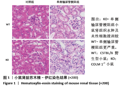
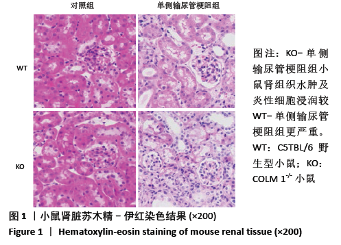
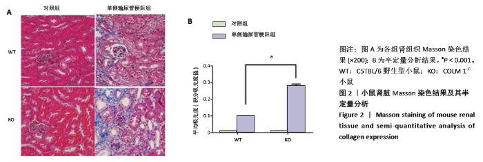
2.1 肾组织形态学变化 苏木精-伊红染色结果显示,KO-对照组和WT-对照组小鼠肾组织形态未见异常,肾小管间质无扩大、无明显炎性细胞浸润。在单侧输尿管梗阻术后,小鼠结扎侧肾组织,KO-单侧输尿管梗阻组和WT-单侧输尿管梗阻组肾间质均出现了不同程度的水肿、存在炎性细胞的浸润,而KO-单侧输尿管梗阻组小鼠肾组织相对于WT-单侧输尿管梗阻组而言,这些症状更为严重(见图1)。Masson染色结果显示,KO-对照组和WT-对照组肾脏,肾小球大小、形态正常,未观察到蓝色胶原纤维沉积。而单侧输尿管梗阻组小鼠肾脏中均有蓝色胶原纤维沉积,出现了明显的纤维化。同时,半定量分析结果显示,与WT-单侧输尿管梗阻组相比,KO-单侧输尿管梗阻组的肾脏胶原沉积面积增加,差异有显著性意义(P < 0.05),见图2。"
"

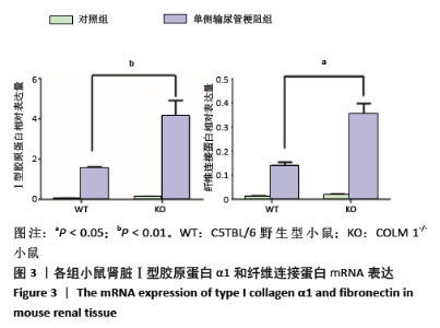
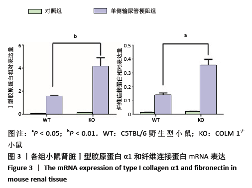
2.2 肾组织细胞外基质主要成分的表达 Ⅰ型胶原蛋白α1和纤维连接蛋白是肾纤维化发生的重要特征分子。qRT-PCR结果表明,单侧输尿管梗阻术后,小鼠组织中Ⅰ型胶原蛋白α1和纤维连接蛋白 mRNA表达量均明显升高,且KO-单侧输尿管梗阻组表达量高于WT-单侧输尿管梗阻组,差异有显著性意义(P < 0.05),见图3。"
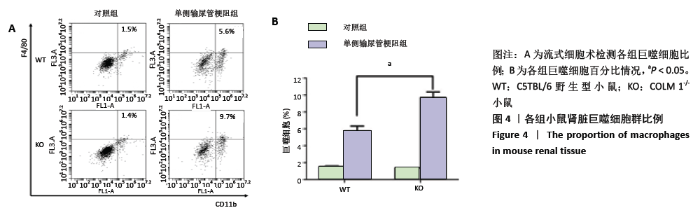
2.3 肾间质巨噬细胞浸润情况 流式结果分析显示,WT-对照组、KO-对照组、WT-单侧输尿管梗阻组、KO-单侧输尿管梗阻组小鼠肾组织中巨噬细胞平均比例分别为1.5%,1.4%,5.6%,9.7%。与对照组相比,单侧输尿管梗阻组肾组织中巨噬细胞增加,KO-单侧输尿管梗阻组巨噬细胞比例最高,较WT-单侧输尿管梗阻组明显增多, 差异有显著性意义(P < 0.05),见图4。"

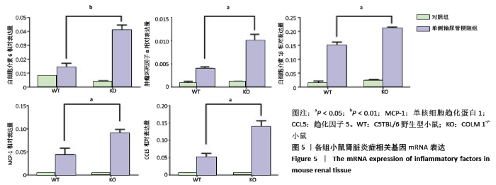
2.4 肾组织中炎症相关细胞因子的表达 qRT-PCR检测炎症因子白细胞介素6、肿瘤坏死因子α、白细胞介素1β 以及趋化因子 CCL5、MCP-1的基因水平表达。结果显示,单侧输尿管梗阻组表达水平较对照组均显著增加。在对照组肾组织中,白细胞介素6、肿瘤坏死因子α、白细胞介素1β、CCL5、MCP-1仅有极少量表达,而KO-单侧输尿管梗阻组白细胞介素6、肿瘤坏死因子α、白细胞介素1β、CCL5、MCP-1 mRNA的表达量均高于WT-单侧输尿管梗阻组,差异有显著性意义(P < 0.05),见图5。以上结果显示,单侧输尿管梗阻术后,KO小鼠出现了更为严重的炎症反应。"

| [1] HUMPHREYS BD. Mechanisms of renal fibrosis. Annu Rev Physiol. 2018;80:309-326. [2] WEBSTER AC, NAGLER EV, MORTON RL, et al. Chronic kidney disease. Lancet. 2017;389(10075):1238-1252. [3] ROMAGNANI P, REMUZZI G, GLASSOCK R, et al. Chronic kidney disease. Nat Rev Dis Primers. 2017;3:17088. [4] RUIZ-ORTEGA M, RAYEGO-MATEOS S, LAMAS S, et al. Targeting the progression of chronic kidney disease. Nat Rev Nephrol. 2020;16:269-288. [5] NOGUEIRA A, PIRES MJ, OLIVEIRA PA. Pathophysiological mechanisms of renal fibrosis: a review of animal models and therapeutic strategies. In Vivo. 2017;31(1):1-22. [6] CHEVALIER RL, FORBES MS, THORNHILL BA. Ureteral obstruction as a model of renal interstitial fibrosis and obstructive nephropathy. Kidney International. 2009;75(11):1145-1152. [7] KLADNEY RD, BULLA GA, GUO L, et al. GP73, a novel Golgi-localized protein upregulated by viral infection. Gene. 2000;249(1-2):53-65. [8] JIANG K, LI W, ZHANG Q, et al. GP73 N-glycosylation at Asn144 reduces hepatocellular carcinoma cell motility and invasiveness. Oncotarget. 2016;7(17):23530-23541. [9] HU L, YAO W, WANG F, et al. GP73 is upregulated by Hepatitis C Virus (HCV) infection and enhances HCV secretion. PLoS ONE. 2014;9(3): e90553. [10] YANG L, LUO P, SONG Q, et al. DNMT1/miR-200a/GOLM1 signaling pathway regulates lung adenocarcinoma cells proliferation. Biomed Pharmacother. 2018;99:839-847. [11] ALLAM MA, ELTIBY DM, NASSAR Y, et al. Studying the value of golgi protein 73 as a serum marker in hepatocellular carcinoma versus alfa feto protein. Nature and Science. 2016;14(1):126-129. [12] KOJIMA S, ENOKIDA H, YOSHINO H, et al. The tumor-suppressive microRNA-143/145 cluster inhibits cell migration and invasion by targeting GOLM1 in prostate cancer. J Hum Genet. 2014;59(2):78-87. [13] YANG S, ZENG C, FANG X, et al, Hepatitis B virus upregulates GP73 expression by activating the HIF-2α signaling pathway. Oncol Lett. 2018; 15(4):5264-5270. [14] YE Q, ZHU W, ZHANG J, et al. GOLM1 modulates EGFR/RTK cell-surface recycling to drive hepatocellular carcinoma metastasis. Cancer Cell. 2016;30:444-458. [15] LIU Y, ZHANG X, ZHOU S, et.al. Knockdown of Golgi phosphoprotein 73 blocks the trafficking of matrix metalloproteinase‐2 in hepatocellular carcinoma cells and inhibits cell invasion. J Cell Mol Med. 2019;23(4): 2399-2409. [16] YANG X, WEI C. LIU N, et al. GP73, a novel TGF-β target gene, provides selective regulation on Smad and non-Smad signaling pathways. Biochim Biophys Acta Mol Cell Res. 2019;1866(4):588-597. [17] ZHANG X, ZHU C, WANG T, et al. GP73 represses host innate immune response to promote virus replication by facilitating MAVS and TRAF6 degradation. PLoS Pathogens. 2017;13(4):e1006321. [18] LIU Y, LIEWEN H, MARKULY M, et al. A review of GOLPH2, an oncogenic protein and novel therapeutic options for GOLPH2 driven tumours. J Translat Sci. 2019;6:1-4. [19] YANG H, LIU G, LIU B, et al. GP73 promotes invasion and metastasis of bladder cancer by regulating the epithelial–mesenchymal transition through the TGF‐β1/Smad2 signalling pathway. J Cell Mol Med. 2018; 22(3):1650-1665. [20] XU R, JI J, ZHANG X, et al. PDGFA/PDGFR α-regulated GOLM1 promotes human glioma progression through activation of AKT. J Exp Clin Cancer Res. 2017;36(1):193. [21] DING X, DENG G, LIU J, et al. GOLM1 silencing inhibits the proliferation and motility of human glioblastoma cells via the Wnt/β-catenin signaling pathway. Brain Res. 2019;1717:117-126. [22] YAN G, RU Y, WU K, GOLM1 promotes prostate cancer progression through activating PI3K‐AKT‐mTOR signaling. Prostate. 2018;78(3): 166-177. [23] LI XM, CAO LL. Identification of GOLM1 as a Positively Regulator of Tumor Metastasis by Regulating MMP13 in Gastric Carcinoma. Cancer Biomark. 2019;26(4):421-430. [24] ZHANG W, KIM H, LV J, et al. Golgi phosphoprotein 2 is a novel regulator of IL-12 production and macrophage polarization. J Immunol. 2018;200(4):1480-1488. [25] LIU Y. Cellular and molecular mechanisms of renal fibrosis. Nat Rev Nephrol. 2011;7(12):684-696. [26] PILLING D, FAN T, HUANG D, et al. Identification of markers that distinguish monocyte-derived fibrocytes from monocytes, macrophages, and fibroblasts. PLoS One. 2009;4(10):e7475. [27] DISTLER JHW, GYÖRFI A, RAMANUJAM M, et al. Shared and distinct mechanisms of fibrosis. Nat Rev Rheumatol. 2019;15:705-730. [28] NOGUEIRA A, PORES MJ, OLIVEIRA P. Pathophysiological mechanisms of renal fibrosis: a review of animal models and therapeutic strategies. In Vivo. 2017;31(1):1-22. [29] BLACK LM, LEVER JM, AGARWAL A. Renal Inflammation and Fibrosis: A Double-edged Sword. J Histochem Cytochem. 2019;67(9):663-681. [30] HUEN SC, Cantley LG. Macrophages in Renal Injury and Repair. Annu Rev Physiol. 2017;79:449-469. [31] KLUTH DC. Pro-resolution properties of macrophages in renal injury. Kidney Int. 2007;72(3):234-236. [32] Huang L, Wang AM, Hao Y, et al. Macrophage Depletion Lowered Blood Pressure and Attenuated Hypertensive Renal Injury and Fibrosis. Front Physiol. 2018;9:473. [33] MARTÍNEZ-KLIMOVA E, APARICIO-TREJO OE, TAPIA E, et al. Unilateral ureteral obstruction as a model to investigate fibrosis-attenuating treatments. Biomo- lecules. 2019;9(4):141. [34] TANG PM, NIKOLIC-PATERSON DJ, LAN H. Macrophages: versatile players in renal inflammation and fibrosis. Nat Rev Nephrol. 2019; 15(3):144-158. [35] LU H, WU L, LIU L.Quercetin ameliorates kidney injury and fibrosis by modulating M1/M2 macrophage polarization. Biochem Pharmacol. 2018;154:203-212. [36] Sato Y, Yanagita M. Immune cells and inflammation in AKI to CKD progression.Am J Physiol Ren Physiol. 2018;315:F1501-F1512. [37] LV W, BOOZ GW, WANG Y, et al. Inflammation and renal fibrosis: recent developments on key signaling molecules as potential therapeutic targets. Eur J Pharmacol. 2018;820:65-76. [38] GRANDE MT, PÉREZ-BARRIOCANAL F, LÓPEZ-NOVOA JM. Role of inflammation in túbulo-interstitial damage associated to obstructive nephropathy.J Inflamm (Lond). 2010;7:19. [39] LIANG H, XU F, WEN X, et al.Interleukin-33 signaling contributes to renal fibrosis following ischemia reperfusion. Eur J Pharmacol. 2017;812:18-27. [40] MAEL-AININ M, ABED A, CONWAY SJ, et al. Inhibition of periostin expression protects against the development of renal inflammation and fibrosis. J Am Soc Nephrol. 2014;25(8):1724-1736. [41] WRIGHT LM, YONG S, PICKEN MM, et al. Decreased survival and hepato-renal pathology in mice with c-terminally truncated GP73 (GOLPH2).Int J Clin Exp Pathol. 2009;2(1):34-47. |
| [1] | Shen Jinbo, Zhang Lin. Micro-injury of the Achilles tendon caused by acute exhaustive exercise in rats: ultrastructural changes and mechanism [J]. Chinese Journal of Tissue Engineering Research, 2021, 25(8): 1190-1195. |
| [2] | Li Jing, Xie Jianshan, Cui Huilin, Cao Ximei, Yang Yanping, Li Hairong. Expression and localization of diacylglycerol kinase zeta and protein kinase C beta II in mouse back skin with different coat colors [J]. Chinese Journal of Tissue Engineering Research, 2021, 25(8): 1196-1200. |
| [3] | Chai Le, Lü Jianlan, Hu Jintao, Hu Huahui, Xu Qingjun, Yu Jinwei, Quan Renfu. Signal pathway variation after induction of inflammatory response in rats with acute spinal cord injury [J]. Chinese Journal of Tissue Engineering Research, 2021, 25(8): 1218-1223. |
| [4] | Geng Qiudong, Ge Haiya, Wang Heming, Li Nan. Role and mechanism of Guilu Erxianjiao in treatment of osteoarthritis based on network pharmacology [J]. Chinese Journal of Tissue Engineering Research, 2021, 25(8): 1229-1236. |
| [5] | Tan Jingyu, Liu Haiwen. Genome-wide identification, classification and phylogenetic analysis of Fasciclin gene family for osteoblast specific factor 2 [J]. Chinese Journal of Tissue Engineering Research, 2021, 25(8): 1243-1248. |
| [6] | Gu Xia, Zhao Min, Wang Pingyi, Li Yimei, Li Wenhua. Relationship between hypoxia inducible factor 1 alpha and hypoxia signaling pathway [J]. Chinese Journal of Tissue Engineering Research, 2021, 25(8): 1284-1289. |
| [7] | Li Cai, Zhao Ting, Tan Ge, Zheng Yulin, Zhang Ruonan, Wu Yan, Tang Junming. Platelet-derived growth factor-BB promotes proliferation, differentiation and migration of skeletal muscle myoblast [J]. Chinese Journal of Tissue Engineering Research, 2021, 25(7): 1050-1055. |
| [8] | Wang Hanyue, Li Furong, Yang Xiaofei, Hu Chaofeng. Direct reprogramming hepatocytes into islet-like cells by efficiently targeting and activating the endogenous genes [J]. Chinese Journal of Tissue Engineering Research, 2021, 25(7): 1056-1063. |
| [9] | Wang Zhengdong, Huang Na, Chen Jingxian, Zheng Zuobing, Hu Xinyu, Li Mei, Su Xiao, Su Xuesen, Yan Nan. Inhibitory effects of sodium butyrate on microglial activation and expression of inflammatory factors induced by fluorosis [J]. Chinese Journal of Tissue Engineering Research, 2021, 25(7): 1075-1080. |
| [10] | Xie Wenjia, Xia Tianjiao, Zhou Qingyun, Liu Yujia, Gu Xiaoping. Role of microglia-mediated neuronal injury in neurodegenerative diseases [J]. Chinese Journal of Tissue Engineering Research, 2021, 25(7): 1109-1115. |
| [11] | Kong Desheng, He Jingjing, Feng Baofeng, Guo Ruiyun, Asiamah Ernest Amponsah, Lü Fei, Zhang Shuhan, Zhang Xiaolin, Ma Jun, Cui Huixian. Efficacy of mesenchymal stem cells in the spinal cord injury of large animal models: a meta-analysis [J]. Chinese Journal of Tissue Engineering Research, 2021, 25(7): 1142-1148. |
| [12] | Shi Yangyang, Qin Yingfei, Wu Fuling, He Xiao, Zhang Xuejing. Pretreatment of placental mesenchymal stem cells to prevent bronchiolitis in mice [J]. Chinese Journal of Tissue Engineering Research, 2021, 25(7): 991-995. |
| [13] | Zheng Xiaolong, He Xiaoming, Gong Shuidi, Pang Fengxiang, Yang Fan, He Wei, Liu Shaojun, Wei Qiushi. Bone turnover characteristics in patients with alcohol-induced osteonecrosis of the femoral head [J]. Chinese Journal of Tissue Engineering Research, 2021, 25(5): 657-661. |
| [14] | Liu Xin, Yan Feihua, Hong Kunhao. Delaying cartilage degeneration by regulating the expression of aquaporins in rats with knee osteoarthritis [J]. Chinese Journal of Tissue Engineering Research, 2021, 25(5): 668-673. |
| [15] | Ma Zetao, Zeng Hui, Wang Deli, Weng Jian, Feng Song. MicroRNA-138-5p regulates chondrocyte proliferation and autophagy [J]. Chinese Journal of Tissue Engineering Research, 2021, 25(5): 674-678. |
| Viewed | ||||||
|
Full text |
|
|||||
|
Abstract |
|
|||||