中国组织工程研究 ›› 2013, Vol. 17 ›› Issue (49): 8563-8569.doi: 10.3969/j.issn.2095-4344.2013.49.016

• 干细胞因子及调控因子 stem cell factors and regulatory factors • 上一篇    下一篇

转化生长因子β1与骨髓间充质干细胞向心肌样细胞的分化

吕  洋1,刘  博2,王海萍1   

  1. 1河北北方学院组织学与胚胎学教研室,河北省张家口市  075000
    2河北北方学院附属第一医院病理科,河北省张家口市  075000
  • 修回日期:2013-09-22 出版日期:2013-12-03 发布日期:2013-12-03
  • 通讯作者: 王海萍,博士,教授,河北北方学院组织学与胚胎学教研室,河北省张家口市 075000 haipingmimi@126.com
  • 作者简介:吕洋★,女,1979年生,河北省张家口市人,回族,2007年解放军军医进修学院毕业,硕士,讲师,主要从事骨髓干细胞向心肌细胞诱导分化的研究。 yyang2bb@163.com
  • 基金资助:

    河北省自然基金资助项目(C2009001022)*;河北省教育厅科学技术研究重点项目(ZH2012001)*;河北北方学院青年基金项目(Q2013027)*

Effect of transforming growth factor beta1 on the differentiation of bone marrow mesenchymal stem cells into cardiomyocyte-like cells in vitro

Lü Yang1, Liu Bo2, Wang Hai-ping1   

  1. 1Department of Histology and Embryology, Hebei North University, Zhangjiakou  075000, Hebei Province, China
    2Department of Pathology, the First Affiliated Hospital of Hebei North University, Zhangjiakou  075000, Hebei Province, China
  • Revised:2013-09-22 Online:2013-12-03 Published:2013-12-03
  • Contact: Wang Hai-ping, M.D., Professor, Department of Histology and Embryology, Hebei North University, Zhangjiakou 075000, Hebei Province, China haipingmimi@126.com
  • About author:Lü Yang★, Master, Lecturer, Department of Histology and Embryology, Hebei North University, Zhangjiakou 075000, Hebei Province, China yyang2bb@163.com
  • Supported by:

     the Natural Science Foundation of Hebei Province, No. C2009001022*; the Scientific Research Projects of the Education Department of Hebei Province, No. ZH2012001*; the Youth Foundation of Hebei North University, No. Q2013027*

摘要:

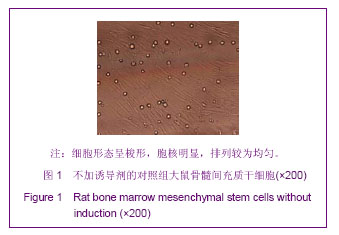
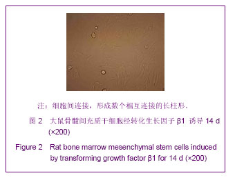
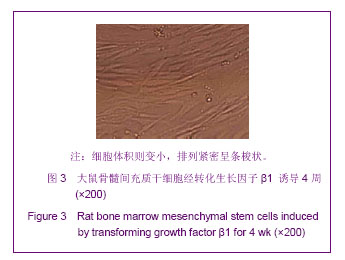
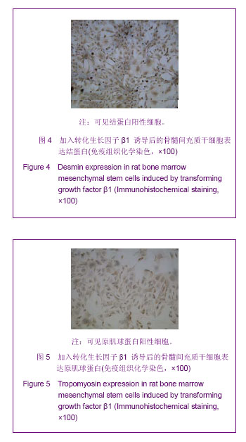
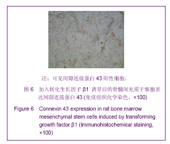
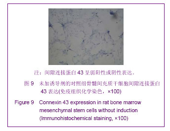
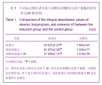
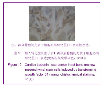
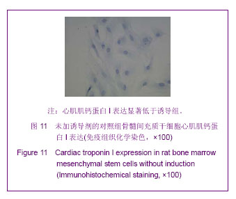
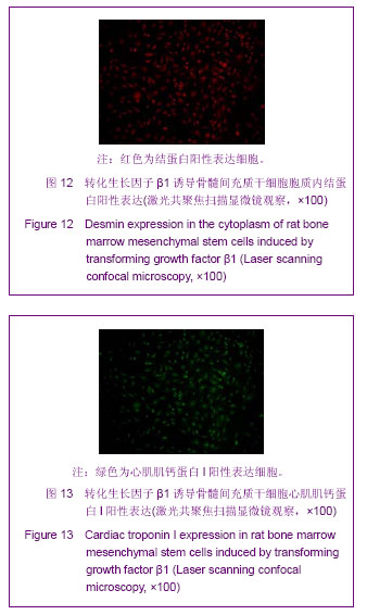
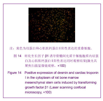
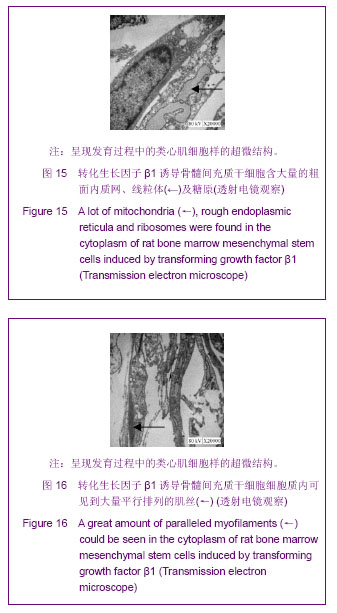
背景:转化生长因子β1是一种具有调控细胞增殖、分化、黏附和凋亡,在生物发育及组织修复过程中具有重要作用的细胞因子。
目的:观察转化生长因子β1对诱导大鼠骨髓间充质干细胞定向分化为心肌样细胞的影响。
方法:取SD大鼠四肢骨骨髓,分离培养骨髓间充质干细胞,应用转化生长因子β1对第2代骨髓间充质干细胞定向诱导,以未诱导的骨髓间充质干细胞做对照。
结果与结论:①加入转化生长因子β1 诱导后的骨髓间充质干细胞培养7 d时,相差显微镜观察多紧密平行排列生长,呈类长柱形,少数呈不规则形;14 d后开始出现细胞间连接,形成数个相互连接的长柱形,28 d时骨髓间充质干细胞体积则变小,排列紧密呈条梭状。②转化生长因子β1 诱导4周后,免疫细胞化学技术检测骨髓间充质干细胞均可见结蛋白、原肌球蛋白及间隙连接蛋白43的阳性细胞,诱导组各指标的积分吸光度值均显著高于对照组(P均< 0.01)。③诱导组心肌肌钙蛋白I呈阳性表达。根据心肌肌钙蛋白I阳性率计算出的心肌样细胞转化率在诱导组显著高于对照组(P=0.000)。④荧光免疫双标技术检测结果显示,经转化生长因子β1诱导4周的骨髓间充质干细胞结蛋白及心肌肌钙蛋白I蛋白均定位于细胞质且呈共表达。⑤透射电镜结果显示,分化细胞的细胞质内可见到平行排列的肌丝,并富含线粒体、糖原和核糖体。提示骨髓间充质干细胞在转化生长因子β1诱导下可定向分化为心肌样细胞。


中国组织工程研究杂志出版内容重点:干细胞;骨髓干细胞;造血干细胞;脂肪干细胞;肿瘤干细胞;胚胎干细胞;脐带脐血干细胞;干细胞诱导;干细胞分化;组织工程


全文链接:

关键词: 干细胞, 干细胞因子及调控因子, 骨髓间充质干细胞, 转化生长因子β1, 心肌样细胞, 心肌细胞, 结蛋白, 原肌球蛋白, 心肌间隙连接蛋白43, 心肌肌钙蛋白I, 省级基金, 干细胞图片文章

Abstract:

BACKGROUND: Transforming growth factor β1 is a cytokine that can regulate cell proliferation, differentiation, adhesion and apoptosis and have an important role in developmental biology and tissue repair process.
OBJECTIVE: To explore the differentiation of rat bone marrow mesenchymal stem cells into cardiomyocyte-like cells in vitro induced by transforming growth factor beta1.
METHODS: Bone marrow mesenchymal stem cells were isolated and cultured from the bone marrow of Sprague-Dawley rats. Then, passage 2 bone marrow mesenchymal stem cells were induced by transforming growth factor beta1. Bone marrow mesenchymal stem cells without induction served as controls.
RESULTS AND CONCLUSION: The morphology of bone marrow mesenchymal stem cells after induced by transforming growth factor beta1 for 7 days became larger and extended. Most of induced cells became long shuttle-shaped and paralleled, and a few arranged irregularly. After 14 days, most cells became rod-shape and touched with adjacent cells to form myotube-shape. Up to 28 days, the induced cells became smaller, showed fusiform shape and arranged tightly. After induced by transforming growth factor beta1 for 4 weeks, bone marrow mesenchymal stem cells could be identified by the positive staining for desmin, tropomyosin, connexin 43 and cardiac troponin I. The integral absorbance values in the induction group were significantly higher than those in the control group (all P < 0.01). According to the positive rate of cardiac troponin I, the conversion rate of cardiomyocyte-like cells in the induction group was significantly higher than that in the control group (P=0.000). Immunofluorescence double-labeling assay showed that after induced by transforming growth factor beta1 for 4 weeks, desmin and cardiac troponin I proteins were localized in the cytoplasm and showed co-expression. Transmission electron microscope showed that the induced cells had a cardiomyocyte-like ultrastructure: a lot of mitochondria, rough endoplasmic reticula and ribosomes were founded in the cytoplasm, and paralleled myofilaments could be seen in the cytoplasm. It is indicated that bone marrow mesenchymal stem cells can differentiate into cardiomyocytes in vitro after induced by transforming growth factor beta1.


中国组织工程研究杂志出版内容重点:干细胞;骨髓干细胞;造血干细胞;脂肪干细胞;肿瘤干细胞;胚胎干细胞;脐带脐血干细胞;干细胞诱导;干细胞分化;组织工程


全文链接:

Key words: stem cells, bone marrow mesenchymal stem cells, transforming growth factor beta1, muscle cells

中图分类号: