中国组织工程研究 ›› 2013, Vol. 17 ›› Issue (53): 9139-9144.doi: 10.3969/j.issn.2095-4344.2013.53.007

• 细胞与组织移植 cell and tissue transplantation • 上一篇    下一篇

FLK-1+胎肝细胞移植治疗急性肝损伤

李  娜1,丁伟荣2,伍柏青3,刘婷婷2,程洪波2,金成豪2   

  1. 1南昌大学医学院,江西省南昌市   330006;江西省人民医院,2血液科,3检验科,江西省南昌市  330006
  • 修回日期:2013-09-19 出版日期:2013-12-31 发布日期:2013-12-31
  • 通讯作者: 金成豪,博士,主任医师,硕士生导师,江西省人民医院血液科,江西省南昌市 330006 jinch227@yahoo.com.cn
  • 作者简介:李娜★,女,1978年生,江西省抚州市人,汉族,2012年南昌大学毕业,硕士,医师,主要从事干细胞生物学研究。 634624912@qq.com. 并列第一作者:丁伟荣☆,男,1975年生,江西省高安市人,汉族,2004年浙江大学毕业,博士,副主任医师,主要从事干细胞定向分化及机制研究。 dingwr97@163.com
  • 基金资助:

    国家自然科学基金项目(30860115)*;江西省自然科学基金项目(2008GZY0084)资助*

Transplantation of fetal liver FLK-1+ cells in treatment of acute liver injury

Li Na1, Ding Wei-rong2, Wu Bo-qing3, Liu Ting-ting2, Cheng Hong-bo2, Jin Cheng-hao2   

  1. 1Medical College of Nanchang University, Nanchang  330006, Jiangxi Province, China; 2Department of Hematology, 3Department of Clinical Laboratories, Jiangxi Provincial People’s Hospital, Nanchang  330006, Jiangxi Province, China
  • Revised:2013-09-19 Online:2013-12-31 Published:2013-12-31
  • Contact: Jin Cheng-hao, Ph.D., Chief physician, Master’s supervisor, Department of Hematology, Jiangxi Provincial People’s Hospital, Nangchang 330006, Jiangxi Province, China inch227@yahoo.com.cn
  • About author:Li Na★, Master, Physician, Medical College of Nanchang University, Nanchang 330006, Jiangxi Province, China 634624912@qq.com Ding Wei-rong☆, M.D., Associate chief physician, Department of Hematology, Jiangxi Provincial People’s Hospital, Nanchang 330006, Jiangxi Province, China dingwr97@163.com Li Na and Ding Wei-rong contributed equally to this work.
  • Supported by:

    the National Natural Science Foundation of China, No. 30860115*; the Natural Science Foundation of Jiangxi Province in China, No. 2008GZY0084*

摘要:

背景:作者先期研究表明,在鼠胚第17-19天胎肝存在一类FLK-1+细胞,表达胚胎干细胞的特征性标志,并具有多向分化功能。

目的:验证胎肝FLK-1+细胞治疗小鼠急性肝损伤的修复作用。
方法:免疫磁珠提取及流式细胞仪检测胎肝FLK-1+细胞;RT-PCR检测FLK-1+细胞Oct-3/4、Rex-1基因;肝细胞生长因子和表皮生长因子诱导FLK-1+细胞向肝细胞分化。腹腔注射四氯化碳建立小鼠急性肝损伤模型并随机分为2组:对照组模型小鼠仅输入生理盐水,实验组模型小鼠尾静脉输注诱导分化FLK-1+细胞(1×106细胞),16 h后两组取血测定肝功能,观察64 h小鼠死亡率。
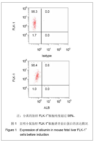
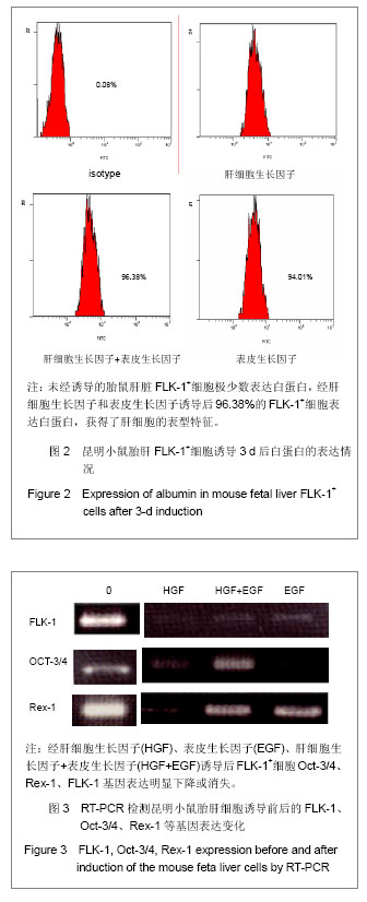
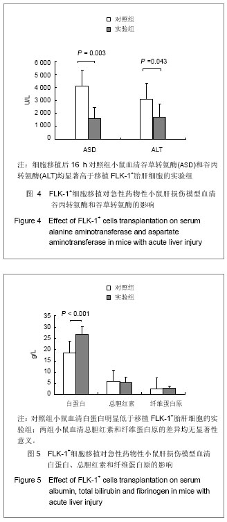
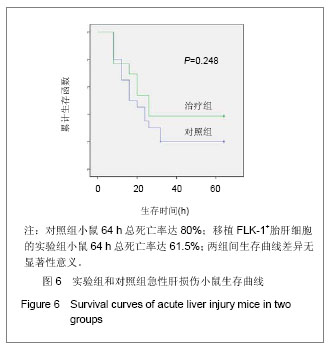
结果与结论:胎鼠肝脏FLK-1+细胞高表达Oct-3/4、Rex-1,白蛋白表达的FLK-1+细胞阳性率为0.6%。经肝细胞生长因子诱导3 d后FLK-1不表达,Oct-3/4和Rex-1表达显著下降或消失。经肝细胞生长因子和表皮生长因子诱导后96.38%的FLK-1+细胞表达白蛋白。诱导3d的FLK-1+细胞移植后16 h小鼠血清谷草转氨酶和谷丙转氨酶显著低于对照组(P < 0.05),血清白蛋白显著高于对照组(P < 0.05);两组血清总胆红素和纤维蛋白原差异无显著性意义(P > 0.05);移植组64 h死亡率为61.5%与对照组(80%)差异无显著性意义(P > 0.05)。说明胎鼠肝脏肝细胞生长因子和表皮生长因子诱导3 d的FLK-1+细胞移植可较好地改善急性肝损伤的肝细胞功能。


中国组织工程研究
杂志出版内容重点:肾移植肝移植移植;心脏移植;组织移植;皮肤移植;皮瓣移植;血管移植;器官移植组织工程


全文链接:

关键词: 器官移植, 细胞移植, 肝功能损伤, FLK-1+细胞, 细胞移植, 肝细胞生长因子, 表皮生长因子, 谷草转氨酶, 谷丙转氨酶, 国家自然科学基金

Abstract:

BACKGROUND: Previous studies of our research group have shown that FLK-1+ cells existed in the mouse fetal liver at embryonic 17-19 days, and they expressed embryonic stem cells and presented multi-differentiation potentials.

OBJECTIVE: To evaluate the therapeutic effect of the transplantation of FLK-1+ cells derived from mouse fetal liver on acute liver injury in mice.
METHODS: The FLK-1+ fraction were enriched from the fetal liver with immunomagnetic beads method and detected by flow cytometry. The Oct-3/4 and Rex-1 genes in FLK-1+ cells were detected by reverse transcription-polymerase chain reaction. The FLK-1+ cells were induced to differentiate into liver cells by hepatocyte growth factor and epidermal growth factor. The model of acute liver injury in mice was established by intraperitoneally injecting carbon tetrachloride 4, and was randomly divided into two groups. In the control group, mouse models with acute liver injury were infused normal saline via tail vein; in the experimental group, mouse models with acute liver injury were infused induced FLK-1+ cells (1×10⁶) via tail vein. The blood was collected at 16 hours and liver functions were detected. The mortality of mice was observed at 64 hours.
RESULTS AND CONCLUSION: The fetal liver FLK-1+ cells highly expressed Oct-3/4 and Rex-1 mRNA, and albumin expressing rate in FLK-1+ cells was 0.6%. After induced by hepatocyte growth factor for 3 days, FLK-1 did not express. After induced by hepatocyte growth factor and epidermal growth factor for 3 days, Oct-3/4 and Rex-1 mRNA expression was significantly reduced or disappeared in FLK-1+ cells, and 96.38% FLK-1+ cells expressed albumin. After FLK-1+ cells induced for 3 days were transplanted to mouse models with acute liver injury for 16 hours, the serum glutamic-oxalacetic transaminease and glutamic-pyruvic transaminase were significantly lower (P < 0.05), but serum albumin was significantly higher than the control group (P < 0.05). However, serum total bilirubin and fibrinogen showed no significant differences between two groups (P > 0.05). The mortality rate in the control group was 80% and the mortality rate in the experimental group was 61.5% at 64 hours, with no significant difference between two groups (P > 0.05). Experimental findings indicate that, transplanting fetal liver FLK-1+ cells induced by hepatocyte growth factor and epidermal growth factor for 3 days can improve liver cells function in mice with acute liver damage.


中国组织工程研究
杂志出版内容重点:肾移植肝移植移植;心脏移植;组织移植;皮肤移植;皮瓣移植;血管移植;器官移植组织工程


全文链接:

Key words: hepatocyte growth factor, epidermal growth factor, aspartate aminotransferase, alanine aminotransferase

中图分类号: