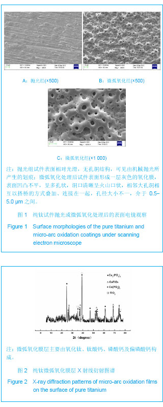
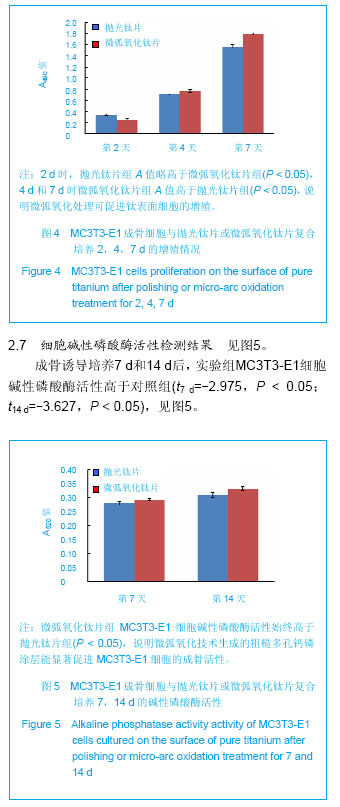
| [1] Yoshiki O.Bioscience and Bioengineering of Titanium Materials. Elsevier Science Ltd, 2007.
[2] Williams DF.On the mechanisms of biocompatibility. Biomaterials. 2008;29(20):2941-2953.
[3] Bose S,Roy M,Das K,et al.Surface modification of titanium for load-bearing applications.J Mater Sci Mater Med.2009; 20: 19-24.
[4] Wei D,Zhou Y,Yang C.Structure, cell response and biomimetic apatite induction of gradient TiO2-based/nano-scale hydrophilic amorphous titanium oxide containing Ca composite coatings before and after crystallization. Colloids Surf B Biointerfaces.2009;74(1):230-237.
[5] Li Y,Lee IS,Cui FZ,et al.The biocompatibility of nanostructured calcium phosphate coated on micro-arc oxidized titanium. Biomaterials. 2008;29(13):2025-2032.
[6] 陈建治,张富强,石玉龙,等.电解液钙磷比对微弧氧化法制备含钙磷氧化物膜结构的影响[J].实用口腔医学杂志,2007,23(2): 249-251.
[7] Huang Y,Wang YJ,Ning CY,et al.Hydroxyapatite coatings produced on commercially pure titanium by micro-arc oxidation.Biomed Mater.2007;2(3): 196-201.
[8] Teng FY,Ko CL,Kuo HN,et al.A comparison of epithelial cells, fibroblasts, and osteoblasts in dental implant titanium topographies.Bioinorg Chem Appl. 2012;2012:687291.
[9] Zhao Z,Chen X,Chen A,et al.Synthesis of bioactive ceramic on the titanium substrate bymicro-arc oxidation.Biomed Mater Res A.2009;90(2):438-445.
[10] Yu S,Yang X,Yang L,et al.Novel technique for preparing Ca- and P-containg ceramic coaing on Ti-6Al-4V by micro-arc oxidation.Biomed Mater Res B Appl Biomater.2007; 83(2): 623-627.
[11] 马臣,王颖慧,曲力.钛合金微弧氧化技术的研究现状及展望[J].中国陶瓷工业,2007,14(1):46-49.
[12] Sul YT,Byon ES,Jeong Y.Biomechanical measurements of calcium-incorporated oxidized implants in rabbit bone:effect of calcium surface chemistry of a novel implant.Clin Implant Dent Relat Res.2004;6(2):101-110.
[13] 李颂,刘耀辉,庞磊.电源频率对铸铝合金微弧氧化陶瓷层的影响[J].材料科学与工艺,2008,16(2):287-289.
[14] 杨斯琴,王焱,宁成云,等.处理时间对纯钛微弧氧化膜层表面性能的影响[J].中华口腔医学杂志,2013,48(1):41-45.
[15] Han Y,Hong SH,Xu KW.Structure and in vitro bioactivity of titania-based films by microarc oxidation.Surf Coat Tech. 2003;168(2-3):249-258.
[16] St-Pierre JP,Gauthier M,Lefebvre LP,et al.Three-dimensional growth of differentiating MC3T3-E1 pre-osteoblasts on porous titanium scaffolds. Biomaterials.2005;26(35):7319-7328.
[17] Rausch-Fan X,Qu Z,Wieland M,et al.Differentiation and cytokine synthesis of human alveolar osteoblasts compared to osteoblast-like cells (MG63) in response to titanium surfaces. Dent Mater.2008;24(1):102-110.
[18] Benoit DS,Schwartz MP,Durney AR,et al.Small functional groups for controlled differentiation of hydrogel-encapsulated human mesenchymal stem cells.Nat Mater.2008;7(10): 816-823.
[19] 陈卓凡.口腔种植治疗的基础研究与临床应用[M].北京:人民军医出版社,2010.
[20] Benoit DS,Schwartz MP,Durney AR,et al.Small functional groups for controlled differentiation of hydrogel-encapsulated human mesenchymal stem cells. Nat Mater. 2008;7(10): 816-823.
[21] Han Y,Chen D,Sun J,et al.UV-enhanced bioactivity and cell response of micro-arc oxidized titania coatings.Acta Biomater. 2008;4(5):1518-1529.
[22] Hao L, Lawrence J. Wettability modification and the subsequent manipulation of protein adsorption on a Ti6Al4V alloy by means of CO2 laser surface treatment. J Mater Sci Mater Med.2007;18(5):807-817.
[23] Watanabe T,Yoshida N.Wettability control of a solid surface by utilizing photocatalysis.Chem Rec.2008;8(5):279-290.
[24] Benoit DS,Schwartz MP,Durney AR,et al.Small functional groups for controlled differentiation of hydrogel-encapsulated human mesenchymal stem cells. Nat Mater.2008;7(10): 816-823.
[25] Rupp F,Scheideler L,Olshanska N,et al.Enhancing surface free energy and hydrophilicity through chemical modification of microstructured titanium implant surfaces.J Biomed Mater Res A.2006;76(2):323-334.
[26] Deng F,Zhang W,Zhang P,et al.Improvement in the morphology of micro-arc oxdised titanium surface: A new process to increase osteoblast response. Mat Sci Eng C-Bio S.2010;1(30):141-147.
[27] Padial-Molina M,Galindo-Moreno P,Fernandez-Barbero JE,et al.Role of wettability and nanoroughness on interactions between osteoblast and modified silicon surfaces.Acta Biomater. 2011;7(2):771-778.
[28] Chen H,Hsiao C,Long H,et al.Micro-arc oxidation of β-titanium alloy: Structural characterization and osteoblast compatibility. Surf Coat Tech.2009; 204(6-7):1126-1131.
[29] Klenke FM,Liu Y,Yuan H,et al.Impact of pore size on the vascularization and osseointegration of ceramic bone substitutes in vivo.J Biomed Mater Res A. 2008;85(3): 777-786.
[30] Han CM,Kim HE,Kim YS,et al.Enhanced biocompatibility of Co-Cr implant material by Ti coating and micro-arc oxidation. J Biomed Mater Res B Appl Biomater. 2009;90(1):165-170.
[31] Kunzler TP,Huwiler C,Drobek T,et al.Systematic study of osteoblast response to nanotopography by means of nanoparticle-density gradients. Biomaterials. 2007;28(33): 5000-5006. |


.jpg)