Chinese Journal of Tissue Engineering Research ›› 2014, Vol. 18 ›› Issue (23): 3627-3632.doi: 10.3969/j.issn.2095-4344.2014.23.004
Previous Articles Next Articles
Expression of vascular endothelial growth factor in bone marrow mesenchymal stem cells under hypoxic conditions
Guo Hui, Zhang Yu-juan, Wei Xiao-guang, Xu Biao, Chen Yong-zhen
- Soochow University, Suzhou 215006, Jiangsu Province, China
-
Revised:2014-04-06Online:2014-06-04Published:2014-06-04 -
Contact:Chen Yong-zhen, Professor, Soochow University, Suzhou 215006, Jiangsu Province, China -
About author:Guo Hui, Master, Soochow University, Suzhou 215006, Jiangsu Province, China -
Supported by:the National Youth Foundation of China, No. 81100448
CLC Number:
Cite this article
Guo Hui, Zhang Yu-juan, Wei Xiao-guang, Xu Biao, Chen Yong-zhen. Expression of vascular endothelial growth factor in bone marrow mesenchymal stem cells under hypoxic conditions[J]. Chinese Journal of Tissue Engineering Research, 2014, 18(23): 3627-3632.
share this article

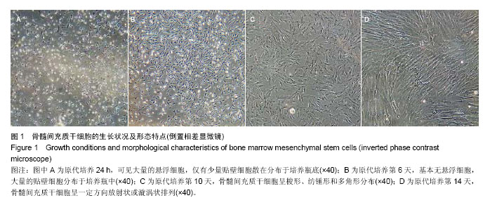
2.1 骨髓间充质干细胞的形态学变化 接种于培养瓶中的原代骨髓间充质干细胞培养24 h后镜下观察有大量悬浮的细胞,仅有少量贴壁细胞散在分布于培养瓶底(图1A);随后6 d连续换液培养后发现悬浮细胞基本清除,只留下贴壁细胞,且贴壁细胞的数量随着培养时间的增加而明显增加,少量的贴壁细胞呈梭形、纺锤形(图1B);7-10 d在培养瓶的边缘部有大量的梭形、纺锤形和多角形的贴壁细胞分布(图1C);14 d可见单层细胞贴壁生长,细胞呈一定方向放射状或漩涡状排列,细胞基本上呈长梭形的成纤维细胞形态(图1D)。消化传代,24 h后传代细胞完全贴壁生长。"
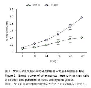
2.2 低氧诱导骨髓间充质干细胞的增殖情况 CCK-8结果显示,两组细胞在6-12 h均处于适应期,细胞数量缓慢的增加。常氧组细胞经过24-36 h快速增殖后,从48 h开始进入增殖平台期,而低氧组细胞的增殖活性在各个时间段内均高于常氧组,且在36 h和48 h细胞增殖更加明显,两者之间差异均有显著性意义(P < 0.05);低氧组培养48 h后,细胞数量持续增加,且明显高于常氧组,至72 h未见明显的细胞数量减少(图2)。"
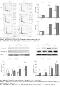
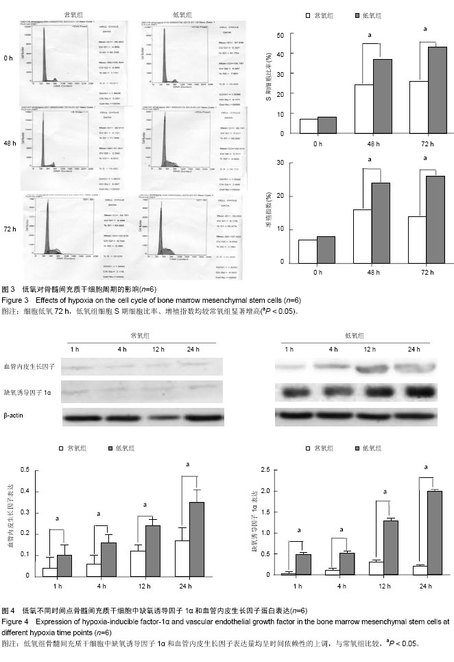
2.3 低氧诱导骨髓间充质干细胞的细胞周期变化 流式细胞术结果显示,随着培养时间的延长,与常氧组比较,低氧组骨髓间充质干细胞的细胞周期有较为明显改变。在培养48 h和72 h后,低氧组处于S期的细胞比例和细胞增殖指数均增加更为明显,两者差异均有显著性意义(P < 0.05,图3)。 2.4 低氧对骨髓间充质干细胞中缺氧诱导因子1α和血管内皮生长因子蛋白含量变化的影响 Western-blot法结果显示,两组内参β-actin的表达比较一致,常氧组缺氧诱导因子1α及血管内皮生长因子仅有少量的表达,而低氧组在低氧的诱导下,缺氧诱导因子1α和血管内皮生长因子表达量均呈时间依赖性的上调,且在24 h时,缺氧诱导因子1α和血管内皮生长因子蛋白表达量均显著增高,差异有显著性意义(P < 0.05,图4)。"
| [1] Pittenger MF, Mackay AM, Beck SC,et al.Multilineage potential of adult human mesenchymal stem cells.Science. 1999;284(5411):143-147. [2] Ranera B, Lyahyai J, Romero A,et al.Immunophenotype and gene expression profiles of cell surface markers of mesenchymal stem cells derived from equine bone marrow and adipose tissue.Vet Immunol Immunopathol. 2011; 144(1-2):147-154. [3] Orlic D, Kajstura J, Chimenti S, et al.Bone marrow cells regenerate infarcted myocardium.Nature. 2001;410(6829): 701-705. [4] Lennon DP, Edmison JM, Caplan AI.Cultivation of rat marrow-derived mesenchymal stem cells in reduced oxygen tension: effects on in vitro and in vivo osteochondrogenesis.J Cell Physiol. 2001;187(3):345-355. [5] Ball SG, Shuttleworth CA, Kielty CM.Vascular endothelial growth factor can signal through platelet-derived growth factor receptors.J Cell Biol. 2007;177(3):489-500. [6] 张戎,张勉,李成华,等.碱性成纤维细胞生长因子和血管内皮生长因子对人牙周膜干细胞体外增殖、迁移和黏附的影响[J].中华口腔医学杂志,2013,48(5):278-284. [7] 崔斌,黄岚,谭虎,等.血管内皮生长因子调节内皮祖细胞生物学功能[J].中华老年多器官疾病杂志,2009,8(3):265-268. [8] Campagnoli C, Roberts IA, Kumar S,et al.Identification of mesenchymal stem/progenitor cells in human first-trimester fetal blood, liver, and bone marrow.Blood. 2001;98(8):2396-2402. [9] Lee OK, Kuo TK, Chen WM,et al.Isolation of multipotent mesenchymal stem cells from umbilical cord blood.Blood. 2004;103(5):1669-1675. [10] Jiang Y, Jahagirdar BN, Reinhardt RL,et al.Pluripotency of mesenchymal stem cells derived from adult marrow.Nature. 2002;418(6893):41-49. [11] Dong J,Uemura T,Kojima H,et al.Application of low-pressure system to sustain in vivo bone formation in osteoblast/porous hydroxyapatite composite.Materials Science and Engineering: C.2001; 17(1): 37-43. [12] Neuhuber B, Swanger SA, Howard L,et al.Effects of plating density and culture time on bone marrow stromal cell characteristics.Exp Hematol. 2008;36(9):1176-1185. [13] Chen ZZ, Van Bockstaele DR, Buyssens N, et al.Stromal populations and fibrosis in human long-term bone marrow cultures.Leukemia. 1991;5(9):772-781. [14] Kawada H, Takizawa S, Takanashi T,et al.Administration of hematopoietic cytokines in the subacute phase after cerebral infarction is effective for functional recovery facilitating proliferation of intrinsic neural stem/progenitor cells and transition of bone marrow-derived neuronal cells.Circulation. 2006;113(5):701-710. [15] Dickhut A, Schwerdtfeger R, Kuklick L,et al.Mesenchymal stem cells obtained after bone marrow transplantation or peripheral blood stem cell transplantation originate from host tissue.Ann Hematol. 2005;84(11):722-727. [16] Sun B, Bai CX, Feng K,et al.Effects of hypoxia on the proliferation and differentiation of CD34(+) hematopoietic stem/progenitor cells and their response to cytokines.Sheng Li Xue Bao. 2000;52(2):143-146. [17] Cipolleschi MG, Dello Sbarba P, Olivotto M.The role of hypoxia in the maintenance of hematopoietic stem cells.Blood. 1993;82(7):2031-2037. [18] Krinner A, Zscharnack M, Bader A,et al.Impact of oxygen environment on mesenchymal stem cell expansion and chondrogenic differentiation.Cell Prolif. 2009;42(4):471-484. [19] Hao Y, Cheng D, Ma Y,et al.The relationship between oxygen concentration, reactive oxygen species and the biological characteristics of human bone marrow hematopoietic stem cells.Transplant Proc. 2011;43(7):2755-2761. [20] Bicheikina NI.Concentration of free oxygen in rabbit brain and bone marrow following administration of chemical radiation-protective agents.Radiobiologiia. 1963;3:898-902. [21] Chen LZ, Yin SM, Zhang XL,et al.Protective effects of human bone marrow mesenchymal stem cells on hematopoietic organs of irradiated mice.Zhongguo Shi Yan Xue Ye Xue Za Zhi. 2012;20(6):1436-1441. [22] Li ZY, Wang CQ, Lu G.Effects of bone marrow mesenchymal stem cells on hematopoietic recovery and acute graft-versus-host disease in murine allogeneic umbilical cord blood transplantation model.Zhonghua Xue Ye Xue Za Zhi. 2011;32(11):786-789. [23] Westra J, Brouwer E, van Roosmalen IA,et al.Expression and regulation of HIF-1alpha in macrophages under inflammatory conditions; significant reduction of VEGF by CaMKII inhibitor. BMC Musculoskelet Disord. 2010;11:61. [24] von Zglinicki T, Saretzki G, Döcke W,et al.Mild hyperoxia shortens telomeres and inhibits proliferation of fibroblasts: a model for senescence?Exp Cell Res. 1995;220(1):186-193. [25] Taylor WG, Camalier RF.Modulation of epithelial cell proliferation in culture by dissolved oxygen.J Cell Physiol. 1982;111(1):21-27. [26] Martin-Rendon E,Wilmot C, Carr C,et al. Hypoxic preconditioning promotes proliferation of mesenchymal stem cells in vitro and does not alter their effects in the infarcted rat heart in vivo.Heart. 2006;92:8. [27] Ziello JE, Jovin IS, Huang Y.Hypoxia-Inducible Factor (HIF)-1 regulatory pathway and its potential for therapeutic intervention in malignancy and ischemia.Yale J Biol Med. 2007;80(2):51-60. [28] Ceradini DJ, Kulkarni AR, Callaghan MJ,et al.Progenitor cell trafficking is regulated by hypoxic gradients through HIF-1 induction of SDF-1.Nat Med. 2004;10(8):858-864. [29] Blancher C, Moore JW, Robertson N,et al.Effects of ras and von Hippel-Lindau (VHL) gene mutations on hypoxia-inducible factor (HIF)-1alpha, HIF-2alpha, and vascular endothelial growth factor expression and their regulation by the phosphatidylinositol 3'-kinase/Akt signaling pathway.Cancer Res. 2001;61(19):7349-7355. [30] Acarregui MJ, Penisten ST, Goss KL,et al.Vascular endothelial growth factor gene expression in human fetal lung in vitro.Am J Respir Cell Mol Biol. 1999;20(1):14-23. [31] Okuyama H, Krishnamachary B, Zhou YF,et al.Expression of vascular endothelial growth factor receptor 1 in bone marrow-derived mesenchymal cells is dependent on hypoxia-inducible factor 1.J Biol Chem. 2006;281(22): 15554-15563. [32] Brogi E, Schatteman G, Wu T,et al.Hypoxia-induced paracrine regulation of vascular endothelial growth factor receptor expression.J Clin Invest. 1996;97(2):469-476. |
| [1] | Lin Qingfan, Xie Yixin, Chen Wanqing, Ye Zhenzhong, Chen Youfang. Human placenta-derived mesenchymal stem cell conditioned medium can upregulate BeWo cell viability and zonula occludens expression under hypoxia [J]. Chinese Journal of Tissue Engineering Research, 2021, 25(在线): 4970-4975. |
| [2] | Pu Rui, Chen Ziyang, Yuan Lingyan. Characteristics and effects of exosomes from different cell sources in cardioprotection [J]. Chinese Journal of Tissue Engineering Research, 2021, 25(在线): 1-. |
| [3] | Hou Jingying, Yu Menglei, Guo Tianzhu, Long Huibao, Wu Hao. Hypoxia preconditioning promotes bone marrow mesenchymal stem cells survival and vascularization through the activation of HIF-1α/MALAT1/VEGFA pathway [J]. Chinese Journal of Tissue Engineering Research, 2021, 25(7): 985-990. |
| [4] | Shi Yangyang, Qin Yingfei, Wu Fuling, He Xiao, Zhang Xuejing. Pretreatment of placental mesenchymal stem cells to prevent bronchiolitis in mice [J]. Chinese Journal of Tissue Engineering Research, 2021, 25(7): 991-995. |
| [5] | Liang Xueqi, Guo Lijiao, Chen Hejie, Wu Jie, Sun Yaqi, Xing Zhikun, Zou Hailiang, Chen Xueling, Wu Xiangwei. Alveolar echinococcosis protoscolices inhibits the differentiation of bone marrow mesenchymal stem cells into fibroblasts [J]. Chinese Journal of Tissue Engineering Research, 2021, 25(7): 996-1001. |
| [6] | Fan Quanbao, Luo Huina, Wang Bingyun, Chen Shengfeng, Cui Lianxu, Jiang Wenkang, Zhao Mingming, Wang Jingjing, Luo Dongzhang, Chen Zhisheng, Bai Yinshan, Liu Canying, Zhang Hui. Biological characteristics of canine adipose-derived mesenchymal stem cells cultured in hypoxia [J]. Chinese Journal of Tissue Engineering Research, 2021, 25(7): 1002-1007. |
| [7] | Geng Yao, Yin Zhiliang, Li Xingping, Xiao Dongqin, Hou Weiguang. Role of hsa-miRNA-223-3p in regulating osteogenic differentiation of human bone marrow mesenchymal stem cells [J]. Chinese Journal of Tissue Engineering Research, 2021, 25(7): 1008-1013. |
| [8] | Lun Zhigang, Jin Jing, Wang Tianyan, Li Aimin. Effect of peroxiredoxin 6 on proliferation and differentiation of bone marrow mesenchymal stem cells into neural lineage in vitro [J]. Chinese Journal of Tissue Engineering Research, 2021, 25(7): 1014-1018. |
| [9] | Zhu Xuefen, Huang Cheng, Ding Jian, Dai Yongping, Liu Yuanbing, Le Lixiang, Wang Liangliang, Yang Jiandong. Mechanism of bone marrow mesenchymal stem cells differentiation into functional neurons induced by glial cell line derived neurotrophic factor [J]. Chinese Journal of Tissue Engineering Research, 2021, 25(7): 1019-1025. |
| [10] | Duan Liyun, Cao Xiaocang. Human placenta mesenchymal stem cells-derived extracellular vesicles regulate collagen deposition in intestinal mucosa of mice with colitis [J]. Chinese Journal of Tissue Engineering Research, 2021, 25(7): 1026-1031. |
| [11] | Pei Lili, Sun Guicai, Wang Di. Salvianolic acid B inhibits oxidative damage of bone marrow mesenchymal stem cells and promotes differentiation into cardiomyocytes [J]. Chinese Journal of Tissue Engineering Research, 2021, 25(7): 1032-1036. |
| [12] | Wang Xianyao, Guan Yalin, Liu Zhongshan. Strategies for improving the therapeutic efficacy of mesenchymal stem cells in the treatment of nonhealing wounds [J]. Chinese Journal of Tissue Engineering Research, 2021, 25(7): 1081-1087. |
| [13] | Wang Shiqi, Zhang Jinsheng. Effects of Chinese medicine on proliferation, differentiation and aging of bone marrow mesenchymal stem cells regulating ischemia-hypoxia microenvironment [J]. Chinese Journal of Tissue Engineering Research, 2021, 25(7): 1129-1134. |
| [14] | Kong Desheng, He Jingjing, Feng Baofeng, Guo Ruiyun, Asiamah Ernest Amponsah, Lü Fei, Zhang Shuhan, Zhang Xiaolin, Ma Jun, Cui Huixian. Efficacy of mesenchymal stem cells in the spinal cord injury of large animal models: a meta-analysis [J]. Chinese Journal of Tissue Engineering Research, 2021, 25(7): 1142-1148. |
| [15] | Chen Junyi, Wang Ning, Peng Chengfei, Zhu Lunjing, Duan Jiangtao, Wang Ye, Bei Chaoyong. Decalcified bone matrix and lentivirus-mediated silencing of P75 neurotrophin receptor transfected bone marrow mesenchymal stem cells to construct tissue-engineered bone [J]. Chinese Journal of Tissue Engineering Research, 2021, 25(4): 510-515. |
| Viewed | ||||||
|
Full text |
|
|||||
|
Abstract |
|
|||||