Chinese Journal of Tissue Engineering Research ›› 2014, Vol. 18 ›› Issue (6): 829-834.doi: 10.3969/j.issn.2095-4344.2014-06-002
Previous Articles Next Articles
Bone marrow mesenchymal stem cells differentiate into neuron-like cells induced by combination of two cytokines
Huang Jian-feng1, Huang Ji-feng2, Zhang Wei-cai2
- 1Department of Orthopaedics, Jingmen Municipal Shihua Hospital, Jingmen 448000, Hubei Province, China; 2Department of Orthopaedics, Wuhan General Hospital of Guangzhou Military Command, Wuhan 430070, Hubei Province, China
-
Revised:2014-01-03Online:2014-02-05Published:2014-02-05 -
Contact:Huang Ji-feng, M.D., Chief physician, Master’s supervisor, Department of Orthopaedics, Wuhan General Hospital of Guangzhou Military Command, Wuhan 430070, Hubei Province, China -
About author:Huang Jian-feng, Associate chief physician, Department of Orthopaedics, Jingmen Municipal Shihua Hospital, Jingmen 448000, Hubei Province, China
CLC Number:
Cite this article
Huang Jian-feng, Huang Ji-feng, Zhang Wei-cai. Bone marrow mesenchymal stem cells differentiate into neuron-like cells induced by combination of two cytokines[J]. Chinese Journal of Tissue Engineering Research, 2014, 18(6): 829-834.
share this article

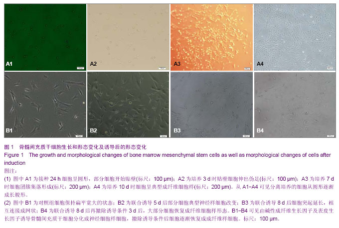
2.1 骨髓间充质干细胞生长情况和形态变化 初接种的原代细胞呈圆形,悬浮状,接种24 h后部分细胞开始贴壁,但仍保持圆形(图1A1);接种3 d后全量换液去除未贴壁细胞,贴壁细胞有伪足伸出,大部分细胞呈多角形或短梭形,折光性变强(图1A2);培养7 d后出现团簇集落样生长并有纤维发出(图1A3);培养10 d后细胞呈成纤维细胞样的长梭形,胞核呈卵圆形,胞浆丰富,彼此融合成片(图1A4)。 2.2 诱导后骨髓间充质干细胞形态的变化 对照组细胞保持扁平宽大的状态(图1B1); 干预组联合诱导5 d后细胞胞浆回缩,呈水滴样,折光性明显增强,部分细胞呈现典型神经细胞样改变,细胞伸出2个或多个突起(图1B2);联合诱导8 d后细胞突起逐渐增多并延长,相互连接成网状,可见细胞核及核仁(图1B3);撤除诱导条件3 d后可见大部分细胞逐渐恢复原来的成纤维细胞样形态(图1B4)。 "
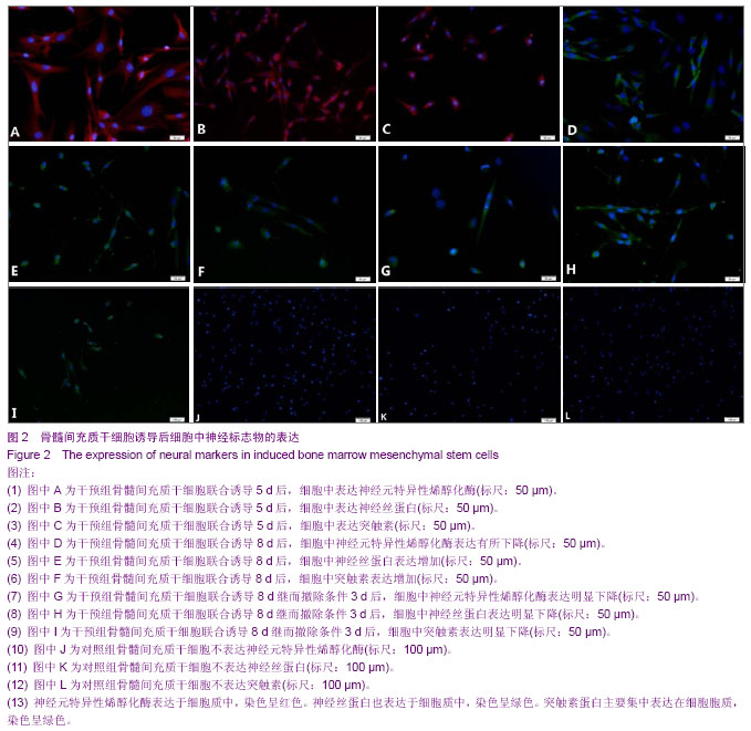
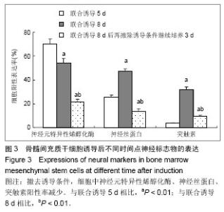
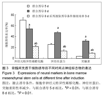
2.3 免疫组化结果进一步确定骨髓间充质干细胞的神经分化 为进一步确定骨髓间充质干细胞是否分化为神经细胞,分别于各观察时相点采用神经元特异标志物神经元特异性烯醇化酶、神经丝蛋白,突触囊泡膜特异性蛋白质突触素进行鉴定[35-37]。免疫组化结果显示,碱性成纤维生长因子及表皮生长因子联合诱导5 d后,大部分有核细胞胞质表达神经元特异性烯醇化酶,染色呈红色(图2A),部分细胞突起延长相互交织成网状。神经丝蛋白是一种神经元特异性的中间纤维,是神经元胞质的主要结构之一[36],其阳性表达部位与神经元特异性烯醇化酶类似,染色呈绿色,主要存在于细胞胞体与突起部位(图2D)。突触素蛋白是存在于突触囊泡膜上的一种糖蛋白质,参与突触发生[37],其表达主要集中在胞质,染色呈绿色(图2G)。联合诱导8 d后,细胞呈典型的神经细胞形态,神经元特异性烯醇化酶的表达有所下降(图2B),神经丝蛋白和突触素的表达上升,主要集中在胞质,甚至是部分突触(图2E,H)。撤除诱导条件3 d后,大部分细胞逐渐恢复原来的成纤维细胞形态,上述3种蛋白的表达量均明显下降(图2C,F,I)。对照组细胞未见3中蛋白阳性表达(图2J,K,L)。各个观察时相点3种蛋白的阳性率:诱导5 d后,神经元特异性烯醇化酶、神经丝蛋白、突触素阳性率表达分别为(70.3±4.2)%,(25.7±1.6)%,(3.6±0.5)%;诱导8 d后,神经元特异性烯醇化酶、神经丝蛋白、突触素阳性率表达分别为(54.1±3.8)%,(47.1±2.3)%,(32.0±2.1)%,与诱导5 d后比较差异有显著性意义(P < 0.01);撤除诱导条件3 d后,神经元特异性烯醇化酶、神经丝蛋白、突触素阳性率表达分别为(21.6±2.3)%,(13.8±1.7)%,(9.4±1.1)%,与诱导5 d、诱导8 d后比较差异均有显著性意义(P < 0.01,图3)。"
"
| [1] Erakat MS, Chuang SK, Shanti RM, et al. Interval between injury and lingual nerve repair as a prognostic factor for success using type I collagen conduit. J Oral Maxillofac Surg. 2013;71(5):833-838.[2] He J, Gu D, Wu X, et al. Major causes of death among men and women in China. N Engl J Med. 2005;353(11):1124-1134.[3] Jiang J, Lv Z, Gu Y, et al. Adult rat mesenchymal stem cells differentiate into neuronal-like phenotype and express a variety of neuro-regulatory molecules in vitro. Neurosci Res. 2010;66(1):46-52. [4] Hom DB. Growth factors in wound healing. Otolaryngol Clin North Am. 1995;28(5):933-953.[5] 姜琨,秦樾,童坦君.erbB-2表达抑制与表皮生长因子刺激对信号转导与转录激活分子及细胞周期蛋白D1的影响[J].北京医科大学学报,1998,30(3):205-208. [6] 王阁,汪思应,许望翔,等.肝部分切除及表皮生长因子迅速诱导PC3基因的表达[J].科学通报,2000,45(20):2005-2009.[7] Leslie CC, McCormick-Shannon K, Shannon JM, et al. Heparin-binding EGF-like growth factor is a mitogen for rat alveolar type II cells. Am J Respir Cell Mol Biol. 1997;16(4): 379-387.[8] Chia CM, Winston RM, Handyside AH. EGF, TGF-alpha and EGFR expression in human preimplantation embryos. Development. 1995;121(2):299-307.[9] Stubbs SC, Hargreave TB, Habib FK. Localization and characterization of epidermal growth factor receptors on human testicular tissue by biochemical and immunohistochemical techniques. J Endocrinol. 1990;125(3):485-492.[10] Hongo M, Itoi M, Yamaguchi N, et al. Distribution of epidermal growth factor (EGF) receptors in rabbit corneal epithelial cells, keratocytes and endothelial cells, and the changes induced by transforming growth factor-beta 1. Exp Eye Res. 1992; 54(1): 9-16.[11] Parelman JJ, Nicolson M, Pepose JS. Epidermal growth factor in human aqueous humor. Am J Ophthalmol. 1990; 109(5):603-604.[12] 赵培林,王慧敏,李肇特.下颌下腺切除对大鼠胃粘膜影响的组织学和组织化学观察[J].解剖学报,1994,25(3):318-321.[13] Calabrò A, Orsini B, Brocchi A, et al. Gastric juice immunoreactive epidermal growth factor levels in patients with peptic ulcer disease. Am J Gastroenterol. 1990;85(4): 404-407.[14] 周欣,黄志华,林汉华. 表皮生长因子与小儿消化性溃疡[J].华中医学杂志,2000,24(2):79-81. [15] 董光龙,王俊义,王为忠,等.表皮生长因子减少腹部辐射肠外营养大鼠肠道细菌移位[J].第四军医大学学报,2000,21(1):76-79.[16] 王涛,王俊义,陈冬利,等. 表皮生长因子对胰腺炎相关蛋白基因表达及胰腺炎的影响[J].第四军医大学学报,2000,21(11):1357- 1360.[17] 孙秀,陈先文,陈生弟.FGF和EGF对神经干细胞增殖及分化的影响[J].中国神经科学杂志,2000,16(2):170-173.[18] Okumura M, Okuda T, Nakamura T, et al. Acceleration of wound healing in diabetic mice by basic fibroblast growth factor. Biol Pharm Bull. 1996;19(4):530-535.[19] Takeuchi K, Takehara K, Tajima K, et al. Impaired healing of gastric lesions in streptozotocin-induced diabetic rats: effect of basic fibroblast growth factor. J Pharmacol Exp Ther. 1997; 281(1):200-207.[20] Satoh H, Asano S, Maeda R, et al. Prevention of gastric ulcer relapse induced by indomethacin in rats by a mutein of basic fibroblast growth factor. Jpn J Pharmacol. 1997;73(3): 229- 241.[21] Rieck P, Denis J, Peters D, et al. Fibroblast growth factor 2, heparin and suramin reduce epithelial ulcer development in experimental HSV-1 keratitis. Graefes Arch Clin Exp Ophthalmol. 1997;235(11):733-740.[22] Nakamura K, Kawaguchi H, Aoyama I, et al. Stimulation of bone formation by intraosseous application of recombinant basic fibroblast growth factor in normal and ovariectomized rabbits. J Orthop Res. 1997;15(2):307-313.[23] Allouche M, Bikfalvi A. The role of fibroblast growth factor-2 (FGF-2) in hematopoiesis. Prog Growth Factor Res. 1995; 6(1):35-48.[24] Kaye DM, Kelly RA, Smith TW. Cytokines and cardiac hypertrophy: roles of angiotensin II and basic fibroblast growth factor. Clin Exp Pharmacol Physiol Suppl. 1996;3: S136-141.[25] Schumacher B, Pecher P, von Specht BU, et al. Induction of neoangiogenesis in ischemic myocardium by human growth factors: first clinical results of a new treatment of coronary heart disease. Circulation. 1998;97(7):645-350.[26] Nakagami Y, Saito H, Matsuki N. Basic fibroblast growth factor and brain-derived neurotrophic factor promote survival and neuronal circuit formation in organotypic hippocampal culture. Jpn J Pharmacol. 1997;75(4):319-326.[27] Müller HW, Junghans U, Kappler J. Astroglial neurotrophic and neurite-promoting factors. Pharmacol Ther. 1995;65(1): 1-18.[28] Stachowiak MK, Moffett J, Maher P, et al. Growth factor regulation of cell growth and proliferation in the nervous system. A new intracrine nuclear mechanism. Mol Neurobiol. 1997;15(3):257-283.[29] Marini AM, Spiga G, Mocchetti I. Toward the development of strategies to prevent ischemic neuronal injury. In vitro studies. Ann N Y Acad Sci. 1997;825:209-219.[30] Shimada J, Fushiki S, Tsujimura A, et al. Fibroblast growth factor-2 expression is up-regulated after denervation in rat lung tissue. Brain Res Mol Brain Res. 1997;49(1-2):295-298.[31] Yoshida K, Toya S. Neurotrophic activity in cytokine-activated astrocytes. Keio J Med. 1997;46(2):55-60.[32] Sieber-Blum M, Zhang JM. Growth factor action in neural crest cell diversification. J Anat. 1997;191 ( Pt 4):493-499.[33] Adolphe M, Parodi AL. Ethical issues in animal experimentation. Bull Acad Natl Med. 2009;193(8): 1803-1804.[34] 胡辉,张伟才,黄继锋,等.新型神经导管复合材料与骨髓间充质干细胞的相容性[J].中国组织工程研究, 2013,17(16):2913-2920.[35] Lamers KJ, Vos P, Verbeek MM, et al. Protein S-100B, neuron-specific enolase (NSE), myelin basic protein (MBP) and glial fibrillary acidic protein (GFAP) in cerebrospinal fluid (CSF) and blood of neurological patients. Brain Res Bull. 2003; 61(3):261-264.[36] Uchida K, Baba H, Maezawa Y, et al. Progressive changes in neurofilament proteins and growth-associated protein-43 immunoreactivities at the site of cervical spinal cord compression in spinal hyperostotic mice. Spine (Phila Pa 1976). 2002;27(5):480-486.[37] Ferreira A, Chin LS, Li L, et al. Distinct roles of synapsin I and synapsin II during neuronal development. Mol Med. 1998;4(1): 22-28.[38] Kim SS, Yoo SW, Park TS, et al. Neural induction with neurogenin1 increases the therapeutic effects of mesenchymal stem cells in the ischemic brain. Stem Cells. 2008;26(9):2217-2228. [39] Wang Y, Zhao Z, Ren Z, et al. Recellularized nerve allografts with differentiated mesenchymal stem cells promote peripheral nerve regeneration. Neurosci Lett. 2012;514(1): 96-101. [40] Wang X, Luo E, Li Y, et al. Schwann-like mesenchymal stem cells within vein graft facilitate facial nerve regeneration and remyelination. Brain Res. 2011;1383:71-80. [41] Ao Q, Fung CK, Tsui AY, et al. The regeneration of transected sciatic nerves of adult rats using chitosan nerve conduits seeded with bone marrow stromal cell-derived Schwann cells. Biomaterials. 2011;32(3):787-796.[42] Moyse E, Segura S, Liard O, et al. Microenvironmental determinants of adult neural stem cell proliferation and lineage commitment in the healthy and injured central nervous system. Curr Stem Cell Res Ther. 2008;3(3): 163- 184.[43] Sun D, Bullock MR, McGinn MJ, et al. Basic fibroblast growth factor-enhanced neurogenesis contributes to cognitive recovery in rats following traumatic brain injury. Exp Neurol. 2009;216(1):56-65. [44] Weiss S, Reynolds BA, Vescovi AL, et al. Is there a neural stem cell in the mammalian forebrain? Trends Neurosci. 1996; 19(9):387-393.[45] Murrey HE, Gama CI,Kalovidouris SA,et al. Protein fucosylation regulates synapsin Ia/Ib expression and neuronal morphology in primary hippocampal neurons. Proc Natl Acad Sci U S A. 2006;103:21-26. |
| [1] | Wang Jian-ji, Yang Long, Li Jing, Sun Qi, Zuo Wei-min, Ren Qi-feng, Sun Yu, Wu Zhan-yu, Zou Qiang, Ma Min-xian, Ye Chuan. Development and application of special-purpose grafter by femoral head decompression combined with bone marrow mesenchymal stem cells transplantation based on three-dimensional printing technology [J]. Chinese Journal of Tissue Engineering Research, 2016, 20(44): 6636-6642. |
| [2] | Zhou Chang-yan, Zhou Qing-huan, Bian Jing, Chen Ke, Chen Wen. Bone marrow mesenchymal stem cells combined with calcium phosphate cement to repair articular cartilage defects in rabbits [J]. Chinese Journal of Tissue Engineering Research, 2015, 19(8): 1195-1199. |
| [3] | Jing Cai-xia, Liu Chang-kui, Tan Xin-ying, Luo Jin-chao, Hu Min. Bone mesenchymal stem cells with allogeneic bone to repair canine mandibular defects: detection of osteogenic ability [J]. Chinese Journal of Tissue Engineering Research, 2015, 19(14): 2138-2143. |
| [4] | Xu Xiang, Yin He-ping. Platelet-rich plasma accelerates the proliferation of bone marrow mesenchymal stem cells [J]. Chinese Journal of Tissue Engineering Research, 2015, 19(14): 2144-2148. |
| [5] | Lv Pin-lei, Su Yue-han, Cao Yun, Wang Zheng. Treatment of osteoarthritis using colony-forming cells in stromal vascular fraction of adipose tissue [J]. Chinese Journal of Tissue Engineering Research, 2015, 19(14): 2149-2154. |
| [6] | Ruan Guang-ping, Yao Xiang, Liu Ju-fen, Wang Jin-xiang, Hu Yuan-yuan, Li Zi-an, Yang Jian-yong, Pang Rong-qing, Pan Xing-hua. Transplantation of human umbilical cord mesenchymal stem cells in the treatment of systemic lupus erythematosus [J]. Chinese Journal of Tissue Engineering Research, 2015, 19(14): 2172-2178. |
| [7] | Jiang Yan-jie, Mao Cheng-gang, Ning Xian-feng, Li Rong, Li Zi-pu. Human umbilical cord mesenchymal stem cells via intramuscular injection influence the expression of cytokines related to dilated cardiomyopathy in rats [J]. Chinese Journal of Tissue Engineering Research, 2015, 19(14): 2179-2185. |
| [8] |
Liu Yu-liang, Li Jun, He Yu-qin, Zhuo Feng, Wei Kai-bin.
Transplantation of human umbilical cord derived-mesenchymal stem cells by different ways for the treatment of spinal cord injury
|
| [9] | Zhao Xiao-jian, Lu Cai-ping, Chu Wei-wei, Zhang Ya-xiao, Zhang Bing, Zhen Qiang, Tan Guo-liang, Wang Ren-feng, Liu Jia-bao, Wu Lin. Bone marrow mesenchymal stem cell transplantation for treatment of emphysema: intravenous versus intratracheal approach [J]. Chinese Journal of Tissue Engineering Research, 2015, 19(14): 2211-2215. |
| [10] | Liang Jian-ji, He Zhi-yong, Liu Kang, Li Xiao-ling, Cheng Wei-min, Yu Xin-ping, Chen Er-dong. Intraarticular injection of autologous bone marrow mesenchymal stem cells for mild-to-moderate osteoarthritis [J]. Chinese Journal of Tissue Engineering Research, 2015, 19(14): 2216-2223. |
| [11] | Du Qing-hua, Cao Jun-kai, Dong Xi-xi, E Ling-ling, Wei Li-jun. Osteogenic differentiation of pluripotent stem cells induced by akermanite extracts [J]. Chinese Journal of Tissue Engineering Research, 2015, 19(14): 2236-2242. |
| [12] | Wu Yan, Huang Lan . Bone morphogenetic protein 9-induced osteogenic differentiation of dental follicle cells in vitro [J]. Chinese Journal of Tissue Engineering Research, 2015, 19(14): 2255-2260. |
| [13] | Rao Li-jia, Li Qi-meng, Li Jin-ling, Xu Qiong. Expression pattern of ten-eleven translocation family during differentiation of human dental pulp cells [J]. Chinese Journal of Tissue Engineering Research, 2015, 19(14): 2261-2266. |
| [14] | Gao Zhuo-yue, Liu Yong-qi, He Jian-xin, Wu Zhi-wei, Luo Ya-li, Su Yun, Zhang Li-ying, Zhang Qi, Wu You-ming, Zhou Ni-na. Regulatory effects of warming yang and invigorating qi treatment on the inflammatory balance and genetic stability of bone marrow mesenchymal stem cells under tumor microenvironment [J]. Chinese Journal of Tissue Engineering Research, 2015, 19(14): 2267-2272. |
| [15] | Han Xiang-zhen, He Hui-yu, Hu Yang, Ba Jiao-jiao, Wang Huan-huan, Mi Xue, Abulizi•Abudula. Recombinant lentiviral vector transfected sheep bone marrow mesenchymal stem cells and osteogenic gene expression changes [J]. Chinese Journal of Tissue Engineering Research, 2014, 18(6): 821-828. |
| Viewed | ||||||
|
Full text |
|
|||||
|
Abstract |
|
|||||