Chinese Journal of Tissue Engineering Research ›› 2014, Vol. 18 ›› Issue (9): 1313-1318.doi: 10.3969/j.issn.2095-4344.2014.09.001

    Next Articles

Alterations in imaging and histopathology after aspiration of nucleus pulposus of rabbit lumbar intervertebral disc

Liu Hang-tao1, Wang Wan-ming2, Lin Zhi-jun1, Chen Guo-xian3, Lin Guo-ye1, Li Ping-sheng1   

  1. 1 Department of Orthopedics, the First Hospital Affiliated to Fuzhou General Hospital of Nanjing Military Area Command of Chinese PLA, Putian 351100, Fujian Province, China; 2 Department of Orthopedics, Fuzhou General Hospital of Nanjing Military Area Command of Chinese PLA, Fuzhou 350025, Fujian Province, China; 3 Department of Orthopedics, Putian Municipal First Hospital, Putian 351100, Fujian Province, China
  • Online:2014-02-26 Published:2014-02-26
  • Contact: Wang Wan-ming, M.D., Chief physician, Master’s supervisor, Department of Orthopedics, Fuzhou General Hospital of Nanjing Military Area Command of Chinese PLA, Fuzhou 350025, Fujian Province, China
  • About author:Liu Hang-tao, Master, Attending physician, Department of Orthopedics, the First Hospital Affiliated to Fuzhou General Hospital of Nanjing Military Area Command of Chinese PLA, Putian 351100, Fujian Province, China
  • Supported by:

    the Putian Municipal Science and Technology Project, No.2010 S09-2

Abstract:

BACKGROUND: It remains unclear what kind of changes in imaging and histopathology would appear in intervertebral disc after removal of nucleus pulposus over time.
OBJECTIVE: To observe the alteration in imaging and histopathology of rabbit lumbar intervertebral disc after nucleus pulposus aspiration.
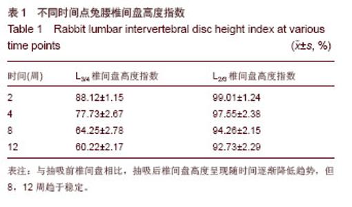
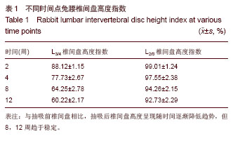
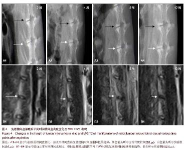
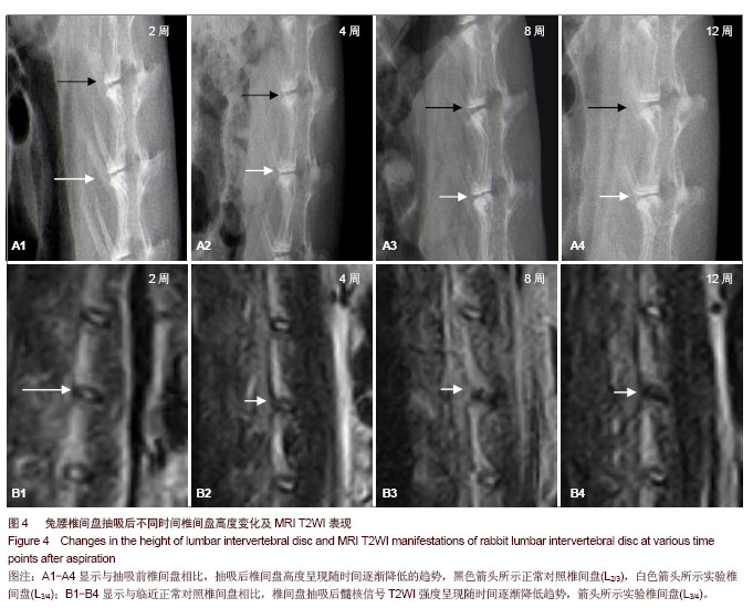
METHODS: A total of 32 Japanese rabbits were subjected to nucleus pulposus aspiration in posterior lateral L 3/4 intervertebral disc with a 21-gauge hypodermic needle. L 2/3 intervertebral disc served as a normal control. At 2, 4, 8 and 12 weeks after aspiration, eight rabbits according to grouping received radiograph in the lateral position of lumbar vertebra. The height of L 3/4 and L 2/3 intervertebral space was measured and intervertebral disc height index was calculated. Rabbits received median sagittal MRI examination and histopathological examination of intervertebral disc.
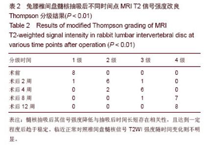
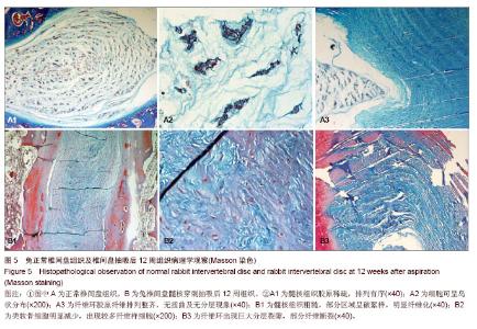
RESULTS AND CONCLUSION: The intervertebral disc height degraded slowly at 2, 4, 8, and 12 weeks after aspiration. However, the difference between 8 and 12 weeks was reduced. Compared with normal control, intervertebral disc height index significantly reduced at various time points (P < 0.05). The nucleus pulposus signal intensity was gradually reduced at 2, 4, 8, and 12 weeks after aspiration, and reached grade 4 of modified Thompson grading criteria at 8 weeks. After aspiration, gelatinous nucleus pulposus of disc slowly exhibited fissures with time, morphous was slowly disordered and showed significant fibrosis performance at 12 weeks. In nucleus pulposus region, more cartilage-like cells were found at 4 weeks after operation. The cells were active. Nucleus pulposus cells decreased significantly. At 8 and 12 weeks after operation, fibroblasts increased in nucleus pulposus region, and the number of cartilage-like cells reduced. The annulus fibrosus gradually became distorted, disordered, with processes and layers, and fiber breakage appeared. These results indicated that after puncturing lateral annulus fibrosus and aspirating the nucleus pulposus, radiograph height of the intervertebral disc and MRI T2-weighted signal intensity gradually reduced. Pathological changes were observed. However, the degeneration alleviated between 8-12 weeks.


中国组织工程研究杂志出版内容重点:人工关节;骨植入物;脊柱骨折;内固定;数字化骨科;组织工程


全文链接:

Key words: intervertebral disk, disk degeneration, radiographic image enhancement, pathology

CLC Number: