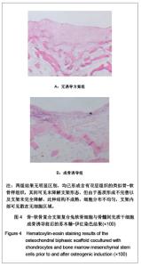
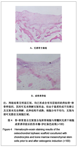
| [1] Yang Q,Peng J,Lu SB,et al.Zhongguo Jiaoxing Waike Zazhi. 2008;16(8):606-609. 杨强,彭江,卢世璧,等. 组织工程骨软骨复合体的研究进展及展望[J].中国矫形外科杂志,2008,16(8):606-609.[2] Nooeaid P,Salih V,Beier JP,et al.Osteochondral tissue engineering: scaffolds,stem cells and applications.J Cell Mol Med.2012; 16(10):1582-4934.[3] Panseri S,Russo A,Cunha C,et al.Osteochondral tissue engineering approaches for articular cartilage and subchondral bone regeneration. Knee Surg Sports Traumatol Arthrosc.2012; 20(6):1182-1191.[4] Lu J,Ke J,Wang L,et al. Zhongguo Zuzhi Gongcheng Yanjiu yu Linchuang Kangfu. 2011;15(19):3421-3426. 吕晶,柯杰,王琳,等. 与软骨细胞共培养和条件培养液诱导骨髓间充质干细胞向软骨细胞分化的比较[J].中国组织工程研究与临床康复,2011,15(19):3421-3426.[5] Tortelli F,Cancedda R.Three-dimensional cultures of osteogenic and chondrogenic cells: a tissue engineering approach to mimic bone and cartilage in vitro. Eur Cell Mater.2009;17:1-14.[6] Rodrigues MT,Lee SJ,Gomes ME,et al.Bilayered constructs aimed at osteochondral strategies: The influence of medium supplements in the osteogenic and chondrogenic differentiation of amniotic fluid-derived stem cells. Acta Biomater.2012;8(7):2795-2806.[7] Castro NJ,Hacking SA,Zhang LG.Recent progress in interfacial tissue engineering approaches for osteochondral defects.Ann Biomed Eng.2012; 40(8):1628-1640.[8] Khanarian NT, Haney NM, Burga RA, et al. A functional agarose-hydroxyapatite scaffold for osteochondral interface regeneration. Biomaterials.2012; 33(21):5247-5258.[9] Deng T,Lv J,Pang J,et al.Construction of tissue-engineered osteochondral composites and repair of large joint defects in rabbit.J Tissue Eng Regen Med.2012 [Epub ahead of print]][10] Castro NJ,Hacking SA,Zhang LG.Recent progress in interfacial tissue engineering approaches for osteochondral defects.Ann Biomed Eng. 2012;40(8):1628-1640.[11] Dominici M,Le BK,Mueller I,et al. Minimal criteria for defining multipotent mesenchymal stromal cells. The International Society for Cellular Therapy position statement. Cytotherapy. 2006;8(4):315-317.[12] Fan H,Tao H,Wu Y,et al.TGF-β3 immobilized PLGA- gelatin/ chondroitin sulfate/hyaluronic acid hybrid scaffold for cartilage regeneration. J Biomed Mater Res A.2010; 95(4):982-992.[13] Situ ZQ,Wu JZ.Xian:Shijie Tushu Chuban Xian Gongsi.2004: 161-162. 司徒镇强,吴军正.细胞培养[M].西安:世界图书出版西安公司, 2004:161 - 162.[14] Nooeaid P,Salih V,Beier JP,et al.Osteochondral tissue engineering: scaffolds, stem cells and applications. J Cell Mol Med.2012 [Epub ahead of print][15] Tampieri A,Sprio S,Sandri M,et al.Mimicking natural bio-mineralization processes: a new tool for osteochondral scaffold development. Trends Biotechnol.2011; 29(10): 526-535.[16] Yang Q,Peng J,Lu SB,et al. Evaluation of an extracellular matrix-derived acellular biphasic scaffold/cell construct in the repair of a large articular high-load-bearing osteochondral defect in a canine model. Chin Med J (Engl). 2011; 124(23): 3930-3938.[17] Fedorovich NE,Schuurman W,Wijnberg HM,et al.Biofabrication of osteochondral tissue equivalents by printing topologically defined, cell-laden hydrogel scaffolds.Tissue Eng Part C Methods. 2012; 18(1):33-44.[18] Mohan N,Dormer NH,Caldwell KL,et al.Continuous gradients of material composition and growth factors for effective regeneration of the osteochondral interface.Tissue Eng Part A. 2011;17(21-22):2845-2855.[19] Haaparanta AM,Haimi S,Ellä V,et al.Porous polylactide/ beta-tricalcium phosphate composite scaffolds for tissue engineering applications. J Tissue Eng Regen Med.2010;4(5): 366-373.[20] Ishimoto Y,Hattori K,Ohgushi H.Articular cartilage regeneration using scaffold. Clin Calcium.2008;18(12): 1775-1780.[21] Rothenberg AR,Ouyang L,Elisseeff JH.Mesenchymal stem cell stimulation of tissue growth depends on differentiation state.Stem Cells Dev.2011; 20(3):405-414.[22] Sundelacruz S,Kaplan DL.Stem cell- and scaffold-based tissue engineering approaches to osteochondral regenerative medicine.Semin Cell Dev Biol. 2009; 20(6):646-655.[23] Brady MA,Sivananthan S,Mudera V,et al. The primordium of a biological joint replacement: Coupling of two stem cell pathways in biphasic ultrarapid compressed gel niches. J Craniomaxillofac Surg.2011;39(5):380-386.[24] Davies JE,Matta R,Mendes VC,et al.Development, characterization and clinical use of a biodegradable composite scaffold for bone engineering in oro-maxillo-facial surgery. Organogenesis.2010; 6(3):161-166.[25] Im GI,Ahn JH,Kim SY,et al.A hyaluronate-atelocollagen/ beta-tricalcium phosphate-hydroxyapatite biphasic scaffold for the repair of osteochondral defects: a porcine study. Tissue Eng Part A. 2010; 16(4):1189-1200.[26] Keeney M, Pandit A. The osteochondral junction and its repair via bi-phasic tissue engineering scaffolds.Tissue Eng Part B Rev.2009; 15(1):55-73.[27] Matsusaki M,Kadowaki K,Tateishi K,et al.Scaffold-free tissue-engineered construct-hydroxyapatite composites generated by an alternate soaking process: potential for repair of bone defects.Tissue Eng Part A. 2009; 15(1):55-63.[28] Getgood AM,Kew SJ,Brooks R,et al. Evaluation of early-stage osteochondral defect repair using a biphasic scaffold based on a collagen-glycosaminoglycan biopolymer in a caprine model. Knee. 2012; 19(4):422-430.[29] Filová E,Jelínek F,Handl M,et al.Novel composite hyaluronan/type I collagen/fibrin scaffold enhances repair of osteochondral defect in rabbit knee. J Biomed Mater Res B Appl Biomater.2008;87(2):415-424.[30] Khanarian NT,Jiang J,Wan LQ,et al.A hydrogel-mineral composite scaffold for osteochondral interface tissue engineering.Tissue Eng Part A.2012; 18(5-6):533-545. |