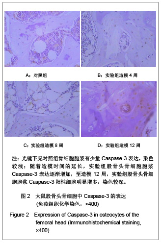
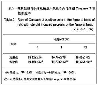
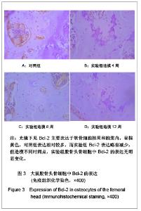
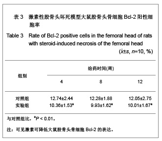
| [1] Kaushik AP, Das A, Cui Q. Osteonecrosis of the femoral head: An update in year 2012. World J Orthop. 2012;3(5):49-57.[2] Chan KL, Mok CC. Glucocorticoid-induced avascular bone necrosis: diagnosis and management. Open Orthop J. 2012; 6:449-457.[3] Sato M, Sugano N, Ohzono K, et al. Apoptosis and expression of stress protein (ORP150, HO1) during development of ischaemic osteonecrosis in the rat. J Bone Joint Surg Br. 2001;83(5):751-759. [4] Weinstein RS, Nicholas RW, Manolagas SC. Apoptosis of osteocytes in glucocorticoid-induced osteonecrosis of the hip. J Clin Endocrinol Metab. 2000;85(8):2907-2912.[5] Youm YS, Lee SY, Lee SH. Apoptosis in the osteonecrosis of the femoral head. Clin Orthop Surg. 2010;2(4):250-255.[6] Weinstein RS. Glucocorticoid-induced osteoporosis and osteonecrosis. Endocrinol Metab Clin North Am. 2012;41(3): 595-611.[7] Zaidi M, Sun L, Robinson LJ, et al. ACTH protects against glucocorticoid-induced osteonecrosis of bone. Proc Natl Acad Sci U S A. 2010;107(19):8782-8787.[8] Kerachian MA, Harvey EJ, Cournoyer D, et al. A rat model of early stage osteonecrosis induced by glucocorticoids. J Orthop Surg Res. 2011;6:62.[9] Li ZR, Zhang NF, Yue DB, et al. Zhonghua Waike Zazhi. 1995; 8(33):485-487. 李子荣,张念非,岳德波,等.激素性股骨头坏死动物模型的诱导和观察[J].中华外科杂志,1995,8(33):485-487.[10] Matsui M, Saito S, Ohzono K, et al. Experimental steroid-induced osteonecrosis in adult rabbits with hypersensitivity vasculitis. Clin Orthop Relat Res. 1992;(277): 61-72.[11] Wei Q, Yang XF, Liu L, et al. Zhongguo Guzhi Shusong Zazhi. 1999;5(2):26-28. 魏青,杨杏芬,刘力,等.激素性股骨头坏死动物模型的建立和评价[J].中国骨质疏松杂志,1999,5(2):26-28.[12] Yamamoto T, Hirano K, Tsutsui H, et al. Corticosteroid enhances the experimental induction of osteonecrosis in rabbits with Shwartzman reaction. Clin Orthop Relat Res. 1995;(316):235-243.[13] Wang JZ, Gao HY, Wang KZ, et al. Effect of epimedium extract on osteoprotegerin and RANKL mRNA expressions in glucocorticoid-induced femoral head necrosis in rats. Nan Fang Yi Ke Da Xue Xue Bao. 2011;31(10):1714-1717.[14] Fransen JH, Dieker JW, Hilbrands LB, et al. Synchronized turbo apoptosis induced by cold-shock. Apoptosis. 2011;16(1): 86-93.[15] Short BG, Zimmerman DM, Schwartz LW. Automated double labeling of proliferation and apoptosis in glutathione S-transferase-positive hepatocytes in rats. J Histochem Cytochem. 1997;45(9):1299-1305.[16] Johnstone RW, Ruefli AA, Lowe SW. Apoptosis: a link between cancer genetics and chemotherapy. Cell. 2002; 108(2): 153-164.[17] Végran F, Boidot R, Solary E, et al. A short caspase-3 isoform inhibits chemotherapy-induced apoptosis by blocking apoptosome assembly. PLoS One. 2011;6(12):e29058.[18] Nicholson DW. Caspase structure, proteolytic substrates, and function during apoptotic cell death. Cell Death Differ. 1999; 6(11):1028-1042.[19] Cotter TG. Apoptosis and cancer: the genesis of a research field. Nat Rev Cancer. 2009;9(7):501-507.[20] Sasi N, Hwang M, Jaboin J, et al. Regulated cell death pathways: new twists in modulation of BCL2 family function. Mol Cancer Ther. 2009;8(6):1421-1429.[21] Ben-Izhak O, Laster Z, Akrish S, et al. The salivary tip of the p53 mutagenesis iceberg: novel insights. Cancer Biomark. 2009;5(1):23-31.[22] Miyashita T, Reed JC. Tumor suppressor p53 is a direct transcriptional activator of the human bax gene. Cell. 1995; 80(2):293-299. [23] Gomes CC, Bernardes VF, Diniz MG, et al. Anti-apoptotic gene transcription signature of salivary gland neoplasms. BMC Cancer. 2012;12:61.[24] Tsang WP, Chau SP, Kong SK, et al. Reactive oxygen species mediate doxorubicin induced p53-independent apoptosis. Life Sci. 2003;73(16):2047-2058.[25] Li J, Huang LY, Li XK, et al. Zhongguo Jiaoxing Waike Zazhi. 2003;11(5):323-324. 李靖,黄鲁豫,李新奎,等.Bcl-2过度表达对大鼠成骨细胞Bax、Caspase-3表达的影响[J]. 中国矫形外科杂志,2003,11(5): 323-324. |