Chinese Journal of Tissue Engineering Research ›› 2013, Vol. 17 ›› Issue (4): 612-619.doi: 10.3969/j.issn.2095-4344.2013.04.008
Previous Articles Next Articles
Finite element analysis of different tibial shaft fracture fixed by Locking compression plate under different movement gait
Li Yun-gang1, 2, Chen Wei-jian2, Li Gui-tao2, Sun Hong-tao2, Yan Jian-hao2, Wang Xiong-chang2, Wang Jing2
- 1 Graduate School, Southern Medical University, Guangzhou 510515, Guangdong Province, China 2 Department of Orthopaedics, Guangdong No.2 Provincial People's Hospital, Southern Medical University, Guangzhou 510317, Guangdong Province, China
-
Received:2012-11-10Revised:2012-11-17Online:2013-01-22Published:2013-03-05 -
Contact:Li Gui-tao, Chief physician, Professor, Master's supervisor, Department of Orthopaedics, Guangdong No.2 Provincial People's Hospital, Southern Medical University, Guangzhou 510317, Guangdong Province, China lgtgk@163.com -
About author:Li Yun-gang★, Studying for master's degree, Graduate School, Southern Medical University, Guangzhou 510515, Guangdong Province, China; Department of Orthopaedics, Guangdong No.2 Provincial People's Hospital, Southern Medical University, Guangzhou 510317, Guangdong Province, China jhym12@yahoo.cn
CLC Number:
Cite this article
Li Yun-gang, Chen Wei-jian, Li Gui-tao, Sun Hong-tao, Yan Jian-hao, Wang Xiong-chang, Wang Jing. Finite element analysis of different tibial shaft fracture fixed by Locking compression plate under different movement gait[J]. Chinese Journal of Tissue Engineering Research, 2013, 17(4): 612-619.
share this article
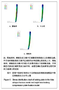
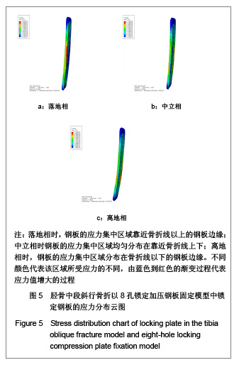
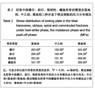
实验成功建立锁定加压钢板固定不同类型胫骨干骨折的有限元模型,并对模型进行负荷加载。经有限元分析计算测得结果如下: 2.1 钢板的受力分布情况 锁定钢板在胫骨中段横行、斜行、粉碎性骨折中的应力分布情况由小到大均为:中立相<落地相<离地相;锁定钢板在胫骨中段螺旋形骨折中应力分布情况由小到大均为:离地相<中立相<落地相;从有限元应力分布云图上可以看出,锁定钢板的应力集中区域位于钢板中间或边缘,呈现中间集中,两端分散的趋势。见图5和表2。"

"
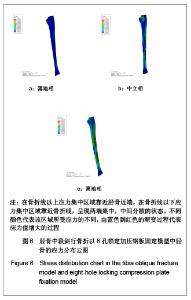
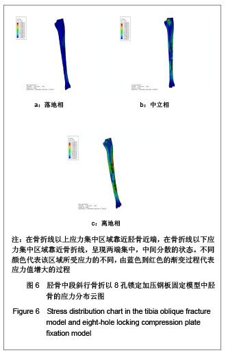
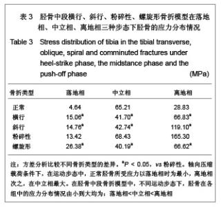
2.2 胫骨的受力分布情况 轴向压缩载荷条件下,在运动步态中,正常胫骨所受应力以落地相时为最小,离地相次之,在中立相最大。在胫骨中段骨折模型中,不同运动步态下,胫骨在各组中的应力分布情况由小到大均为:落地相<中立相<离地相,见图6和表3。"
"

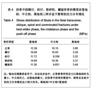
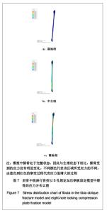
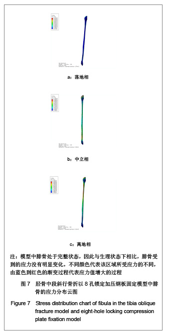
从胫骨的有限元应力分布云图上可以看出,在骨折线以上应力集中区域靠近胫骨近端,在骨折线以下应力集中区域靠近骨折线,呈现两端集中,中间分散的状态。粉碎性骨折组无论胫骨还是锁定钢板的受力均较横行、斜行及螺旋形骨折差异有显著性意义(P < 0.05),且后三者差异无显著性意义(P > 0.05)。 2.3 腓骨的受力分布情况 轴向压缩载荷条件下,在运动步态中,正常腓骨所受应力以离地相时为最小,中立相次之,在落地相最大。 在胫骨中段骨折模型中,腓骨在各组中的应力分布情况由小到大均为:离地相<中立相<落地相,见表4和图7。"

"
| [1] Frigg R. Locking Compression Plate (LCP). An osteosynthesis plate based on the Dynamic Compression Plate and the Point Contact Fixator (PC-Fix). Injury. 2001;32 Suppl 2:63-66.[2] Cao Q, Ma BT. Zhongguo Jiaoxing Waike Zazhi. 2008;16(12): 930-931. 曹清,马宝通.锁定加压钢板的临床应用[J].中国矫形外科杂志, 2008,16(12):930-931. [3] Zou J, Zhang Q. Zhonghua Chuangshang Guke Zazhi. 2009; 11(10):981-984. 邹剑,张长青.锁定钢板治疗四肢骨折的研究进展[J].中华创伤骨科杂志,2009,11(10):981-984. [4] Agarwala S, Shah SB. Staple versus locking compression plate fixation after lateral closing wedge high tibial osteotomy. J Orthop Surg (Hong Kong). 2008;16(3):303-307.[5] Hasenboehler E, Rikli D, Babst R. Locking compression plate with minimally invasive plate osteosynthesis in diaphyseal and distal tibial fracture: a retrospective study of 32 patients. Injury. 2007;38(3):365-370. [6] Sod GA, Mitchell CF, Hubert JD, et al. In vitro biomechanical comparison of locking compression plate fixation and limited-contact dynamic compression plate fixation of osteotomized equine third metacarpal bones. Vet Surg. 2008; 37(3):283-288.[7] Heiney JP, Barnett MD, Vrabec GA, et al. Distal femoral fixation: a biomechanical comparison of trigen retrograde intramedullary (i.m.) nail, dynamic condylar screw (DCS), and locking compression plate (LCP) condylar plate. J Trauma. 2009;66(2):443-449.[8] Aguila AZ, Manos JM, Orlansky AS, et al. In vitro biomechanical comparison of limited contat dynamic compression plate and locking compression plate. Vet Comp Orthop Traumatol. 2005;18(4):220-226.[9] Tang SL, Zhang WB, Yan HB, et al. Linchuang Guke Zazhi. 2009;12(2):165-168. 唐少龙,张文斌,颜海波,等.解剖型钢板与锁定加压钢板治疗胫骨下段粉碎性骨折比较[J].临床骨科杂志,2009,12(2):165-168. [10] Stepanovi? Z, Zivkovi? M, Vulovi? S, et al. High, open wedge tibial osteotomy: finite element analysis of five internal fixation modalities. Vojnosanit Pregl. 2011;68(10):867-871.[11] Saikia K, Bhuyan S, Bhattacharya T, et al. Internal fixation of fractures of both bones forearm: Comparison of locked compression and limited contact dynamic compression plate. Indian J Orthop. 2011;45(5):417-421.[12] Haaland PJ, Sjöström L, Devor M, et al. Appendicular fracture repair in dogs using the locking compression plate system: 47 cases. Vet Comp Orthop Traumatol. 2009;22(4):309-315. [13] Carpenter RS, Galuppo LD, Simpson EL, et al. Clinical evaluation of the locking compression plate for fetlock arthrodesis in six thoroughbred racehorses. Vet Surg. 2008; 37(3):263-268.[14] Levin SM, Nelson CO, Botts JD, et al. Biomechanical evaluation of volar locking plates for distal radius fractures. Hand (N Y). 2008;3(1):55-60. [15] Bae JH, Oh JK, Oh CW, et al. Technical difficulties of removal of locking screw after locking compression plating. Arch Orthop Trauma Surg. 2009;129(1):91-95.[16] Zhong H, Liu LH, Zhu ZM. Zhongguo Gu yu Guanjie Sunshang Zazhi. 2011(3):213-216. 钟华,刘立华,朱智敏.MIPPO技术下LCP锁定和加压固定后应力遮挡效应的有限元研究[J].中国骨与关节损伤杂志,2011(3): 213-216.[17] Hinterhofer C, Haider H, Apprich V, et al. Development of a twenty-one-component finite element distal hind limb model: stress and strain in bovine digit structures as a result of loading on different floorings. J Dairy Sci. 2009;92(3): 972-979.[18] Wong C, Mikkelsen P, Hansen LB, et al. Finite element analysis of tibial fractures. Dan Med Bull. 2010;57(5):A4148.[19] Wieding J, Souffrant R, Fritsche A, et al. Finite element analysis of osteosynthesis screw fixation in the bone stock: an appropriate method for automatic screw modelling. PLoS One. 2012;7(3):e33776. [20] Levine DG, Richardson DW. Clinical use of the locking compression plate (LCP) in horses: a retrospective study of 31 cases (2004-2006). Equine Vet J. 2007;39(5):401-406.[21] Blecha LD, Zambelli PY, Ramaniraka NA, et al. How plate positioning impacts the biomechanics of the open wedge tibial osteotomy; a finite element analysis. Comput Methods Biomech Biomed Engin. 2005;8(5):307-313.[22] Zhongguo Xuesheng Tizhi yu Jiankang Yanjiu Zu. Beijing: Higher Education Press. 2002. 中国学生体制与健康研究组.2000年中国学生体质与健康调研报告[M].北京:高等教育出版社,2002.[23] Lu CH, Yu B, Chen HQ, et al. Nanfang Yike Daxue Xuebao. 2010;30(10):2273-2276. 卢昌怀,余斌,陈辉强,等.正常步态下距骨三维有限元模型的建立及应力分析[J].南方医科大学学报,2010,30(10):2273-2276. [24] Lu CH. Guangzhou: Southern Medical University. 2011. 卢昌怀.距骨数值仿真模型的建立及有限元分析[D].广州:南方医科大学,2011.[25] Xiong Y, Zhao YF, Gong XS, et al. Chuangshang Waike Zazhi. 2009;11(2):155. 熊雁,赵玉峰,龚宪生,等.有限接触动力加压接骨板固定股骨干骨折的三维有限元分析[J].创伤外科杂志,2009,11(2):155. [26] MacLeod AR, Pankaj P, Simpson AH. Does screw-bone interface modelling matter in finite element analyses? J Biomech. 2012;45(9):1712-1716. [27] Xiong Y, Zhao YF, Gong XS, et al. Zhonghua Chuangshang Zazhi. 2009;25(3):245-248. 熊雁,赵玉峰,龚宪生,等.点接触锁定接骨板固定股骨干骨折的三维有限元分析[J].中华创伤杂志,2009,25(3):245-248. [28] Miyoshi S, Takahashi T, Ohtani M, et al. Analysis of the shape of the tibial tray in total knee arthroplasty using a three dimension finite element model. Clin Biomech (Bristol, Avon). 2002;17(7):521-525.[29] Zhang YG, Wang Y. Zhonghua Guke Zazhi. 2010;30(9): 892-898. 张银光,王岩.单柄和四桩设计的膝关节胫骨假体基座生物力学特点的三维有限元分析[J].中华骨科杂志,2010,30(9):892-898. [30] Giddings VL, Beaupré GS, Whalen RT, et al. Calcaneal loading during walking and running. Med Sci Sports Exerc. 2000;32(3):627-634.[31] Hu XJ, Li FW, Wang H, et al. Zhonghua Chuangshang Guke Zazhi. 2011;13(7):666-670. 胡新佳,林博文,王华,等.胫骨中段骨折钢板螺钉固定的有限元分析[J].中华创伤骨科杂志,2011,13(7):666-670. [32] Wall LB, Brodt MD, Silva MJ, et al. The effects of screw length on stability of simulated osteoporotic distal radius fractures fixed with volar locking plates. J Hand Surg Am. 2012;37(3): 446-453. [33] Brekelmans WA, Poort HW, Slooff TJ. A new method to analyse the mechanical behaviour of skeletal parts. Acta Orthop Scand. 1972;43(5):301-317.[34] Xu R, Wu Y, Jia KB, et al. Zhongguo Yixue Yingxiang Jishu. 2002;18(09):952-954. 徐瑞,吴勇,贾克斌,等.数字医学影影像与通信的重要标准-DICOM标准[J].中国医学影像技术,2002,18(09):952-954. |
| [1] | Zheng Xuying, Hu Hongcheng, Xu Libing, Han Jianmin, Di Ping. Stress magnitude and distribution in two-piece cement-retained zirconia implants under different loading conditions and with varying internal connection shapes [J]. Chinese Journal of Tissue Engineering Research, 2026, 30(8): 1979-1987. |
| [2] | Zhong Caihong, Xiao Xiaoge, Li Ming, Lin Jianhong, Hong Jing. Biomechanical mechanism of sports-related patellar tendinitis [J]. Chinese Journal of Tissue Engineering Research, 2026, 30(6): 1417-1423. |
| [3] | Xu Hao, Ding Lu, Li Xiao. Mechanical effect of mechanical wear of abutment screws on the Morse taper connection implant system: a three-dimensional finite element analysis [J]. Chinese Journal of Tissue Engineering Research, 2026, 30(6): 1375-1383. |
| [4] | Wei Bo, Qiu Jiangang. Double lactate threshold exercise training: development context, basic connotation, application effect and mechanism of action [J]. Chinese Journal of Tissue Engineering Research, 2026, 30(4): 964-974. |
| [5] | Yu Xinlin, Chen Huiyu, Wang Yingying, Guo Weizhong, Feng Bin Lin Chengshou, Lin Wang. Finite element analysis of internal fixation with new retrograde intramedullary nail on lateral femur condyle for distal type A2 femur fractures [J]. Chinese Journal of Tissue Engineering Research, 2026, 30(3): 546-552. |
| [6] | Zhao Jingang, Liu Liping, Chen Jianwei, . Finite element analysis comparing lumbar fusion and artificial intervertebral disc replacement [J]. Chinese Journal of Tissue Engineering Research, 2026, 30(3): 553-560. |
| [7] | Wang Meng, Lu Tan, Li Minjie, Liu Zhicheng, Guo Xiaoyong. Finite element analysis of stress distribution of anchors at different implantation depths under different bone density conditions in rotator cuff tears [J]. Chinese Journal of Tissue Engineering Research, 2026, 30(3): 561-569. |
| [8] | Li Xiaomin, Tian Xiangdong, Wang Chaolu. High tibial osteotomy on a single plane: femorofibular angle as a reference marker for mechanical axis correction [J]. Chinese Journal of Tissue Engineering Research, 2026, 30(3): 570-576. |
| [9] | Ma Jingbo, Yang Guangnan, Liu Jiang, Jiang Qiang, Zhang Hanshuo, Han Jiaheng, Ding Yu. Endoscopic lumbar canal decompression for upper lumbar spinal stenosis: a comparison of biomechanical stability of three surgical models [J]. Chinese Journal of Tissue Engineering Research, 2026, 30(3): 577-585. |
| [10] | Abudusalamu·Tuoheti, Xiao Yang, Wang Yixi, Musitapa·Mijiti, Chen Qihao, Maimaitiming·Saiyiti, Guo Hailong, Paerhati·Rexiti. Effects of three internal fixation techniques on biomechanics of adjacent segment degeneration in lumbar interbody fusion [J]. Chinese Journal of Tissue Engineering Research, 2026, 30(3): 586-595. |
| [11] | Xu Xinbao, Chen Feiyang, Chen Yinbing, Zhang Feixiang, Lyu Shujun, Cui Haidong, Chen Zhigang. Univariate and multivariate regression analysis of femoral neck shortening after cannulated screw fixation in femoral neck fractures [J]. Chinese Journal of Tissue Engineering Research, 2026, 30(3): 620-625. |
| [12] | Liao Long, Zhao Zepeng, Li Zongyuan, Yu Qinglong, Zhang Tao, Tang Jinyuan, Ye Nan, Xu Han, Shi Bo. Establishment and validation of a model for femoral head necrosis after internal fixation of femoral neck fracture using logistic regression and SHAP analysis [J]. Chinese Journal of Tissue Engineering Research, 2026, 30(3): 626-633. |
| [13] | Wang Peng, Li Zhijun, Zhang Shaojie, Wu Yimin. Intervertebral disc rehydration after posterior lumbar dynamic internal fixation [J]. Chinese Journal of Tissue Engineering Research, 2026, 30(3): 711-720. |
| [14] | Shang Depeng, Wei Haiyu, Yang Fan. Finite element analysis for three different types of internal screw fixation in treatment of severe lumbar 1 vertebral body fractures [J]. Chinese Journal of Tissue Engineering Research, 2026, 30(3): 537-545. |
| [15] | Cheng Yanan, Yu Jiazhi, Liu Yinchang, Wu Jie, Yu Tong, Wang Lu, Li Xiaoguang. Three-dimensional finite element analysis of molar distalization with clear aligners with different thicknesses and edges [J]. Chinese Journal of Tissue Engineering Research, 2026, 30(2): 310-318. |
| Viewed | ||||||
|
Full text |
|
|||||
|
Abstract |
|
|||||