Chinese Journal of Tissue Engineering Research ›› 2026, Vol. 30 ›› Issue (35): 9355-9364.doi: 10.12307/2026.404
Previous Articles Next Articles
Xiao Ban Tong Mai Fang regulates autophagy via targeting miR-126-3p: bioinformatics analysis for prevention and treatment of atherosclerosis
Cao Shan1, Wang Yanxi2, Duan Kaixuan3, Qi Xiang3, Wang Yuhan4
- 1School of Medicine, 2School of Nursing, 3School of Traditional Chinese Medicine, Henan University of Chinese Medicine, Zhengzhou 450046, Henan Province, China; 4Third Affiliated Hospital of Henan University of Chinese Medicine, Zhengzhou 450008, Henan Province, China
-
Received:2025-07-18Revised:2025-11-10Online:2026-12-18Published:2026-04-30 -
Contact:Cao Shan, School of Medicine, Henan University of Chinese Medicine, Zhengzhou 450046, Henan Province, China -
About author:Cao Shan, PhD, Professor, Doctoral supervisor, School of Medicine, Henan University of Chinese Medicine, Zhengzhou 450046, Henan Province, China -
Supported by:Henan Provincial Natural Science Foundation, No. 242300421295 (to CS); Cui Yingmin National Famous Traditional Chinese Medicine Expert Inheritance Studio Construction Project, No. [2022]75 (to CS); Henan Provincial Science and Technology Key Project, No. 232102310434 (to CS); Henan Provincial Major Special Project on Traditional Chinese Medicine Research, No. 2022ZYZD20 (to CS); Henan Provincial Key Project on Traditional Chinese Medicine Research, No. 2023ZY1031 (to CS); Research Project on the Management of Traditional Chinese Medicine Culture in Henan Province, No. TCM2025041 (to CS)
CLC Number:
Cite this article
Cao Shan, Wang Yanxi, Duan Kaixuan, Qi Xiang, Wang Yuhan. Xiao Ban Tong Mai Fang regulates autophagy via targeting miR-126-3p: bioinformatics analysis for prevention and treatment of atherosclerosis[J]. Chinese Journal of Tissue Engineering Research, 2026, 30(35): 9355-9364.
share this article
Add to citation manager EndNote|Reference Manager|ProCite|BibTeX|RefWorks

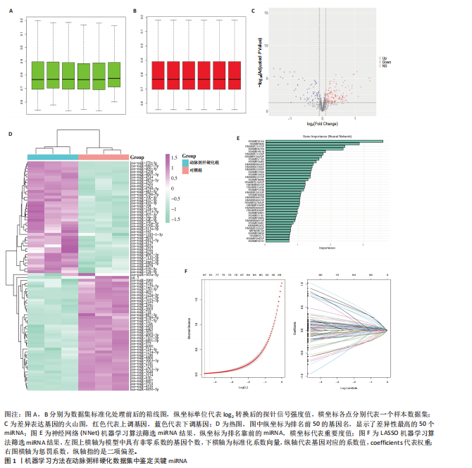
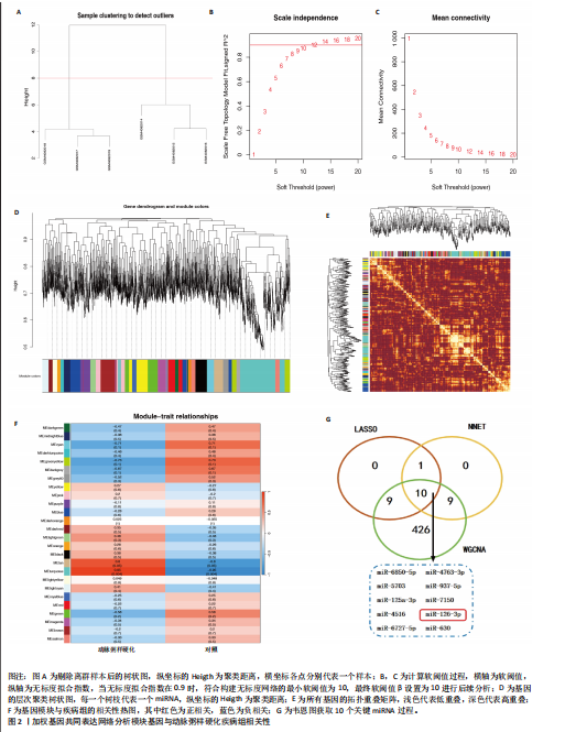
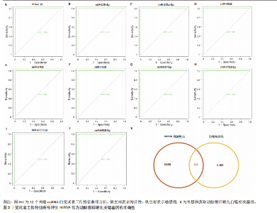
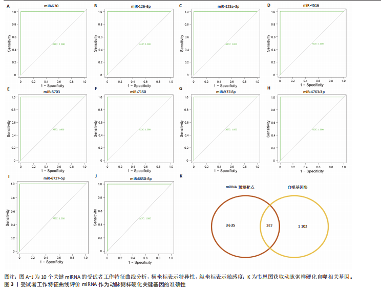
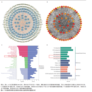
2.1 生物信息学分析学结果 2.1.1 机器学习筛选miRNA结果 研究在GSE137580数据集中总共鉴定出207个显著差异的差异表达基因,由此推测这些基因在动脉粥样硬化病理进程中发挥着一定作用。进一步结合LASSO和神经网络机器学习算法鉴定出排名前20的miRNA,基于鉴定结果进行下一步分析,见图1。 2.1.2 加权基因共同表达网络的构建以及关键miRNA的获取 基于加权基因共表达网络分析GSE137580数据集基因中与动脉粥样硬化疾病组的相关的模块基因,软阈值β设置为10,最终鉴定出28个目标模块,见图2A-E。通过热图展示出每个模块和动脉粥样硬化疾病组的相关性,结果发现绿松石模块(包含454个基因)与动脉粥样硬化疾病组高度正相关(r=0.95,P=0.004),见图2F。将以上454个模块基因与机器学习鉴定出的miRNA取交集获取miR-6850-5p,miR-4763-3p,miR-937-5p,miR-7150,miR-126-3p,miR-630,miR-5703,miR-125a-3p,miR-4516,miR-6727-5p等10个关键miRNA,见图2G。ROC结果分析显示,以上10个miRNA的AUC值均高于0.9,这表"

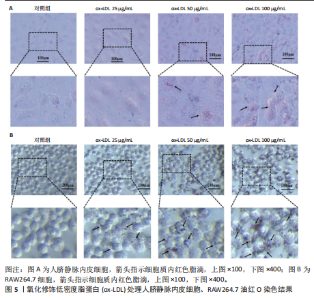
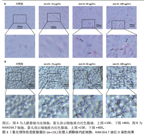
明以上基因作为动脉粥样硬化关键基因具有一定准确性。结合文献查阅以及前期基础[22-24],此研究最终选miR-126-3p进行实验验证,见图3A-J。 2.1.3 miRNA潜在调控基因的预测与富集分析 使用miRDB数据库预测到10个关键miRNA的3 892个潜在调控基因,将潜在调控基因与GeneCards数据库检索到的1 359个自噬基因集取交集获得257个自噬相关基因,见图3K。最后,对交集基因进行PPI分析并导出排名前80的核心基因进行富集分析,见图4A,B。GO富集分析结果显示,核心基因涉及到细胞对氧化应激的反应、凋亡信号通路的调节、自噬的调节、MAPK级联的正调控、细胞-基质接合、吞噬泡组装位点、自噬体、泛素蛋白连接酶结合、泛素样蛋白连接酶结合、转录共调节因子结合等生物学过程。KEGG富集分析结果显示,核心基因涉及到叉头盒O类转录因子(Forkhead box O,FoxO)信号通路、自噬通路、磷脂酰肌醇3激酶-蛋白激酶B(phosphatidylinositol 3-kinase–protein kinase B,PI3K-Akt)信号通路、腺苷酸活化蛋白激酶(AMP-activated protein kinase,AMPK)信号通路、MAPK信号通路、雷帕霉素靶蛋白(mechanistic target of rapamycin,mTOR)信号通路等。根据结果推测MAPK信号通路可能受到关键miRNA的调控,并通过介导自噬在动脉粥样硬化疾病进程中发挥着重要作用,因此,此次研究基于MAPK信号通路开展实验验证,见图4C,D。 2.2 油红“O”染色鉴定细胞泡沫化模型 油红“O”染色实验结果发现,100 μg/mL的氧化修饰低密度脂蛋白干预人脐静脉内皮细胞和RAW264.7细胞24 h后,相较于其他剂量组,人脐静脉内皮细胞和RAW264.7细胞的体积有所增大,并出现脂质沉积、细胞破裂、细胞泡沫化的病理现象。表明100 μg/mL的氧化修饰"
"
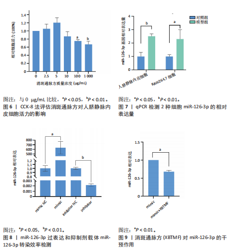
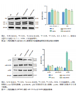
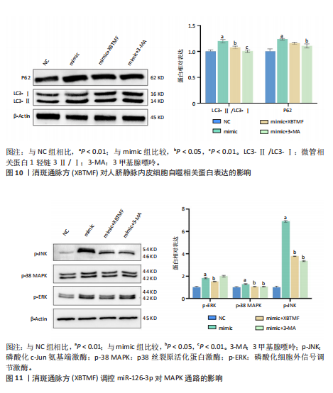
低密度脂蛋白的干预可构建人脐静脉内皮细胞和RAW264.7细胞泡沫化模型,后续研究将使用该浓度构建模型,见图5。 2.3 消斑通脉方对人脐静脉内皮细胞活力的影响 分析结果发现,消斑通脉方干预人脐静脉内皮细胞的浓度超过10 μg/mL时,细胞活力开始下降,尤其100 μg/mL及以上浓度,抑制效果显著(P < 0.05),且呈剂量依赖性,结合实验后续研究选用10 μg/mL的消斑通脉方干预细胞,见图6。 2.4 miR-126-3p在动脉粥样硬化细胞泡沫化模型中的表达 QPCR检测氧化修饰低密度脂蛋白干预后两组细胞中miR-126-3p的表达。与对照组比较,氧化修饰低密度脂蛋白干预后人脐静脉内皮细胞中miR-126-3p表达水平上调2.5倍(P < 0.01),RAW264.7细胞miR-126-3p表达水平上调2.3倍(P < 0.05),见图7。 2.5 消斑通脉方干预人脐静脉内皮细胞miR-126-3p的表达 首先构建miR-126-3p过表达和抑制载体,qPCR检测miR-126-3p转染效率。结果显示,与NC组比较,mimic组miR-126-3p表达水平显著升高(P < 0.01),inhibitor组miR-126-3p表达水平显著降低(P < 0.05),见图8。由上文可知,氧化修饰低密度脂蛋白干预后miR-126-3p表达上调,因此以miR-126-3p mimic模拟氧化修饰低密度脂蛋白的干预效果,进而检测中药消斑通脉方的疗效。消斑通脉方干预后miR-126-3p表达水平显著降低(P < 0.01),见图9。 2.6 消斑通脉方调控miR-126-3p对人脐静脉内皮细胞自噬的影响 应用Western Blot检测蛋白相对表达量发现,与NC组比较,mimic组微管相关蛋白1轻链3Ⅱ/Ⅰ(Microtubule-associated protein 1 light chain 3,LC3-Ⅱ/LC3-Ⅰ)、P62蛋白表达显著升高(P < 0.01);与mimic组比较,mimic+消斑通脉方组LC3-Ⅱ/LC3-Ⅰ蛋白表达显著降低(P < 0.05);mimic+3-MA组LC3-Ⅱ/LC3-Ⅰ和P62蛋白表达显著降低(P < 0.05),见图10。 2.7 消斑通脉方调控miR-126-3p对MAPK通路的影响 应用Western Blot检测蛋白相对表达量发现,与NC组比较,mimic组磷酸化细胞外信号调节激酶(phosphorylated extracellular signal-regulated kinase,p-ERK)、磷酸化p38丝裂原活化蛋白激酶(phosphorylated p38 MAPK,p-p38 MAPK)、磷酸化c-Jun氨基端激酶(phosphorylated c-Jun N-terminal kinase,p-JNK)蛋白表达水平均显著升高(P < 0.01);联合消斑通脉方进行干预,与mimic组比较,mimic+消斑通脉方组p-ERK、p-38MAPK、p-JNK蛋白表达水平均显著降低(P < 0.01);联合自噬抑制剂3-MA进行干预,蛋白表达趋势与消斑通脉方组相近,见图11。"

"

"
"
| [1] 刘明波, 王增武, 樊静, 等. 《中国心血管健康与疾病报告2023》要点解读[J]. 中国心血管病研究,2024,22(7):577-593. [2] TABARES-GUEVARA JH, VILLA-PULGARIN JA, HERNANDEZ JC. Atherosclerosis: immunopathogenesis and strategies for immunotherapy. Immunotherapy. 2021;13(14): 1231-1244. [3] 卜军, 陈章炜, 崔晓通, 等. 中国成人代谢异常与心血管疾病防治[J]. 上海医学,2020, 43(3):129-164. [4] MAKI KC, KIRKPATRICK CF, CHEELEY MK, et al. Statin-Associated Muscle Symptoms: Identification and Recommendations for Management. Curr Atheroscler Rep. 2024; 27(1):5. [5] RASTEGAR TF, KHAN IA, CHRISTOPHER-STINE L. Decoding the Intricacies of Statin-Associated Muscle Symptoms. Curr Rheumatol Rep. 2024; 26(7):260-268. [6] HLATKY MA, GONZALEZ PE, MANSON JE, et al. Statin-Associated Muscle Symptoms Among New Statin Users Randomly Assigned to Vitamin D or Placebo. JAMA Cardiol. 2023; 8(1):74-80. [7] HONG D, LEE J, LEE H, et al. Cost-Effectiveness of Intravascular Imaging-Guided Complex PCI: Prespecified Analysis of RENOVATE-COMPLEX-PCI Trial. Circ Cardiovasc Qual Outcomes. 2024;17(3):e010230. [8] MEHAFFEY J H, HAYANGA J, KAWSARA M, et al. Contemporary Coronary Artery Bypass Grafting vs Multivessel Percutaneous Coronary Intervention. Ann Thorac Surg. 2023;116(6):1213-1220. [9] 袁一顺, 刘中勇. 基于“脾气散精”理论探讨内质网应激与动脉粥样硬化的相关性及中医药干预策略[J]. 中国老年学杂志, 2025,45(9):2285-2290. [10] 栾玉洁, 袁晨露, 陈子真, 等. 基于积证理论运用攻、消、补、散法分期干预动脉粥样硬化的思路[J]. 中医杂志,2025,66(7): 685-689. [11] 张静怡, 陈纪烨, 于宗良, 等. 瘀毒脉积与泛血管疾病诊疗思路探析[J].中国中西医结合杂志,2025,45(2):235-240. [12] 王阶, 李军, 董艳, 等. 泛血管疾病的中医内涵及防治策略[J]. 中国实验方剂学杂志, 2025,31(7):1-14. [13] 动脉粥样硬化中西医防治专家共识(2021年)[J]. 中国中西医结合杂志,2022,42(3): 287-293. [14] 曹盼夏, 彭紫凝, 刘珊珊, 等. 腺苷酸活化蛋白激酶介导巨噬细胞脂肪酸氧化:中药防治动脉粥样硬化的途径[J]. 中国组织工程研究,2025,29(18):3906-3914. [15] 马奕敏, 苏和, 郑晨宏. 中医药及相关疗法干预动脉粥样硬化炎性因子TNF-α、IL-6的研究进展[J]. 辽宁中医杂志,2024, 51(11):217-220. [16] 梁清芝, 陈正涛, 周若然, 等. 内质网应激在动脉粥样硬化中的作用及中医药调控研究进展[J]. 中国实验方剂学杂志,2024, 30(5):226-235. [17] 张艺嘉, 樊珂, 崔小数, 等. 崔应珉辨治动脉粥样硬化经验[J]. 中医学报,2020,35(11): 2376-2379. [18] 曹珊, 张艺嘉, 白杨, 等. 基于消斑通脉方抗动脉粥样硬化作用机制的网络药理学分析和体外实验验证[J]. 吉林大学学报(医学版), 2024,50(4):925-938. [19] CAO Y, JING P, YU L, et al. miR-214-5p/C1QTNF1 axis enhances PCV2 replication through promoting autophagy by targeting AKT/mTOR signaling pathway. Virus Res. 2023;323:198990. [20] 李帅帅, 于红红, 田维毅. 中医药通过调节microRNAs干预动脉粥样硬化研究进展[J]. 时珍国医国药,2021,32(8):1968-1971. [21] JAKUBOWSKI H, WITUCKI L. Homocysteine Metabolites, Endothelial Dysfunction, and Cardiovascular Disease. Int J Mol Sci. 2025; 26(2):746. [22] ZHU K, LIU C, GUO X, et al. Exosomal miR-126-3p: Potential protection against vascular damage by regulating the SLC7A5/mTOR Signalling pathway in human umbilical vein endothelial cells. Scand J Immunol. 2024;99(4): e13354. [23] CHEN W, CUI F, FAN J, et al. The Diagnostic and Prognostic Value of Circulating miR-126-3p and miR-145-5p in Coronary Artery Calcification Lesions. Catheter Cardiovasc Interv. 2025;106(2):780-791. [24] LEISTNER DM, BOECKEL JN, REIS SM, et al.Transcoronary gradients of vascular miRNAs and coronary atherosclerotic plaque characteristics. Eur Heart J. 2016;37(22): 1738-1749. [25] PANGHALIA A, SINGH V. Machine learning approaches for predicting the small molecule-miRNA associations: a comprehensive review. Mol Divers. 2025;29(4):3825-3856. [26] HAN GS, GAO Q, PENG LZ, et al. Hessian Regularized L2,1-Nonnegative Matrix Factorization and Deep Learning for miRNA-Disease Associations Prediction. Interdiscip Sci. 2024;16(1):176-191. [27] MONACO C, MCNAMARA CA, SLUTTER B, et al.Immunotherapy for atherosclerosis.Physiol Rev. 2025;105(4):2141-2230. [28] WANG H, TIAN Q, ZHANG R, et al. Nobiletin alleviates atherosclerosis by inhibiting lipid uptake via the PPARG/CD36 pathway. Lipids Health Dis. 2024;23(1):76. [29] 张青, 杜高辉, 魏宇淼.支架内新生动脉粥样硬化的研究进展[J]. 华中科技大学学报(医学版),2024,53(4):545-551. [30] CAO H, JIA Q, YAN L, et al. Quercetin Suppresses the Progression of Atherosclerosis by Regulating MST1-Mediated Autophagy in ox-LDL-Induced RAW264.7 Macrophage Foam Cells. Int J Mol Sci. 2019;20(23):6093. [31] 刘芳. 基于TLR4/MyD88/NF-κB通路的炎症调控作用探讨益气养阴活血化痰方抗动脉粥样硬化的效应机制研究[D]. 南京:南京中医药大学,2022. [32] ZHAO D, LIU J, WANG M, et al. Epidemiology of cardiovascular disease in China: current features and implications. Nat Rev Cardiol. 2019;16(4):203-212. [33] KARERE GM, GLENN JP, LI G, et al. Potential miRNA biomarkers and therapeutic targets for early atherosclerotic lesions. Sci Rep. 2023; 13(1):3467. [34] ZAPATA-MARTINEZ L, AGUILA S, DE LOS RA, et al. Inflammatory microRNAs in cardiovascular pathology: another brick in the wall. Front Immunol. 2023;14:1196104. [35] CHEN X, CAO Y, GUO Y, et al. microRNA-125b-1-3p mediates autophagy via the RRAGD/mTOR/ULK1 signaling pathway and mitigates atherosclerosis progression. Cell Signal. 2024; 118:111136. [36] WANG C, LIU C, SHI J, et al.Nicotine exacerbates endothelial dysfunction and drives atherosclerosis via extracellular vesicle-miRNA. Cardiovasc Res. 2023;119(3):729-742. [37] HUANG P, HE XY, XU M. The Role of miRNA-146a and Proinflammatory Cytokines in Carotid Atherosclerosis. Biomed Res Int. 2020; 2020:6657734. [38] ZENG P, YANG J, LIU L, et al.ERK1/2 inhibition reduces vascular calcification by activating miR-126-3p-DKK1/LRP6 pathway. Theranostics. 2021;11(3):1129-1146. [39] WASSAIFI S, KAEFFER B, ZARROUK S. Cellular Phenotypic Transformation During Atherosclerosis: The Potential Role of miRNAs as Biomarkers. Int J Mol Sci. 2025; 26(5):2083. [40] MARTINEZ-ARROYO O, ORTEGA A, FLORES-CHOVA A, et al. High miR-126-3p levels associated with cardiovascular events in a general population. Eur J Intern Med. 2023; 113:49-56. [41] LIN X, OUYANG S, ZHI C, et al. Focus on ferroptosis, pyroptosis, apoptosis and autophagy of vascular endothelial cells to the strategic targets for the treatment of atherosclerosis. Arch Biochem Biophys. 2022;715:109098. [42] GROS F, MULLER S. The role of lysosomes in metabolic and autoimmune diseases. Nat Rev Nephrol. 2023;19(6):366-383. [43] MARTINET W, DE MEYER GR. Autophagy in atherosclerosis: a cell survival and death phenomenon with therapeutic potential. Circ Res. 2009;104(3):304-317. [44] MIAO J, ZANG X, CUI X, et al. Autophagy, Hyperlipidemia, and Atherosclerosis. Adv Exp Med Biol. 2020;1207:237-264. [45] ZHANG Y, WENG J, HUAN L, et al. Mitophagy in atherosclerosis: from mechanism to therapy. Front Immunol. 2023;14:1165507. [46] HU G, YUAN Z, WANG J. Autophagy inhibition and ferroptosis activation during atherosclerosis: Hypoxia-inducible factor 1alpha inhibitor PX-478 alleviates atherosclerosis by inducing autophagy and suppressing ferroptosis in macrophages. Biomed Pharmacother. 2023;161:114333. [47] LEE JS, KIM YH, JHUN J, et al. Oxidized LDL Accelerates Cartilage Destruction and Inflammatory Chondrocyte Death in Osteoarthritis by Disrupting the TFEB-Regulated Autophagy-Lysosome Pathway. Immune Netw. 2024;24(3):e15. [48] 粟裕冬, 张敬, 李慧歆, 等. 中医药调控MAPK通路防治疾病的研究进展[J].中国处方药, 2023,21(10):182-188. [49] YAN Y, DAI T, GUO M, et al. A review of non-classical MAPK family member, MAPK4: A pivotal player in cancer development and therapeutic intervention. Int J Biol Macromol. 2024;271(Pt 2):132686. [50] QI XM, CHEN G. p38gamma MAPK Inflammatory and Metabolic Signaling in Physiology and Disease. Cells. 2023;12(13): 1674. [51] WANG X, LIU R, LIU D. The Role of the MAPK Signaling Pathway in Cardiovascular Disease: Pathophysiological Mechanisms and Clinical Therapy.Int J Mol Sci. 2025;26(6):2667. [52] 霍宏磊, 曹灿, 刘笑迎, 等. MAPK信号通路在动脉粥样硬化中的作用及中药干预研究进展[J]. 现代中西医结合杂志,2024, 33(24):3499-3504. [53] LUO G, XIANG L, XIAO L. Quercetin alleviates atherosclerosis by suppressing oxidized LDL-induced senescence in plaque macrophage via inhibiting the p38MAPK/p16 pathway. J Nutr Biochem. 2023;116:109314. [54] CHU T, WANG Y, WANG S, et al. Kaempferol regulating macrophage foaming and atherosclerosis through Piezo1-mediated MAPK/NF-kappaB and Nrf2/HO-1 signaling pathway. J Adv Res. 2024;17:S2090-1232(24) 00535-6. [55] HUANG R, SUN Y, LIU R, et al. ZeXieYin formula alleviates atherosclerosis by inhibiting the MAPK/NF-kappa B signaling pathway in APOE-/- mice to attenuate vascular inflammation and increase plaque stability. J Ethnopharmacol.2024;327:117969. [56] 刘继东, 樊程程, 王天朗, 等. 化瘀祛痰方调控MAPK通路对AS小鼠主动脉巨噬细胞的影响及机制研究[J]. 时珍国医国药, 2024,35(6):1357-1360. [57] CAPETINI VC, QUINTANILHA BJ, DE OLIVEIRA DC, et al. Blood orange juice intake modulates plasma and PBMC microRNA expression in overweight and insulin-resistant women: impact on MAPK and NFkappaB signaling pathways. J Nutr Biochem. 2023;112:109240. [58] ZHAO C, SUN G, LI Y, et al. Forkhead box O3 attenuates osteoarthritis by suppressing ferroptosis through inactivation of NF-kappaB/MAPK signaling. J Orthop Translat. 2023;39:147-162. [59] ZHOU Y, MING J, LI Y, et al. Exosomes derived from miR-126-3p-overexpressing synovial fibroblasts suppress chondrocyte inflammation and cartilage degradation in a rat model of osteoarthritis. Cell Death Discov. 2021;7(1):37. [60] LEE S, LEE D H, LEE JP, et al. Rosuvastatin activates autophagy via inhibition of the Akt/mTOR axis in vascular smooth muscle cells. Korean J Physiol Pharmacol. 2025;29(1):117-126. [61] ZHANG X, QIN Y, WAN X, et al. Rosuvastatin exerts anti-atherosclerotic effects by improving macrophage-related foam cell formation and polarization conversion via mediating autophagic activities. J Transl Med. 2021;19(1):62. |
| [1] | Jia Jinwen, Airefate·Ainiwaer, Zhang Juan. Effects of EP300 on autophagy and apoptosis related to allergic rhinitis in rats [J]. Chinese Journal of Tissue Engineering Research, 2026, 30(6): 1439-1449. |
| [2] | You Huijuan, Wu Shuzhen, Rong Rong, Chen Liyuan, Zhao Yuqing, Wang Qinglu, Ou Xiaowei, Yang Fengying. Macrophage autophagy in lung diseases: two-sided effects [J]. Chinese Journal of Tissue Engineering Research, 2026, 30(6): 1516-1526. |
| [3] | Lyu Guoqing, Aizimaitijiang·Rouzi, Xiong Daohai. Irisin inhibits ferroptosis in human articular chondrocytes: roles and mechanisms [J]. Chinese Journal of Tissue Engineering Research, 2026, 30(6): 1359-1367. |
| [4] | Liu Kexin, , Hao Kaimin, Zhuang Wenyue, , Li Zhengyi. Autophagy-related gene expression in pulmonary fibrosis models: bioinformatic analysis and experimental validation [J]. Chinese Journal of Tissue Engineering Research, 2026, 30(5): 1129-1138. |
| [5] | Hu Jing, Zhu Ling, Xie Juan, Kong Deying, Liu Doudou. Autophagy regulates early embryonic development in mice via affecting H3K4me3 modification [J]. Chinese Journal of Tissue Engineering Research, 2026, 30(5): 1147-1155. |
| [6] | Wang Yuhe, Xie Tianyu, Ma Shijia, Wang Yujiao, Li Mengting, Xie Daojun. Regulatory effects of Yuping Shen’an Granules on neuronal autophagy in a mouse model of insomnia [J]. Chinese Journal of Tissue Engineering Research, 2026, 30(35): 9217-9230. |
| [7] | Su Xu, Zhang Xiaoxi, Yang Yaqing, Fu Zhenyi, Liu Jiaxin. Mechanism by which non-apoptotic regulated cell death induces neuronal injury in ischemic stroke [J]. Chinese Journal of Tissue Engineering Research, 2026, 30(35): 9281-9293. |
| [8] | Ma Runqiu, Yang Huixia, Li Xuer, Bai Zhigang, Li Guizhong, Hao Yinju, Ma Shengchao, Jiang Yideng. Mechanism of glucocorticoid-induced mitochondrial dysfunction in osteoblasts in steroid-induced osteonecrosis of the femoral head [J]. Chinese Journal of Tissue Engineering Research, 2026, 30(34): 8845-8851. |
| [9] | Yang Huaqun, Abudouainijiang·Abulimiti, Wang Fazheng, Maimaitishawutiaji·Maimaiti, Li Simi, Muhetaer·Maimaitirexiati. Weighted gene co-expression network analysis combined with machine learning identifies autophagy and senescence signature genes in osteoarthritis chondrocytes [J]. Chinese Journal of Tissue Engineering Research, 2026, 30(34): 8889-8898. |
| [10] | Yuan Jingjing, Zhang Xiaomin, Du Pengyang, Wang Weifeng. Mazdutide improves cognitive function in APP/PS1/Tau triple transgenic mice [J]. Chinese Journal of Tissue Engineering Research, 2026, 30(34): 8962-8969. |
| [11] | Yang Yunhong, Guo Lihua, Tang Han, Lin Lvping, Kuang Hongjun, Zhao Hong. Proteomic analysis of the mechanism of moxibustion intervention in a rat model of atopic dermatitis [J]. Chinese Journal of Tissue Engineering Research, 2026, 30(29): 7581-7591. |
| [12] | Li Zhengpeng, Shao Weigang, Zeng Hao, Xiang Kelin, Zhang Botao, Zou Shunyi, Chen Sheng, Qi Wen. Osteoarthritis characteristic genes and prediction of targeted food-medicine homology traditional Chinese medicine: bioinformatics analysis and kinetic simulation [J]. Chinese Journal of Tissue Engineering Research, 2026, 30(29): 7739-7748. |
| [13] | Yan Yuge, Wang Yanxi, Qi Xiang, Cao Shan, Zou Xiaoyan, Liu Yujuan. Screening biomarkers for premature ovarian insufficiency based on cellular senescence and endoplasmic reticulum stress with experimental validation [J]. Chinese Journal of Tissue Engineering Research, 2026, 30(28): 7447-7455. |
| [14] | Hu Jie, He Hui, Ma Fengyu, Shen Xiaotian, Yuan Zhangqin, Liang Ting, Han Fengxuan. 10-Hydroxy-2-decenoic acid facilitates osteogenic differentiation via the enhancement of autophagy and antioxidant capacity [J]. Chinese Journal of Tissue Engineering Research, 2026, 30(25): 6433-6445. |
| [15] | Li Yiwei, Luo Zongming, Rong Yifa, Jiang Kai, Zhang Jiahao, Lu Bowen, Li Gang. Druggable gene and single cell analyses reveal potential therapeutic targets for osteoporosis [J]. Chinese Journal of Tissue Engineering Research, 2026, 30(25): 6654-6660. |
| Viewed | ||||||
|
Full text |
|
|||||
|
Abstract |
|
|||||