[1] PANG EQ, DOUGLASS N, KAMAL RN. Association of Lunate Morphology With Carpal Instability in Scapholunate Ligament Injury. Hand (N Y). 2018;13(4):418-422.
[2] LANGER MF, OECKENPÖHLER S, BREITER S, et al. [Anatomy and biomechanics of the scaphoid]. Orthopade. 2016;45(11):926-937.
[3] 杨伟铎, 康兵, 陈少伟, 等. 手舟骨骨折内固定装置的应用现状[J]. 世界复合医学,2023,9(8):186-190.
[4] 常紫东, 邹宾, 郭旺, 等. 三维CT成像技术在腕舟骨骨折诊治中的应用[J]. 国际骨科学杂志,2022,43(6):344-347.
[5] DUFOUR J, CHRISTEN T, BECCE F, et al. Three-dimensional CT for the diagnosis and management of bipartite scaphoids: a report of four cases in three patients. J Hand Surg Eur. 2022;47(3):264-269.
[6] 沈重成, 王鼎予, 方璇, 等. 微CT构建人手舟骨骨内血管高精度三维模型[J]. 解剖学报,2020,51(4):557-560.
[7] TYSVER T, JAWA A. Fractures in brief: scaphoid fractures. Clin Orthop Relat Res. 2010;468(9):2553-2555.
[8] MORSY M, SABBAGH MD, VAN ALPHEN NA, et al. The Vascular Anatomy of the Scaphoid: New Discoveries Using Micro-Computed Tomography Imaging. J Hand Surg Am. 2019;44(11):928-938.
[9] 张旭林, 徐永清, 何晓清, 等.手舟骨骨内动脉的数字解剖学研究及其临床意义[J]. 中国临床解剖学杂志,2020,38(3):259-262.
[10] KÄMMERLING N, TESSELAAR E, BOOIJ R, et al. A comparative study of image quality and diagnostic confidence in diagnosis and follow-up of scaphoid fractures using photon-counting detector CT and energy-integrating detector CT. Eur J Radiol. 2024;173:111383.
[11] RONG C, ZHANG Q, ZHU S, et al. 3D printed guide-assisted percutaneous screw fixation for minimally displaced scaphoid waist fractures with delayed diagnosis or presentation. BMC Musculoskelet Disord. 2024;25(1):127.
[12] BENEDIKT S, ZELGER P, HORLING L, et al. Deep Convolutional Neural Networks Provide Motion Grading for High-Resolution Peripheral Quantitative Computed Tomography of the Scaphoid. Diagnostics (Basel). 2024;14(5):568.
[13] 曹冉, 于亚东, 于晓飞, 等. 3D打印部分手舟骨置换术的初步临床应用[J]. 河北医科大学学报,2024,45(1):9-12.
[14] 张旭林, 徐永清, 何晓清, 等. 手舟骨腰部骨折3种内固定方式的有限元分析[J]. 中国临床解剖学杂志,2019,37(5):553-558.
[15] 任京天, 李世昌, 王中宇, 等. 机器人导航辅助下联合3D-C型臂微创手术治疗手舟骨骨折的临床疗效分析[J]. 骨科,2024,15(5): 417-420.
[16] 张必欢, 蔡兴博, 王斌, 等. 个性化3D打印钛合金舟骨部分置换术后有限元模型的建立及生物力学分析[J]. 中国临床解剖学杂志, 2024,42(3):293-303.
[17] 沈强, 张旭, 杨晓亮, 等.腕背中、远动脉弓第二交通支带蒂骨瓣治疗舟骨骨折不愈合的疗效[J].武警医学,2024,35(5):369-373.
[18] 雷建锋, 王战京, 武文琦, 等.Micro-CT探测器不同视野采集对影像学参数的影响[J].现代仪器与医疗,2024,30(4):51-55.
[19] 刘卫华, 唐曦, 王智, 等. Micro CT三维重建手舟骨相关显微影像解剖学数据测量[J]. 中国组织工程研究,2014,18(22):3537-3541.
[20] 赵俊豪, 罗祥耕, 朱俊俊. 缺损手舟骨三维形状预测及有限元生物力学评估[J]. 医用生物力学,2024,39(S1):19.
[21] HUNTINGTON LS, MANDALESON A, HIK F, et al. Measurement of Scaphoid Bone Microarchitecture: A Computed Tomography Imaging Study and Implications for Screw Placement. J Hand Surg Am. 2020; 45(12):1185.e1-1185.e8.
[22] BEVERS MSAM. Application of high-resolution peripheral quantitative computed tomography in a clinical setting. Maastricht: Maastricht University, 2024.
[23] KHULLAR M, CHAUDHARY P, SINGH S. Morphological And Morphometric Study Of Dry Scaphoid Bone In The North Indian Population. Int J Anat Res. 2019;7(21):6361-6369.
[24] KEKLIKOGLOU K, ARVANITIDIS C, CHATZIGEORGIOU G, et al. Micro-CT for Biological and Biomedical Studies: A Comparison of Imaging Techniques. J Imaging. 2021;7(9):172.
[25] EL-GIZAWY AS, MA X, PFEIFFER F, et al. Characterization of microarchitectures, stiffness and strength of human trabecular bone using micro-computed tomography (Micro-CT) scans. BioMed. 2023; 3(1):89-100.
[26] LI K, MA R, XU B, et al. Osteonecrosis of the Femoral Head in People Living With Human Immunodeficiency Virus: A Micro-Computed Tomography Study. Open Forum Infect Dis. 2023;11(1):ofad660.
[27] KAROBARI MI, BATUL R, KHAN M, et al. Micro computed tomography (Micro-CT) characterization of root and root canal morphology of mandibular first premolars: a systematic review and meta-analysis. BMC Oral Health. 2024;24(1):1.
[28] MOLINO G, MONTALBANO G, PONTREMOLI C, et al. Imaging techniques for the assessment of the bone osteoporosis-induced variations with particular focus on micro-CT potential. Appl Sci. 2020;10(24):8939.
[29] YANG X, WANG Q, YAN C, et al. A dual-functional strontium-decorated titanium implants that guides the immune response for osseointegration of osteoporotic rats. Colloids Surf B Biointerfaces. 2024;233:113643.
[30] AKHTER MP, RECKER RR. High resolution imaging in bone tissue research-review. Bone. 2021;143:115620.
[31] WANG Y, WU Z, LI C, et al. Effect of bisphosphonate on bone microstructure, mechanical strength in osteoporotic rats by ovariectomy. BMC Musculoskelet Disord. 2024;25(1):725.
[32] LEE SB, KIM HJ, CHUN JM, et al. Osseous microarchitecture of the scaphoid: Cadaveric study of regional variations and clinical implications. Clin Anat. 2012;25(2):203-211.
[33] OFTADEH R, PEREZ-VILORIA M, VILLA-CAMACHO JC, et al. Biomechanics and mechanobiology of trabecular bone: a review. Journal of biomechanical engineering, J Biomech Eng. 2015;137(1): 0108021-01080215.
[34] ÖHMAN-MÄGI C, HOLUB O, WU D, et al. Density and mechanical properties of vertebral trabecular bone-A review. JOR Spine. 2021; 4(4):e1176.
[35] CHAMMAÏ Y, BRAX M. Medium-term comparison of results in obese patients and non-obese hip prostheses with Metha® short stem. Eur J Orthop Surg Traumatol. 2015;25(3):503-508.
[36] CHAVASSIEUX P, CHAPURLAT R. Interest of Bone Histomorphometry in Bone Pathophysiology Investigation: Foundation, Present, and Future. Front Endocrinol (Lausanne). 2022;13:907914.
[37] ZHU X, MEI J, NI M, et al. [General anatomy and image reconstruction analysis of the proximal femoral trabecular structures]. Zhongguo Xiu Fu Chong Jian Wai Ke Za Zhi. 2019;33(10):1254-1259.
[38] 袁真, 闵珺, 王恺, 等. 杜仲黄酮类3种药物成分治疗大鼠骨质疏松的比较研究[J]. 中国骨质疏松杂志,2018,24(2):244-248.
[39] WANG J, WU X, DUAN Y. Magnesium Lithospermate B Protects against Lipopolysaccharide-Induced Bone Loss by Inhibiting RANKL/RANK Pathway. Front Pharmacol. 2018;9:64.
[40] BARRETT JM, MCKINNON C, CALLAGHAN JP. Cervical spine joint loading with neck flexion. Ergonomics. 2020;63(1):101-108.
[41] 钟毅征, 黄培镇, 蔡群斌, 等.影响骨小梁微有限元模型最大应力的骨微结构指标[J]. 中国组织工程研究,2023,27(9):1313-1318.
[42] ALOMARI AH, WILLE ML, LANGTON CM. Bone volume fraction and structural parameters for estimation of mechanical stiffness and failure load of human cancellous bone samples; in-vitro comparison of ultrasound transit time spectroscopy and X-ray μCT. Bone. 2018;107: 145-153.
[43] MCPHEE S, KERSHAW LE, DANIEL CR, et al. QCT-based computational bone strength assessment updated with MRI-derived ‘hidden’ microporosity. J Mech Behav Biomed Mater. 2023;147:106094.
[44] MAQUER G, MUSY SN, WANDEL J, et al. Bone volume fraction and fabric anisotropy are better determinants of trabecular bone stiffness than other morphological variables. J Bone Miner Res. 2015;30(6): 1000-1008.
[45] DING M, OVERGAARD S. 3-D microarchitectural properties and rod- and plate-like trabecular morphometric properties of femur head cancellous bones in patients with rheumatoid arthritis, osteoarthritis, and osteoporosis. Orthop Translat. 2021;28:159-168.
[46] 范春燕, 常林宝, 胡淑丽. 中等强度运动训练结合仙灵骨葆胶囊对去势大鼠骨代谢的影响[J]. 医用生物力学,2021,36(6):978-983.
[47] 吴文强, 夏宸渝, 谢鑫炎, 等. 姜黄素对衰老相关骨质疏松小鼠骨代谢平衡的影响[J]. 中南药学,2021,19(6):1108-1113.
[48] 江亚, 詹俊锋, 王林, 等. 雷公藤内酯对睾丸切除雄性大鼠骨丢失的保护作用实验研究[J]. 中国骨质疏松杂志,2018,24(11): 433-1437.
[49] 陈继铭, 吴晓静, 刘田丰, 等. 水飞蓟素对四氯化碳致小鼠肝损伤和骨代谢的影响[J]. 中国组织工程研究,2021,25(8):1224-1228.
[50] 文才, 周黄君, 叶思娴, 等. 口腔种植骨愈合期内骨小梁分形维度化的初步研究[J]. 口腔医学研究,2022,38(4):335-339.
[51] CARVALHO BF, DE CASTRO JGK, DE MELO NS, et al. Fractal dimension analysis on CBCT scans for detecting low bone mineral density in postmenopausal women. Imaging Sci Dent. 2022;52(1):53-60.
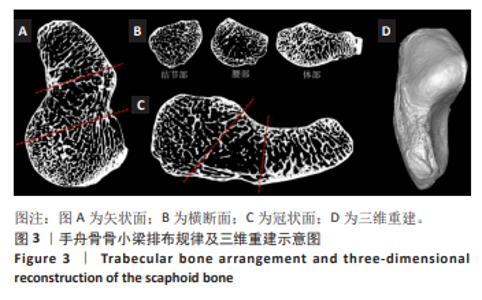
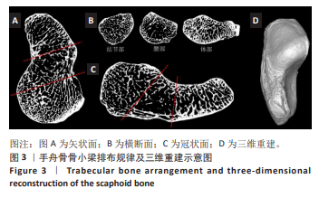
|