Chinese Journal of Tissue Engineering Research ›› 2026, Vol. 30 ›› Issue (21): 5556-5564.doi: 10.12307/2026.359
Previous Articles Next Articles
X-ray imaging and finite element analysis of the L6-S1 intervertebral disc in rats under abnormal forward-flexed posture
He Miao1, Wu Gang1, 2, 3, Zhang Xuxing3
- 1School of Sports Medicine, Wuhan Sports University, Wuhan 430079, Hubei Province, China; 2Center of Orthopedic Diagnosis and Treatment, Hubei Provincial Hospital of Traditional Chinese Medicine, Wuhan 430074, Hubei Province, China; 3First Clinical Medicine College of Hubei University of Chinese Medicine, Wuhan 430060, Hubei Province, China
-
Accepted:2025-06-30Online:2026-07-28Published:2026-03-05 -
Contact:Wu Gang, MD, Associate chief physician, Master’s supervisor, School of Sports Medicine, Wuhan Sports University, Wuhan 430079, Hubei Province, China; Center of Orthopedic Diagnosis and Treatment, Hubei Provincial Hospital of Traditional Chinese Medicine, Wuhan 430074, Hubei Province, China; First Clinical Medicine College of Hubei University of Chinese Medicine, Wuhan 430060, Hubei Province, China -
About author:He Miao, Master candidate, School of Sports Medicine, Wuhan Sports University, Wuhan 430079, Hubei Province, China -
Supported by:Hubei Provincial Administration of Traditional Chinese Medicine Chinese Medicine Research Project, No. ZY2023M004 (to WG)
CLC Number:
Cite this article
He Miao, Wu Gang, Zhang Xuxing. X-ray imaging and finite element analysis of the L6-S1 intervertebral disc in rats under abnormal forward-flexed posture[J]. Chinese Journal of Tissue Engineering Research, 2026, 30(21): 5556-5564.
share this article
Add to citation manager EndNote|Reference Manager|ProCite|BibTeX|RefWorks
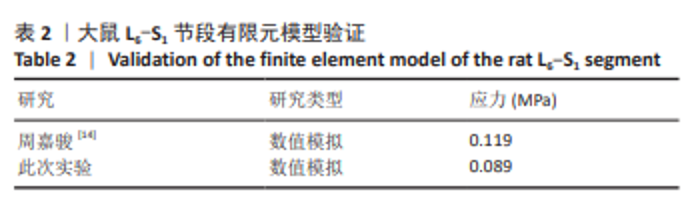
2.1 大鼠L6-S1节段有限元模型建立和验证结果 经过Mimics蒙版分割、Geomagic Wrap优化、SolidWorks构建几何、ANSYS Workbench划分网格等操作后,最终生成462 617个中间节点、355 073个单元,模拟了大鼠脊柱L6-S1节段的几何结构与材料属性(图2K)。 在模型验证过程中,此次研究构建的正常大鼠L6-S1节段在前屈5°载荷下的计算结果与既往研究的结果相接近[14],见表2,表明此模型有效,可以用于后续研究。"
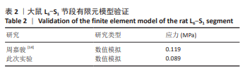
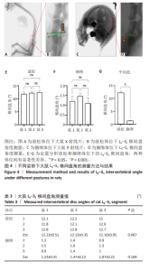
2.2 有限元中模拟异常前屈姿势的载荷设置:椎间盘角测量及变化值 对大鼠大体X射线片观察可知,放松姿势下椎间盘呈现较明显的前方厚、后方薄趋势,捆绑姿势下椎间盘前厚高度差异缩小明显。对X射线片进行如图4A-D所示的椎间盘角测量,结果如下:放松状态下,3只大鼠L6-S1椎间盘角分别为(12.23±0.51)°,(12.13±0.35)°,(12.10±0.95)°;捆绑状态下,L6-S1椎间盘角分别为(1.23±0.31)°,(1.47±0.12)°,(1.07±0.21)°,见表3。统计分析显示,放松体位下,3只大鼠间椎间盘角差异无显著性意义(P=0.967),见图4E,具有可比性;捆绑体位下,3只大鼠间数据同样无显著性差异(P=0.169),见图4F,表明捆绑造模具有良好的一致性。进一步分析发现,相较于放松状态,捆绑操作显著降低了L6-S1椎间盘角,差异有显著性意义(P < 0.001),见图4G。 基于实验测量,放松状态与捆绑状态下椎间盘角变化量分别为11.00°,10.67°和11.03°,平均角度变化量约为10.90°。文献复核结果表明,王田等[18]报道的L5-S1椎间盘角变化量为10.74°,与此次研究测得的大鼠L6-S1椎间盘角变化量高度相似,进一步支持捆绑造模的合理性。因而,此次研究基于实验数据与文献证据,最终将有限元模型的前屈加载角度确定为10°。"

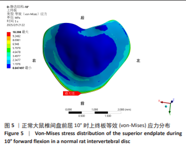
2.3 上终板应力及分布 在L6-S1椎间盘前屈10°姿势下,椎间盘上终板的最大等效应力为10.398 MPa,分布在终板与L6椎骨下表面骨质交界的前侧区域。中部区域等效应力较小且分布较均匀,最小值为0.047 MPa,见图5。"

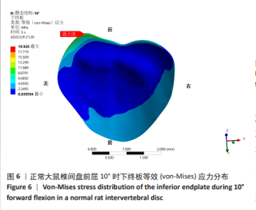
2.4 下终板应力及分布 在L6-S1椎间盘前屈10°姿势下,椎间盘下终板的最大等效应力为19.928 MPa,分布在终板与S1椎骨上表面交界的前侧区域。在左前与右后方向及中部区域等效应力最小且均匀分布,最小值为0.040 MPa,见图6。"

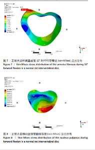
2.5 纤维环应力及分布 在L6-S1椎间盘前屈10°姿势下,椎间盘纤维环最大应力为6.819 MPa,分布在纤维环与下终板上表面交界的后侧偏左区域。左前与右后方向上小范围区域等效应力较小,最小值为0.126 MPa,见图7。 2.6 髓核应力及分布 在L6-S1椎间盘前屈10°姿势下,椎间盘髓核最大应力为0.104 MPa,分布在髓核与下终板上表面交界的前侧区域,等效应力最小值为0.018 MPa,整体应力分布较为均匀,见图8。"

| [1] ANDREW JAMES B, UZAIR A, HARSHA HARALURU J, et al. The prevalence of lumbar disc degeneration in symptomatic younger patients: A study of MRI scans. J Clin Orthop Trauma. 2020;11(5): 932-936. [2] THEO V, RYAN MB, BRAD B, et al. Global, regional, and national incidence, prevalence, and years lived with disability for 301 acute and chronic diseases and injuries in 188 countries, 1990–2013: a systematic analysis for the Global Burden of Disease Study 2013. Lancet. 2015; 386(9995):743-800. [3] ISMA LIZA MOHD I, SEONG LIN T, NURUL HUDA MOHD N, et al. Discogenic Low Back Pain: Anatomy, Pathophysiology and Treatments of Intervertebral Disc Degeneration. J Mol Sci. 2022;24(1):208. [4] IAN AFS, JAMES CI. Mechanical Conditions That Accelerate Intervertebral Disc Degeneration: Overload Versus Immobilization. Spine (Phila Pa 1976). 2004;29(23):2724-2732. [5] PYNT J, MACKEY MG, HIGGS J. Kyphosed seated postures: extending concepts of postural health beyond the office. J Occup Rehabil. 2008;18(1):35-45. [6] ZANOLA RL, DONIN CB, BERTOLINI GRF, et al. Biomechanical repercussion of sitting posture on lumbar intervertebral discs: A systematic review. J Bodyw Mov Ther. 2024;38:384-390. [7] KAI H, MAXIMILIAN L, PHILIPP E, et al. Patient-related risk factors and lifestyle factors for lumbar degenerative disc disease: a systematic review. Neurochirurgie. 2023;69(5):101482. [8] 陈钵, 秦大平, 张晓刚,等. 有限元分析法在脊柱生物力学中的研究进展 [J].中国疼痛医学杂志,2020,26(3):208-211+216. [9] 肖广润, 杨建东, 林升元,等. 有限元分析在脊柱外科中的应用及研究进展 [J]. 国际骨科学杂志,2020,41(6):347-351. [10] 于群. 基于有限元法的脊髓型颈椎病不同内固定方式的生物力学分析研究[D].济南: 山东师范大学,2024. [11] JAUMARD NV, LEUNG J, GOKHALE AJ, et al. Relevant Anatomic and Morphological Measurements of the Rat Spine: Considerations for Rodent Models of Human Spine Trauma. Spine (Phila Pa 1976). 2015;40(20):E1084-1092. [12] 岳静. 增材制造个体化定制腰椎人工椎板的研发及其生物力学与生物学验证[D].长春:吉林大学,2023. [13] 金凤. 针刀配合手法治疗腰椎退变的大鼠实验研究及生物力学分析[D].上海:复旦大学,2011. [14] 周嘉骏. 大鼠尾部椎间盘在异常弧度下的退行性改变及有限元分析[D].苏州:苏州大学,2016. [15] 张硕, 刘正. Bryan人工颈椎间盘角对颈椎病疗效的影响[J].颈腰痛杂志,2023,44(1):36-38. [16] 白文媛, 顾洪生, 廖振华,等. 正常成人腰椎间盘相关参数的测量和意义[J]. 中国临床解剖学杂志,2013,31(5):505-510. [17] 姜锦鹏, 顾洪生, 刘伟强,等. 正常成人颈椎间盘相关参数测量及意义[J]. 中国临床解剖学杂志,2013,31(1):32-36. [18] 王田, 胡玲玲, 王郑兴,等. 不同椅背倾角下的午睡姿势对人体脊柱曲度的影响[J]. 家具,2022,43(6):7-10+43. [19] LIU X, HOU Y, SHI H, et al. A retrospective cohort study on the significance of preoperative radiological evaluation of lumbar degenerative diseases for surgical reference. Quant Imaging Med Surg. 2023;13(8):5100-5108. [20] GONGHUAN Y, YU W, YIXIN Z, et al. Rapid health transition in China, 1990–2010: findings from the Global Burden of Disease Study 2010. Lancet. 2013;381(9882):1987-2015 [21] YURUBE T. Proteoglycan Dysfunction as a Key Hallmark of Intervertebral Disc Degeneration: Commentary on “Proteoglycan Dysfunction: A Common Link Between Intervertebral Disc Degeneration and Skeletal Dysplasia”. Neurospine. 2024;21(1):179-181. [22] TONGZHOU L, BO G, JINLANG Z, et al. Constructing intervertebral disc degeneration animal model: A review of current models. Front Surg. 2023;9:1089244. [23] WAWROSE RA, COUCH BK, DOMBROWSKI M, et al. Percutaneous lumbar annular puncture: A rat model to study intervertebral disc degeneration and pain-related behavior. JOR Spine. 2022;5(2): e1202. [24] LINDBLOM K. Experimental ruptures of intervertebral discs in rats’ tails; a preliminary report. J Bone Joint Surg Am. 1952;34-a(1):123-128. [25] XIE W, HUANG Z, HUANG Z, et al. A mouse coccygeal intervertebral disc degeneration model with tail-looping constructed using a suturing method. Animal Model Exp Med. 2025. doi: 10.1002/ame2.12501. [26] JI Y, ZHU P, ZHANG L, et al. A novel rat tail disc degeneration model induced by static bending and compression. Animal Model Exp Med. 2021;4(3):261-267. [27] 田子扬, 李展春. 椎间盘退变体内与体外模型的研究进展 [J]. 中国矫形外科杂志,2025,33(8):707-711. [28] MACLEAN JJ, LEE CR, GRAD S, et al. Effects of immobilization and dynamic compression on intervertebral disc cell gene expression in vivo. Spine (Phila Pa 1976). 2003;28(10):973-981. [29] NAKAMURA T, IRIBE T, ASOU Y, et al. Effects of compressive loading on biomechanical properties of disc and peripheral tissue in a rat tail model. Eur Spine J. 2009;18(11):1595-1603. [30] IATRIDIS JC, MENTE PL, STOKES IA, et al. Compression-induced changes in intervertebral disc properties in a rat tail model. Spine (Phila Pa 1976). 1999;24(10):996-1002. [31] BAILEY AS, ADLER F, MIN LAI S, et al. A comparison between bipedal and quadrupedal rats: do bipedal rats actually assume an upright posture? Spine (Phila Pa 1976). 2001;26(14):E308-313. [32] JIN LY, YIN HL, XU YQ, et al. Long-term whole-body vibration induces degeneration of intervertebral disc and facet joint in a bipedal mouse model. Front Bioeng Biotechnol. 2023;11:1069568. [33] 孙孝先, 白雪, 刘孟敏,等. 双上肢去势联合椎间盘刺破诱导建立大鼠椎间盘退变模型[J]. 中国组织工程研究,2023,27(35):5616-5621. [34] 崔力扬, 刘尚礼, 丁悦, 等. 大鼠腰椎间盘针刺退变模型的建立 [J]. 中国矫形外科杂志,2007(13):1008-1011. [35] 武圣达, 闫晓东, 张舒, 等. 高载荷作用对大鼠椎间盘退行性变的影响 [J]. 中国矫形外科杂志,2019,27(10):921-925. [36] 刘盾. 去双前肢直立诱导大鼠腰椎间盘退变模型的建立及骨痹合剂干预机制研究 [D]. 昆明:云南中医药大学,2014. [37] LI H, YAN JZ, CHEN YJ, et al. Non-invasive quantification of age-related changes in the vertebral endplate in rats using in vivo DCE-MRI. J Orthop Surg Res. 2017;12(1):169. [38] MURIUKI MG, HAVEY RM, VORONOV LI, et al. Effects of motion segment level, Pfirrmann intervertebral disc degeneration grade and gender on lumbar spine kinematics. J Orthop Res. 2016;34(8):1389-1398. [39] HILL R. A theory of the yielding and plastic flow of anisotropic metals. R Soc Lond. 1948;193(1033):281-297. [40] LUO C, JIANG T, TIAN S, et al. Finite element analysis of lumbar spine with different backpack positions in parachuting landing. Comput Methods Biomech Biomed Engin. 2021;24(15):1679-1686. [41] JIN LY, YIN HL, XU YQ, et al. Long-term whole-body vibration induces degeneration of intervertebral disc and facet joint in a bipedal mouse model. Front Bioeng Biotechnol. 2023;11:1069568. [42] PAN CC, LEE CH, CHEN KH, et al. Comparative Biomechanical Analysis of Unilateral, Bilateral, and Lateral Pedicle Screw Implantation in Oblique Lumbar Interbody Fusion: A Finite Element Study. Bioengineering (Basel). 2023;10(11):1238. [43] NATARAJAN RN, KE JH, ANDERSSON GB. A model to study the disc degeneration process. Spine (Phila Pa 1976). 1994;19(3):259-265. [44] LEE TC, TAYLOR D. Bone remodelling: should we cry Wolff? Ir J Med Sci. 1999;168(2):102-105. [45] WANG J, ISHIMOTO T, MATSUZAKA T, et al. Adaptive enhancement of apatite crystal orientation and Young’s modulus under elevated load in rat ulnar cortical bone. Bone. 2024;181:117024. [46] CAMILO LA, LAURA ABW, DAISUKE K, et al. Variation in cross‐sectional shape and biomechanical properties of the bat humerus under Wolff’s law. Anat Rec (Hoboken). 2021;304(9):1937-1952. [47] BARAK MM. Cortical and Trabecular Bone Modeling and Implications for Bone Functional Adaptation in the Mammalian Tibia. Bioengineering (Basel). 2024;11(5):514. [48] CHENG X, TIAN W, YANG J, et al. Engineering approaches to manipulate osteoclast behavior for bone regeneration. Mater Today Bio. 2024;26: 101043. [49] LI H, TANG Y, LIU Z, et al. Lumbar instability remodels cartilage endplate to induce intervertebral disc degeneration by recruiting osteoclasts via Hippo-CCL3 signaling. Bone Res. 2024;12(1):34. [50] CHRISTIAN W, GEHRCHEN PM, THOMAS K. Can Experimental Data in Humans Verify the Finite Element-Based Bone Remodeling Algorithm?. Spine (Phila Pa 1976). 2008;33(26):2875-2880. [51] RUFF C, HOLT B, TRINKAUS E. Who’s afraid of the big bad Wolff: “Wolff’s law” and bone functional adaptation. Am J Phys Anthropol. 2006;129(4):484-498. [52] YU Y, XU C. Correlation between sagittal morphology of lower lumbar end plate and degenerative changes in patients with lumbar disc herniation. J Craniovertebr Junction Spine. 2024;15(3):298-302. [53] ARUL PN, RISHI MK, AJOY PS, et al. Intervertebral disc degeneration and vertebral end plate damage in acute lumbar disc herniation. Indian Spine J. 2023;6(2):118-124. |
| [1] | Liu Wenlong, Dong Lei, Xiao Zhengzheng, Nie Yu. Finite element analysis of tibial prosthesis loosening after fixed-bearing unicompartmental knee arthroplasty for osteoporosis [J]. Chinese Journal of Tissue Engineering Research, 2026, 30(9): 2191-2198. |
| [2] | Zheng Wangyang, Fei Ji, Yang Di, Zhao Lang, Wang Lingli, Liu Peng, Li Haiyang. Finite element analysis of the force changes of the supraspinatus tendon and glenohumeral joint during the abduction and flexion of the humerus [J]. Chinese Journal of Tissue Engineering Research, 2026, 30(9): 2199-2207. |
| [3] | Cai Qirui, Dai Xiaowei, Zheng Xiaobin, Jian Sili, Lu Shaoping, Liu Texi, Liu Guoke, Lin Yuanfang. Mechanical effects of Long’s traction orthopedic method on cervical functional units: quantitative analysis of biomechanical model of head and neck [J]. Chinese Journal of Tissue Engineering Research, 2026, 30(9): 2208-2216. |
| [4] | Rao Jingcheng, Li Yuwan, Zheng Hongbing, Xu Zhi, Zhu Aixiang, Shi Ce, Wang Bing, Yang Chun, Kong Xiangru, Zhu Dawei. Biomechanical differences between the new proximal femoral stable intramedullary nail and traditional intramedullary nail#br# [J]. Chinese Journal of Tissue Engineering Research, 2026, 30(9): 2217-2225. |
| [5] | Chen Long, Wang Xiaozhen, Xi Jintao, Lu Qilin. Biomechanical performance of short-segment screw fixation combined with expandable polyetheretherketone vertebral body replacement in osteoporotic vertebrae [J]. Chinese Journal of Tissue Engineering Research, 2026, 30(9): 2226-2235. |
| [6] | Yan Xiangning, Chen Lei, Chen Yonghuan, Wang Chao, Li Xiaosheng. Influence of different depths and loads on knee joint mechanics and peripheral muscle force characteristics during squatting [J]. Chinese Journal of Tissue Engineering Research, 2026, 30(9): 2236-2247. |
| [7] | Zhang Nan, Meng Qinghua, Bao Chunyu. Characteristics and clinical application of ankle joint finite element models [J]. Chinese Journal of Tissue Engineering Research, 2026, 30(9): 2343-2349. |
| [8] | Zhu Xiaolong, Zhang Wei, Yang Yang. Visualization analysis of research hotspots and cutting-edge information in the field of intervertebral disc regeneration and repair [J]. Chinese Journal of Tissue Engineering Research, 2026, 30(9): 2391-2402. |
| [9] | Xinjiang Branch of China Trauma Rescue & Treatment Association. Expert consensus on diagnosis and treatment of brucellar osteoarthritis [J]. Chinese Journal of Tissue Engineering Research, 2026, 30(9): 2403-2412. |
| [10] | Wen Fayan, Li Yan, Qiang Tianming, Yang Chen, Shen Linming, Li Yadong, Liu Yongming. Unilateral biportal endoscopic technology for treatment of lumbar degenerative diseases: global research status and changing trends [J]. Chinese Journal of Tissue Engineering Research, 2026, 30(9): 2380-2390. |
| [11] | Cheng Qisheng, Julaiti·Maitirouzi, Xiao Yang, Zhang Chenwei, Paerhati·Rexiti. Finite element analysis of novel variable-diameter screws in modified cortical bone trajectory of lumbar vertebrae [J]. Chinese Journal of Tissue Engineering Research, 2026, 30(9): 2162-2171. |
| [12] | Wu Hongxu, Liu Xuanyu, Wang Taoyu, Wang Shiyao, Cheng Jingyi, Zhang Mingwen, Zhang Yinxia, Liu Zhihua, Wang Xiaojie. Finite element simulation of scoliosis with muscle unit introduction: verification of correction effect under bidirectional load [J]. Chinese Journal of Tissue Engineering Research, 2026, 30(9): 2172-2181. |
| [13] | Liu Jiafu, Ren Ruxia, Liao Zhouwei, Zhou Xiali, Wu Yihong, Zhang Shaoqun. Three-dimensional finite element analysis of cervical spine biomechanical characteristics in a rat model of cervical vertigo [J]. Chinese Journal of Tissue Engineering Research, 2026, 30(9): 2182-2190. |
| [14] | Chen Huiting, Zeng Weiquan, Zhou Jianhong, Wang Jie, Zhuang Congying, Chen Peiyou, Liang Zeqian, Deng Weiming. Tail anchoring technique of vertebroplasty in treatment of osteoporotic vertebral compression fractures with intravertebral cleft: a finite element analysis [J]. Chinese Journal of Tissue Engineering Research, 2026, 30(9): 2145-2152. |
| [15] | Zeng Xuan, Weng Rui, Ye Shicheng, Tang Jiadong, Mo Ling, Li Wenchao. Two lumbar rotary manipulation techniques in treating lumbar disc herniation: a finite element analysis of biomechanical differences [J]. Chinese Journal of Tissue Engineering Research, 2026, 30(9): 2153-2161. |
| Viewed | ||||||
|
Full text |
|
|||||
|
Abstract |
|
|||||