Chinese Journal of Tissue Engineering Research ›› 2013, Vol. 17 ›› Issue (28): 5177-5183.doi: 10.3969/j.issn.2095-4344.2013.28.013
Previous Articles Next Articles
Long-term effect of quad helix expansion of dental arch
Wang Feng, Lu Huai-xiu, Lin Song-shan
- Department of Stomatology, Naval General Hospital of PLA, Beijing 100048, China
-
Online:2013-07-09Published:2013-07-29 -
Contact:Lu Huai-xiu, M.D., Associate chief physician, Department of Stomatology, Naval General Hospital of PLA , Beijing 100048, China Luhuaix123@sina.com -
About author:Wang Feng☆, M.D., Associate chief physician, Department of Stomatology, Naval General Hospital of PLA, Beijing 100048, China wolfwang2003@yahoo.com.cn -
Supported by:National Natural Science Foundation of China, No. 81070837*

CLC Number:
Cite this article
Wang Feng, Lu Huai-xiu, Lin Song-shan. Long-term effect of quad helix expansion of dental arch[J]. Chinese Journal of Tissue Engineering Research, 2013, 17(28): 5177-5183.
share this article
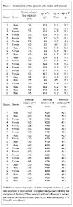
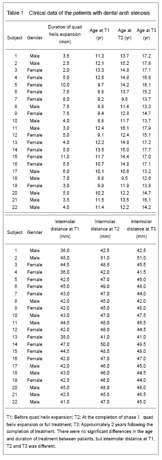
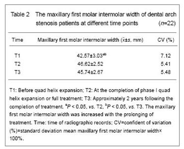
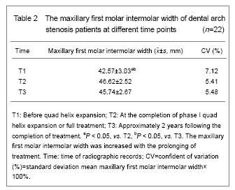
Data analysis and clinical data of the subjects All the 22 patients were included in the analysis. A repeated measures analysis of variance model showed the interaction of time, age, duration of quad helix treatment, the interaction of time and duration had no statistically significant effect on intermolar distance (P > 0.05), but the duration (T1, T2, T3) had a highly significant effect on intermolar distance (P < 0.05) (Table 1). The increasing of intermolar width of the dental arch stenosis patients after dental arch expansion The result showed that the duration (T1, T2, T3) had a significant effect on intermolar distance (P < 0.05). The two sample t-test showed there was statistically significant increasing in intermolar distance from T1 to T2 (P < 0.05), there was no significant difference in intermolar distance from T2 to T3 (P > 0.05), and there was a statistically significant increasing in intermolar distance from T1 to T3 (P < 0.05, Table 2)."
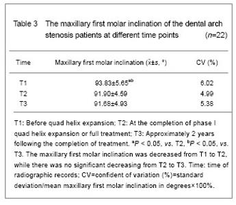
Decreasing of maxillary first molar inclination of the dental arch stenosis patients after expansion For results of inclination measurement (Table 3), a general linear model showed that duration had a statistically significant effect on angle (P < 0.05). The two sample t-test showed that there was a significant decreasing in inclination from T1 to T2 (P < 0.05). There was no significant difference in inclination from T2 to T3 (P > 0.05). However, there was a significant decreasing in inclination from T1 to T3 (P < 0.05). Thus, the results showed that the maxillary first molar inclinations from T1 to T2 were decreased, the curve of Wilson was diminished with treatment. However, the intermolar distances were increased from T1 to T2. There was no significant decrease in maxillary first inclination or intermolar distance between T2 to T3."
"

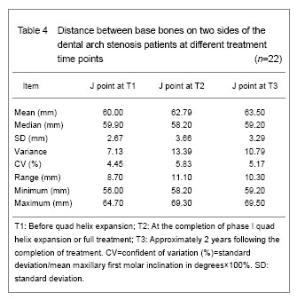
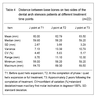
Increasing of distance between base bones on two sides of dental arch stenosis patients after expansion Evaluation of J point measurements (Table 4) using the Wilcoxon Rank Test at the significance level of a=0.5 indicated that there was a statistically significant increasing from T1 to T2 (P < 0.05) and from T1 to T3 (P < 0.05). There was no statistically significant change from T2 to T3 (P > 0.05)."
| [1]Li H, Wang CL, Su YY, et al. Clinical evaluation on rapid maxillary expansion and maxillary protraction in treating Class Ⅲ malocclusion by Johnston analysis. Linchuang Kouqing Yixue Zazhi. 2008;24(10):611-613.[2]Kong FZ, Jing HY, Geng L, et al. Cranio-occlusional changes of skeletal class Ⅲ malocclusion in different aged patients treated with bonded maxillary expansion and maxillary protraction. Zhongguo Zuzhi Gongcheng Yanjiu yu Linchuang Kangfu. 2008;12(30):5824-5828.[3]Li MH, Zhang XY. Dental and skeletal changes after maxillary expansion in adults. Beijing Kouqing Yixue. 2012;20(4):217-219.[4]Ran YD, Zhang H. Skeletal Ⅲ malocclusion orthopedics after PAR index evaluation. Yixue Yanjiu yu Jiaoyu. 2010; 27(4):35-37.[5]Jiang WH. Advances in Biomechanics of Maxillary Transverse Expansion. Kouqiang Hemian Waike Zazhi. 2010;20(3):219-221.[6]Wang S, Wang L, Zhao CY, et al. The effective therapy in the unilateral posterior crossbite treatment. Linchuang Kouqiang Yixue Zazhi. 2007;23(12):743-745.[7]Pan HH, Huang Y, Yang Z, et al. 3-D finite element study on rapid maxillary expansion using implant anchorage. Sichuan Daxue Xuebao: Yixue Ban. 2011;42(6): 831-834.[8]Yu YL, Tang GH, Gong FF, et al. A comparison of rapid palatal expansion and Damon appliance on non-extraction correction of dental crowding. Shanghai Kouqiang Yixue. 2008;17(3):237-242.[9]Tang T. Application of rapid expansion combined with bow in front of traction in skeletal class Ⅲ malocclusion. Yiyao Luntan Zazhi. 2011;32(23):114-115.[10]Zhao ML, Liu FL, Li YM. Clinical application of the rapid maxillary expansion with face mask therapy on the treatment of skeletal Class III malocclusion in the permanent dentition stage. Zhongguo Meirong Yixue. 2010;19(4):74-76.[11]Hu YQ, Xue D, Zheng LL, et al. Analysis of the effect of maxillary protraction combined with Hass rapid maxillary expansion in the treatment of osteal anterior crossbite malocclusion. Zhongguo Shiyong Kouqiang Ke Zazhi. 2012;5(2):113-115.[12]Honme Y, Motoyoshi M, Shinohara A, et al. Efficient palatal expansion with a quadhelix appliance: an in vitro study using an experimental dental arch model. Eur J Orthod. 2012;34(4):442-446. [13]Guo L, Wang YY, Wang TY, et al. The clinical investigation of Quad Helix appliance in adult orthodontics. Zhongguo Yiyao Daobao. 2008;5(14):106-115. [14]Cheng HJ. Clinical research of modified Hyrax expander application for maxillary narrow dental arch. Kouqiang Yixue. 2008;28(1):23-24. [15]Cepera F, Torres FC, Scanavini MA, et al. Effect of a low-level laser on bone regeneration after rapid maxillary expansion. Am J Orthod Dentofacial Orthop. 2012;141(4): 444-450. [16]Sun FY, Zhang Y. Clinical effect of removable lingual arch plus auxiliary spring for dental arch expansion. Nan Fang Yi Ke Da Xue Xue Bao. 2007;27(4):546-547.[17]Gao X, Lin HP. The Design and application of a new modified removable QuadHelix. Zhongguo Yiyao Zhinan. 2010;34(8):202-204.[18]Cheng P. Clinical application of removable Quad-Helix appliance in the expansion of adult maxillary arch. Linchuang Yixue Gongcheng. 2009;16(9):67-68.[19]Jiang ST, Wang H, An ZJ, et al. The fabrication and clinical application of semi-fixed mandibular lingual arch expansion appliance. Hua Xi Kou Qiang Yi Xue Za Zhi. 2010;28(4): 455-456.[20]Sonnesen L, Bakke M. Bite force in children with unilateral crossbite before and after orthodontic treatment. A prospective longitudinal study. Eur J Orthod. 2007;29(3): 310-313.[21]Donohue VE, Marshman LA, Winchester LJ. A clinical comparison of the quadhelix appliance and the nickel titanium (tandem loop) palatal expander: a preliminary, prospective investigation. Eur J Orthod. 2004;26(4): 411-420.[22]McNally MR, Spary DJ, Rock WP. A randomized controlled trial comparing the quadhelix and the expansion arch for the correction of crossbite. J Orthod. 2005;32(1):29-35.[23]Chen LL, You QL, Yang YM. Non-extraction treatment in borderline Class Ⅲ malocclusion. Shanghai Kouqiang Yixue. 2011;20(1):83-87.[24]Wu QQ, Zhao GZ, Ke J, et al. The clinical application of improved Quadhelix appliance--110 case reports of complex malocclusion. Yati Yasui Yazhou Bing Zazhi. 2012;22(6):357-363.[25]Wang HY, Li H. Clinical analysis of effects of rapid expansion and maxillary protraction on treatment of premature skeletal class Ⅲ malocclusion. Kouqiang Yixue. 2012;32(5):303-305.[26]Chen Z, Liu HL. Articulation induced compliance following dental arch change with rapid maxillary expansion. Zhongguo Zuzhi Gongcheng Yanjiu yu Linchuang kangfu. 2007;11(22):4416-4417.[27]Li X, Shi H. Treatment effect of maxillary protraction on skeletal class III high angle cases in mixed dentition. Linchuang Junyi Zazhi. 2011;39(3):539-541.[28]Li W, Lin J. Dental arch width stability after quadhelix and edgewise treatment in complete unilateral cleft lip and palate. Angle Orthod. 2007;77(6):1067-1072.[29]Qin HL. The effects of rapid maxillary expansion on maxillofacial structures and function. Guoji Kouqiang Yixue Zazhi. 2012;39(1):132-135.[30]Zhu K, Yu YL, Hou FC. Clinical stability of rapid maxillary expansion followed by fixed appliances. Kouqiang Yixue. 2012;32(2):100-102.[31]Yu HY. Clinical outcome of rapid maxillary expansion followed by fixed appliances. Qingdao Yiyao Weisheng. 2011;43(3):177-179.[32]Ding B, Sun P. Soft tissue changes in skeletal Class III patients treated with maxillary protraction. Zhongguo Meirong Yixue. 2010;19(6):888-890.[33]Lu YJ, Chang SH. The study on the effect of maxillary protraction in skeletal Class Ⅲ subjects. Zhonghua Kouqiang Yixue Yanjiu Zazhi: Dianzi Ban. 2012;6(1):93-96.[34]Qian H, Duan YZ, Jin ZL, et al. Study of changes in perioral muscle pressures following rapid maxillary expansion treatment and retention. Zhongguo Meirong Yixue. 2010; 19(2):254-256.[35]Qiu YL, Du FZ, Kapika F, et al. Effects of protraction and rapid maxillary expansion on the sagittal pharyngeal dimensions and tongue posture in Class Ⅲ malocclusion subjects. Shiyong Kouqiang Yixue Zazhi. 2011;27(3): 365-368.[36]Holberg C, Holberg N, Schwenzer K, et al. Biomechanical analysis of maxillary expansion in CLP patients. Angle Orthod. 2007;77(2):280-287.[37]Zhao Y, Armine K, Elizabeth G, et al. Cone-Beam CT evaluation of the changes of upper second premolars after rapid palatal expansion with Hyrax appliances. Kouqiang Yixue Yanjiu. 2009;25(4):455-458.[38]Wei ZL. Effect of rapid maxillary expansion on the level of IL-1β and MMP-8 in gingival crevicular fluid. Kouqiang Yixue Yanjiu. 2011;27(6):484-487.[39]Geng HX, Guo XJ, Liu X. Effects of surgically assisted rapid maxillary expansion in correction of maxillary transverse deficiency. Linchuang Kouqiang Yixue Zazhi. 2012;28(8): 483-485.[40]Pietilä I, Pietilä T, Pirttiniemi P, et al. Orthodontists' views on indications for and timing of orthodontic treatment in Finnish public oral health care. Eur J Orthod. 2008;30(1):46-51. [41]Guo J, Zhang QF, Li GF, et al. Establishment of a rat midpalatal suture expansion model. Zhongguo Zuzhi Gongcheng Yanjiu yu Linchuang Kangfu. 2010;14(2): 272-275.[42]Wang XH, Ye HF. Clinical study on rapid maxillary expansion accompanied with the use of self-ligating system appliance. Zhongwai Yiliao. 2010;36(1):22-23. [43]Scarfe WC, Farman AG, Sukovic P. Clinical applications of cone-beam computed tomography in dental practice. J Can Dent Assoc. 2006;72(1):75-80. [44]Kau CH, Richmond S, Palomo JM, et al. Three-dimensional cone beam computerized tomography in orthodontics. J Orthod. 2005;32(4):282-293.[45]Habersack K, Karoglan A, Sommer B, et al. High-resolution multislice computerized tomography with multiplanar and 3-dimensional reformation imaging in rapid palatal expansion. Am J Orthod Dentofacial Orthop. 2007; 131(6):776-781. |
| [1] | Li Zhifei, Han Bin, Liu Qiuli, Zhang Zhanming, Wei Haokai, Zuo Kuangshi, Zhang Yisheng. Cervical motion characteristics in patients with cervical spondylotic radiculopathy based on motion capture technology [J]. Chinese Journal of Tissue Engineering Research, 2026, 30(9): 2286-2293. |
| [2] | Pan Hongfei, Zhuang Zhenbing, Xu Baiyun, Yang Zhangyang, Lin Kairui, Zhan Bingqing, Lan Jinghan, Gao Heng, Zhang Nanbo, Lin Jiayu. Inhibitory effects of different concentrations of auranofin on M1 macrophage function and its therapeutic potential in diabetic wound healing [J]. Chinese Journal of Tissue Engineering Research, 2026, 30(6): 1390-1397. |
| [3] | Peng Zhiwei, Chen Lei, Tong Lei. Luteolin promotes wound healing in diabetic mice: roles and mechanisms [J]. Chinese Journal of Tissue Engineering Research, 2026, 30(6): 1398-1406. |
| [4] | Lyu Xiaofan, Huang Yi, Ding Liucheng . Mitochondrial mechanism and intervention therapy in diabetic cystopathy [J]. Chinese Journal of Tissue Engineering Research, 2026, 30(6): 1508-1515. |
| [5] | Cao Xinyan, Yu Zifu, Leng Xiaoxuan, Gao Shiai, Chen Jinhui, Liu Xihua. Effect of repetitive transcranial magnetic stimulation and transcranial direct current stimulation on motor function and gait in children with cerebral palsy: a network meta-analysis [J]. Chinese Journal of Tissue Engineering Research, 2026, 30(6): 1539-1548. |
| [6] | Guo Ying, Tian Feng, Wang Chunfang. Potential drug targets for the treatment of rheumatoid arthritis: large sample analysis from European databases [J]. Chinese Journal of Tissue Engineering Research, 2026, 30(6): 1549-1557. |
| [7] | Chen Ju, Zheng Jinchang, Liang Zhen, Huang Chengshuo, Lin Hao, Zeng Li. Effect and mechanism of beta-caryophyllene in mice with osteoarthritis [J]. Chinese Journal of Tissue Engineering Research, 2026, 30(6): 1341-1347. |
| [8] | Wen Guangwei, Zhen Yinghao, Zheng Taikeng, Zhou Shuyi, Mo Guoye, Zhou Tengpeng, Li Haishan, Lai Yiyi. Effects and mechanisms of isoginkgetin on osteoclastogenesis [J]. Chinese Journal of Tissue Engineering Research, 2026, 30(6): 1348-1358. |
| [9] | Li Linzhen, Jiao Hongzhuo, Chen Weinan, Zhang Mingzhe, Wang Jianlong, Zhang Juntao. Effect of icariin-containing serum on lipopolysaccharide-induced inflammatory damage in human chondrocytes [J]. Chinese Journal of Tissue Engineering Research, 2026, 30(6): 1368-1374. |
| [10] | Sun Yajie, Zhao Xinchen, Bo Shuangling. Spatiotemporal expression of bone morphologic protein 7 in mouse kidney development [J]. Chinese Journal of Tissue Engineering Research, 2026, 30(5): 1156-1161. |
| [11] | Li Haojing, Wang Xin, Song Chenglin, Zhang Shengnan, Chen Yunxin. Therapeutic efficacy of extracorporeal shock wave therapy in the upper trapezius muscle area combined with exercise control training in patients with chronic non-specific neck pain [J]. Chinese Journal of Tissue Engineering Research, 2026, 30(5): 1162-1170. |
| [12] | Liu Yu, Lei Senlin, Zhou Jintao, Liu Hui, Li Xianhui. Mechanisms by which aerobic and resistance exercises improve obesity-related cognitive impairment [J]. Chinese Journal of Tissue Engineering Research, 2026, 30(5): 1171-1183. |
| [13] | Yu Huifen, Mo Licun, Cheng Leping. The position and role of 5-hydroxytryptamine in the repair of tissue injury [J]. Chinese Journal of Tissue Engineering Research, 2026, 30(5): 1196-1206. |
| [14] | Wang Zhengye, Liu Wanlin, Zhao Zhenqun. Advance in the mechanisms underlying miRNAs in steroid-induced osteonecrosis of the femoral head [J]. Chinese Journal of Tissue Engineering Research, 2026, 30(5): 1207-1214. |
| [15] | Bu Yangyang, Ning Xinli, Zhao Chen. Intra-articular injections for the treatment of osteoarthritis of the temporomandibular joint: different drugs with multiple combined treatment options [J]. Chinese Journal of Tissue Engineering Research, 2026, 30(5): 1215-1224. |
| Viewed | ||||||
|
Full text |
|
|||||
|
Abstract |
|
|||||