Chinese Journal of Tissue Engineering Research ›› 2024, Vol. 28 ›› Issue (25): 4034-4040.doi: 10.12307/2024.180
Previous Articles Next Articles
Cell-of-origin for heterotopic ossification induced by bone morphogenetic protein 4 in skeletal muscle
Yu Yangyi, Lian Qiang, Wu Jianqun, Zhang Xuan, Ren Jinke, Li Guangheng
- Shenzhen Key Laboratory of Musculoskeletal Tissue Reconstruction and Function Restoration, Department of Adult Joint Reconstruction and Orthopedic Surgery, Shenzhen People’s Hospital (Second Clinical Medical College of Jinan University, First Affiliated Hospital, Southern University of Science and Technology), Shenzhen 510515, Guangdong Province, China
-
Received:2023-04-27Accepted:2023-04-28Online:2024-09-08Published:2023-11-24 -
Contact:Li Guangheng, PhD, Chief physician, Shenzhen Key Laboratory of Musculoskeletal Tissue Reconstruction and Function Restoration, Department of Adult Joint Reconstruction and Orthopedic Surgery, Shenzhen People’s Hospital (Second Clinical Medical College of Jinan University, First Affiliated Hospital, Southern University of Science and Technology), Shenzhen 510515, Guangdong Province, China -
About author:Yu Yangyi, PhD, Attending physician, Shenzhen Key Laboratory of Musculoskeletal Tissue Reconstruction and Function Restoration, Department of Adult Joint Reconstruction and Orthopedic Surgery, Shenzhen People’s Hospital (Second Clinical Medical College of Jinan University, First Affiliated Hospital, Southern University of Science and Technology), Shenzhen 510515, Guangdong Province, China Lian Qiang, Shenzhen Key Laboratory of Musculoskeletal Tissue Reconstruction and Function Restoration, Department of Adult Joint Reconstruction and Orthopedic Surgery, Shenzhen People’s Hospital (Second Clinical Medical College of Jinan University, First Affiliated Hospital, Southern University of Science and Technology), Shenzhen 510515, Guangdong Province, China -
Supported by:General Project of National Natural Science Foundation of China, Nos. 81472136, 82172463 (to LGH)
CLC Number:
Cite this article
Yu Yangyi, Lian Qiang, Wu Jianqun, Zhang Xuan, Ren Jinke, Li Guangheng. Cell-of-origin for heterotopic ossification induced by bone morphogenetic protein 4 in skeletal muscle[J]. Chinese Journal of Tissue Engineering Research, 2024, 28(25): 4034-4040.
share this article
Add to citation manager EndNote|Reference Manager|ProCite|BibTeX|RefWorks
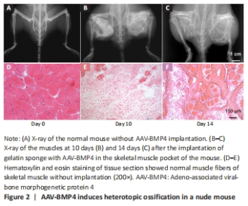
AAV-BMP4 induces heterotopic ossification in nude mouse The gelatin sponge adsorbed with the 15 μL AAV-BMP4 virus solution was embedded in the skeletal muscle pocket of the nude mouse bilaterally. In 2 weeks, the heterotopic ossification was established locally (Figure 2A–C). The process of heterotopic ossification was observed through endochondral bone formation (Figure 2D–F). "

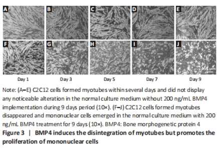
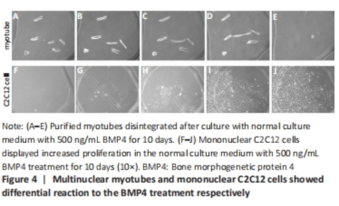
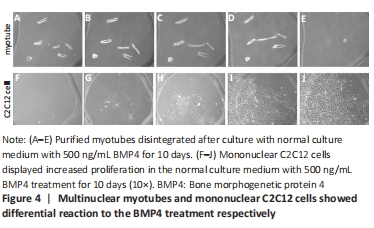
BMP4 induces the disintegration of multinuclear myotubes and promotes the proliferation of mononuclear myogenic cells After C2C12 formed myotubes on Day 9 (Figure 3A–E), BMP4 treatment led to the disintegration of multinuclear myotubes, but promoted the proliferation of mononuclear cells (Figure 3F–J, Figure 4). In addition, according to our previous studies, BMP4 also promotes the osteogenesis of myogenic cells[23]."

"

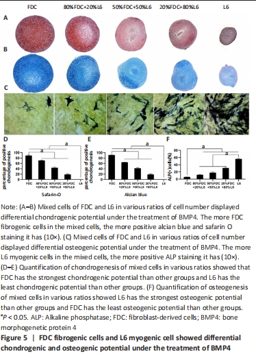
Firborgenic cells displayed more prone to chondrogenic differentiation, while myognic cells showed more readily to osteogenic differentiation As shown in Figure 5A–B, mixed cells of FDC and L6 in various ratios of cell number displayed differential chondrogenic potential under the treatment of BMP4. The more FDC fibrogenic cells in the mixed cells, the more positive alcian blue and safarin O staining it had (Figure 5D–E). Mixed cells of FDC and L6 in various ratios of cell number displayed differential osteogenic potential under the treatment of BMP4. As shown in Figure 5C–F, the more L6 myogenic cells in the mixed cells, the more positive ALP staining it had. "
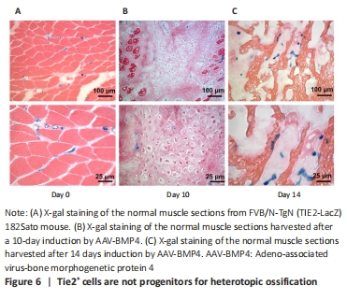
Tie2+ cells are not progenitors for heterotopic ossification in our study Next, we investigated the fate of endothelial cells during heterotopic ossification. Tie2+ is predominantly expressed by vascular endothelial cells, and is widely used as a marker for endothelial cells[23]. Tie2-Cre transgenic mice have been used by several scientists for lineage tracing to understand the progression of heterotopic ossification[24]; they reported that Tie2+ cells could differentiate into chondrocytes or osteocytes during heterotopic ossification induced by BMP4. However, different conclusions were reached in our study. In our experiment, FVB/N-TgN (TIE2-LacZ) 182Sato mice was purchased from The Jackson Laboratory (Bar Harbor, ME) and used express β-galactosidase driven by the endothelial-specific TIE2 promoter. As Tie2-Cre transgenic mice were reported to have nonspecific recombination, the FVB/N-TgN (TIE2-LacZ) 182Sato mouse was used to study the role of Tie2+ cells in the heterotopic ossification process. We embedded the AAV-BMP4 sponge into the muscle of the FVB/N-TgN (TIE2-LacZ) 182Sato transgenic mice. On days 10 and 14, the ectopic bone formed in the muscle was harvested for X-gal staining (Figure 6). Without AAV-BMP4 injection, endothelial cells were distributed between the myofibers (Figure 6A). Round staining indicated that these Tie2+ cells were distributed in the blood vessel. After 10 days of induction, chondrocytes were observed in the muscles (Figure 6B). However, these chondrocytes were not stained with X-gal, indicating that Tie2+ cells were not the original cells for chondrocytes in heterotopic ossification. However, we observed X-gal-stained cells along the chondrocytes and between the myofibers. After 14 days of induction, bone trabeculae were observed (Figure 6C); however, there were no Tie2+ cells present, although stained cells were observed in the bone marrow-like region. Overall, using the transgenic mouse, we found that endothelial cells, which were Tie2 positive, were not progenitors for heterotopic ossification, but were involved in the process. "

| [1] MEYERS C, LISIECKI J, MILLER S, et al. Heterotopic ossification: a comprehensive review. JBMR Plus. 2019;3(4):e10172. [2] YANG YS, KIM JM, XIE J, et al. Suppression of heterotopic ossification in fibrodysplasia ossificans progressiva using AAV gene delivery. Nat Commun. 2022; 13(1):6175. [3] KAN C, DING N, YANG J, et al. BMP-dependent, injury-induced stem cell niche as a mechanism of heterotopic ossification. Stem Cell Res Ther. 2019;10(1):14. [4] LEES-SHEPARD JB, STOESSEL SJ, CHANDLER JT, et al. An anti-ACVR1 antibody exacerbates heterotopic ossification by fibro-adipogenic progenitors in fibrodysplasia ossificans progressiva mice. J Clin Invest. 2022;132(12):e153795. [5] TSANG KY, CHEAH KS. The extended chondrocyte lineage: implications for skeletal homeostasis and disorders. Curr Opin Cell Biol. 2019;61:132-140. [6] HUANG Y, WANG X, ZHOU D, et al. Macrophages in heterotopic ossification: from mechanisms to therapy. NPJ Regen Med. 2021;6(1):70. [7] SHAHRIYARI M, ISLAM MR, SAKIB SM, et al. Engineered skeletal muscle recapitulates human muscle development, regeneration and dystrophy. J Cachexia Sarcopenia Muscle. 2022;13(6):3106-3121. [8] FENG H, XING W, HAN Y, et al. Tendon-derived cathepsin K-expressing progenitor cells activate Hedgehog signaling to drive heterotopic ossification. J Clin Invest. 2020;130(12):6354-6365. [9] ZHANG Q, ZHOU D, WANG H, et al. Heterotopic ossification of tendon and ligament. J Cell Mol Med. 2020;24(10):5428-5437. [10] EGHBALI-FATOURECHI GZ, LAMSAM J, FRASER D, et al. Circulating osteoblast-lineage cells in humans. N Engl J Med. 2005;352(19):1959-1966. [11] NESTI LJ, JACKSON WM, SHANTI RM, et al. Differentiation potential of multipotent progenitor cells derived from war-traumatized muscle tissue. J Bone Joint Surg Am. 2008;90(11):2390-2398. [12] UEZUMI A, FUKADA S, YAMAMOTO N, et al. Mesenchymal progenitors distinct from satellite cells contribute to ectopic fat cell formation in skeletal muscle. Nat Cell Biol. 2010;12(2):143-152. [13] DROUIN G, COUTURE V, LAUZON MA, et al. Muscle injury-induced hypoxia alters the proliferation and differentiation potentials of muscle resident stromal cells. Skelet Muscle. 2019;9(1):18. [14] LAVASANI M, LU A, THOMPSON SD, et al. Isolation of muscle-derived stem/progenitor cells based on adhesion characteristics to collagen-coated surfaces. Methods Mol Biol. 2013;976:53-65. [15] TU B, YU B, WANG W, et al. Inhibition of IL-17 prevents the progression of traumatic heterotopic ossification. J Cell Mol Med. 2021;25(16):7709-7719. [16] FELIX-ILEMHENBHIO F, PICKERING GAE, KISS-TOTH E, et al. Pathophysiology and emerging molecular therapeutic targets in heterotopic ossification. Int J Mol Sci. 2022;23(13):6983. [17] WOSCZYNA MN, BISWAS AA, COGSWELL CA, et al. Multipotent progenitors resident in the skeletal muscle interstitium exhibit robust BMP-dependent osteogenic activity and mediate heterotopic ossification. J Bone Miner Res. 2012; 27(5):1004-1017. [18] LEES-SHEPARD JB, YAMAMOTO M, BISWAS AA, et al. Activin-dependent signaling in fibro/adipogenic progenitors causes fibrodysplasia ossificans progressiva. Nat Commun. 2018;9(1):471. [19] SHEN F, SHI Y. Recent advances in single-cell view of mesenchymal stem cell in osteogenesis. Front Cell Dev Biol. 2021;9:809918. [20] CAPLAN AI. Mesenchymal stem cells. J Orthop Res. 1991;9(5):641-650. [21] LI G, PENG H, CORSI K, et al. Differential effect of BMP4 on NIH/3T3 and C2C12 cells: implications for endochondral bone formation. J Bone Miner Res. 2005; 20(9):1611-1623. [22] LI G, ZHENG B, MESZAROS LB, et al. Identification and characterization of chondrogenic progenitor cells in the fascia of postnatal skeletal muscle. J Mol Cell Biol. 2011;3(6):369-377. [23] WERLEIN C, ACKERMANN M, STARK H, et al. Inflammation and vascular remodeling in COVID-19 hearts. Angiogenesis. 2023;26(2):233-248. [24] PRADOS B, DEL TORO R, MACGROGAN D, et al. Heterotopic ossification in mice overexpressing Bmp2 in Tie2+ lineages. Cell Death Dis. 2021;12(8):729. [25] CHAN NT, LEE MS, WANG Y, et al. CTR9 drives osteochondral lineage differentiation of human mesenchymal stem cells via epigenetic regulation of BMP-2 signaling. Sci Adv. 2022;8(46):eadc9222. [26] ALFAQIH MS, TARAWAN VM, SYLVIANA N, et al. Effects of vitaminutes d on satellite cells: a systematic review of in vivo studies. Nutrients. 2022;14(21):4558. [27] ROSINA M, LANGONE F, GIULIANI G, et al. Osteogenic differentiation of skeletal muscle progenitor cells is activated by the DNA damage response. Sci Rep. 2019; 9(1):5447. [28] RAUCH A, MANDRUP S. Transcriptional networks controlling stromal cell differentiation. Nat Rev Mol Cell Biol. 2021;22(7):465-482. [29] CONTRERAS O, ROSSI FMV, THERET M. Origins, potency, and heterogeneity of skeletal muscle fibro-adipogenic progenitors-time for new definitions. Skelet Muscle. 2021;11(1):16. [30] ALESSI WOLKEN DM, IDONE V, HATSELL SJ, et al. The obligatory role of activin a in the formation of heterotopic bone in fibrodysplasia ossificans progressiva. Bone. 2018;109:210-217. |
| [1] | Yang Yufang, Yang Zhishan, Duan Mianmian, Liu Yiheng, Tang Zhenglong, Wang Yu. Application and prospects of erythropoietin in bone tissue engineering [J]. Chinese Journal of Tissue Engineering Research, 2024, 28(9): 1443-1449. |
| [2] | Wei Juan, Li Ting, Huan Mengting, Xie Ying, Xie Zhouyu, Wei Qingbo, Wu Yunchuan. Mechanism by which static exercise improves insulin resistance in skeletal muscle of type 2 diabetes [J]. Chinese Journal of Tissue Engineering Research, 2024, 28(8): 1271-1276. |
| [3] | Shen Jiangyong, He Xi, Tang Yuting, Wang Jianjun, Liu Jinyi, Chen Yuanyuan, Wang Xinyi, Liu Tong, Sun Haoyuan. RAS-selective lethal small molecule 3 inhibits the fibrosis of pathological scar fibroblasts [J]. Chinese Journal of Tissue Engineering Research, 2024, 28(8): 1168-1173. |
| [4] | Wang Ji, Zhang Min, Li Wenbo, Yang Zhongya, Zhang Long. Effect of aerobic exercise on glycolipid metabolism, skeletal muscle inflammation and autophagy in type 2 diabetic rats [J]. Chinese Journal of Tissue Engineering Research, 2024, 28(8): 1200-1205. |
| [5] | Kong Jianda, Mu Yujing, Zhu Lei, Li Zhilin, Chen Shijuan. Mechanism of satellite cell regulation and its role in ecological niche signaling during skeletal muscle regeneration [J]. Chinese Journal of Tissue Engineering Research, 2024, 28(7): 1105-1111. |
| [6] | Wang Wen, Zheng Pengpeng, Meng Haohao, Liu Hao, Yuan Changyong. Overexpression of Sema3A promotes osteogenic differentiation of dental pulp stem cells and MC3T3-E1 [J]. Chinese Journal of Tissue Engineering Research, 2024, 28(7): 993-999. |
| [7] | Yang Yifeng, Huang Jian, Ye Nan, Wang Lin. Ischemia-reperfusion injury in total knee arthroplasty [J]. Chinese Journal of Tissue Engineering Research, 2024, 28(6): 955-960. |
| [8] | Shen Feiyan, Yao Jixiang, Su Shanshan, Zhao Zhongmin, Tang Weidong. Knockdown of circRNA WD repeat containing protein 1 inhibits proliferation and induces apoptosis of chondrocytes in knee osteoarthritis [J]. Chinese Journal of Tissue Engineering Research, 2024, 28(4): 499-504. |
| [9] | Liu Zhiyang, Fu Zeting, Xia Yu, Ding Haili. The role of BMAL1 and MyoD in exercise-induced skeletal muscle damage [J]. Chinese Journal of Tissue Engineering Research, 2024, 28(4): 510-515. |
| [10] | Qiao Hujun, Wang Guoxiang. Evaluation of rat osteoarthritis chondrocyte models induced by interleukin-1beta [J]. Chinese Journal of Tissue Engineering Research, 2024, 28(4): 516-521. |
| [11] | Wei Yuanxun, Chen Feng, Lin Zonghan, Zhang Chi, Pan Chengzhen, Wei Zongbo. The mechanism of Notch signaling pathway in osteoporosis and its prevention and treatment with traditional Chinese medicine [J]. Chinese Journal of Tissue Engineering Research, 2024, 28(4): 587-593. |
| [12] | Zhu Zhiqi, Yuan Sijie, Zhang Zilin, Ji Shijie, Meng Mingsong, Yan Anming, Han Jing. Mechanism underlying the effect of Liuwei Dihuang Pill on osteolysis and osteogenesis induced by titanium particles [J]. Chinese Journal of Tissue Engineering Research, 2024, 28(3): 392-397. |
| [13] | Zhang Shudong, Huang Yilin, Yao Qi. Punicalagin treats postmenopausal osteoporosis by promoting osteogenesis [J]. Chinese Journal of Tissue Engineering Research, 2024, 28(26): 4101-4105. |
| [14] | Yang Zongrui, Ge Haiya, Shi Jinyu, Wang Zhengming, Wang Yuanyuan, Li Zhengyan, Du Guoqing, Zhan Hongsheng. Characteristic changes in morphology and function of skeletal muscles in a rat model of “tendon off-position” [J]. Chinese Journal of Tissue Engineering Research, 2024, 28(26): 4170-4177. |
| [15] | Luo Shanchao, Tang Jiren. Effect of hesperetin on inflammatory degeneration of chondrocytes by inhibiting oxidative stress [J]. Chinese Journal of Tissue Engineering Research, 2024, 28(26): 4184-4188. |
| Viewed | ||||||
|
Full text |
|
|||||
|
Abstract |
|
|||||