Chinese Journal of Tissue Engineering Research ›› 2024, Vol. 28 ›› Issue (15): 2338-2345.doi: 10.12307/2024.250
Previous Articles Next Articles
Mechanism of black phosphorus regulating oxidative stress-inflammation cascade in retarding intervertebral disc degeneration
Kou Yu, Gu Yong, Chen Liang
- First Affiliated Hospital of Soochow University, Suzhou 215000, Jiangsu Province, China
-
Received:2022-12-13Accepted:2023-02-14Online:2024-05-28Published:2023-09-19 -
Contact:Chen Liang, Professor, Doctoral supervisor, Chief physician, First Affiliated Hospital of Soochow University, Suzhou 215000, Jiangsu Province, China -
About author:Kou Yu, Master candidate, First Affiliated Hospital of Soochow University, Suzhou 215000, Jiangsu Province, China -
Supported by:National Natural Science Foundation of China, No. 81972078, No. 82072438 (to CL)
CLC Number:
Cite this article
Kou Yu, Gu Yong, Chen Liang. Mechanism of black phosphorus regulating oxidative stress-inflammation cascade in retarding intervertebral disc degeneration[J]. Chinese Journal of Tissue Engineering Research, 2024, 28(15): 2338-2345.
share this article
Add to citation manager EndNote|Reference Manager|ProCite|BibTeX|RefWorks
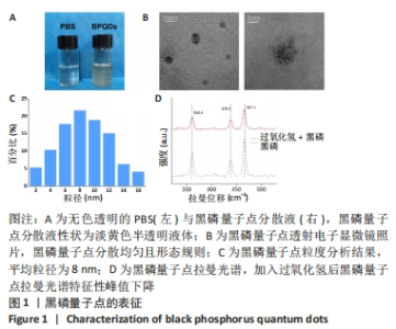
2.1 黑磷量子点表征结果 通过超声液相剥离法成功制备黑磷量子点,如图1A所示,制得的黑磷量子点分散液性状为淡黄色半透明液体,与典型黑磷量子点分散液性状一致[32]。透射电镜下黑磷量子点分散均匀且形态规则,见图1B。粒度分析仪测得黑磷量子点平均粒径为8 nm,见图1C。如图1D所示,黑磷量子点中拉曼散射光谱的代表峰约为平面外声子模(A1g)358.4 cm-1、平面内模(B2g)438.2 cm-1和(A2g)467.1 cm-1,与黑磷的典型拉曼光谱一致[33],加入过氧化氢后黑磷量子点拉曼光谱特征性峰值下降,表明黑磷量子点在与过氧化氢的反应中被消耗[24]。"

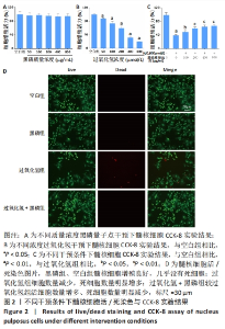
2.2 黑磷量子点体外细胞实验结果 2.2.1 黑磷量子点生物相容性及保护髓核细胞增殖活性能力 CCK-8实验结果显示,黑磷量子点质量浓度在100,200,400,800 μg/mL时,髓核细胞的增殖活性与空白组无明显差异(P > 0.05),见图2A,进一步验证了黑磷量子点良好的生物相容性。过氧化氢浓度在50,100,200,400,800 μmol/L时,髓核细胞的增殖活性受到抑制,且这一作用随过氧化氢浓度的增高更加明显,见图2B。使用黑磷量子点与过氧化氢共同干预髓核细胞,相较空白组,过氧化氢的干预显著降低了髓核细胞的增殖活性,当黑磷量子点质量浓度在100,200,400,800 μg/mL时髓核细胞增殖活性有所恢复,黑磷量子点质量浓度在100,200 μg/mL时细胞增殖活性升高(P < 0.05,P < 0.01);黑磷量子点质量浓度在200,400,800 μg/mL时3组细胞增殖活性无明显差异(P > 0.05),见图2C。活/死染色结果显示,黑磷组、空白组髓核细胞增殖良好,几乎没有死细胞,说明黑磷量子点生物相容性良好,对细胞无毒性作用;过氧化氢组细胞数量减少,死细胞数量明显增多;黑磷+过氧化氢组较过氧化氢干预组活细胞数量增多、死细胞数量明显减少,见图2D,说明黑磷量子点在氧化应激环境下对髓核细胞具有保护作用。"

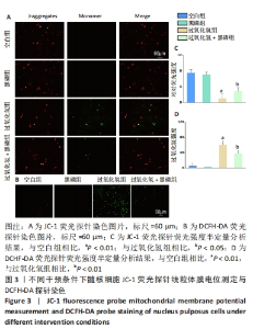
2.2.2 黑磷量子点调控氧化应激导致的髓核细胞线粒体膜电位降低 线粒体膜电位下降标志着细胞凋亡的早期。通过JC-1荧光探针检测髓核细胞线粒体膜电位,结果显示,过氧化氢组线粒体膜电位较空白组降低(P < 0.01),黑磷+过氧化氢组线粒体膜电位较过氧化氢组升高(P < 0.05),而空白组及黑磷组线粒体膜电位无差异(P > 0.05),见图3A、C。 2.2.3 黑磷量子点对细胞内活性氧清除效果 DCHF-DA荧光染料可被细胞内活性氧氧化,形成具有绿色荧光的2’,7’二氯荧光素。根据荧光强度定量分析结果,发现过氧化氢组绿色荧光强度最高,黑磷+过氧化氢组绿色荧光强度显著降低,而空白组与黑磷组绿色荧光强度极低,见图3B、D。这些结果表明,黑磷量子点可以保护细胞免受过氧化氢诱导的细胞内氧化应激。"

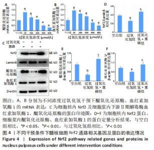
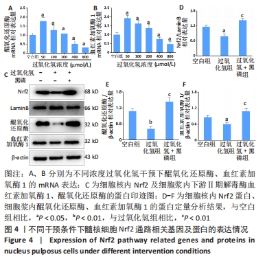
2.2.4 黑磷量子点促进Nrf2核转移及其下游酶谱 RT-qPCR检测结果显示,与空白组比较,50,100,200 μmol/L的过氧化氢增加了髓核细胞内血红素加氧酶1和醌氧化还原酶的mRNA水平(P < 0.05),当过氧化氢浓度达到400 μmol/L以上时,髓核细胞内血红素加氧酶1和醌氧化还原酶的mRNA水平显著下降(P < 0.05),见图4A、B,说明在该浓度过氧化氢下髓核细胞内Nrf2通路活性下降,其下游的Ⅱ期解毒酶基因表达下降。Nrf2的核转位是Nrf2/ARE信号通路激活的标志[34]。实验提取了细胞核蛋白以检测Nrf2的激活,如图4C-F所示,与空白组相比,细胞毒性浓度的过氧化氢干预显著抑制了Nrf2蛋白的核转位,血红素加氧酶1和醌氧化还原酶的蛋白表达也被过氧化氢处理显著抑制;然而,与单独过氧化氢处理相比,黑磷量子点与过氧化氢共同处理后细胞中的Nrf2蛋白核转位显著增加,血红素加氧酶1和醌氧化还原酶的蛋白表达也显著增加。这些结果表明,黑磷量子点有助于Nrf2核转位,提高Ⅱ期解毒酶的表达。"
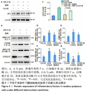
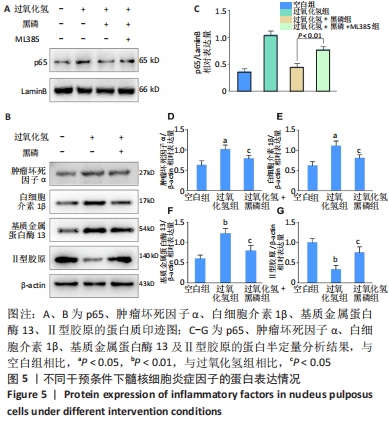
2.2.5 黑磷量子点通过激活Nrf2通路抑制核因子κB炎症通路 核因子κB通路是椎间盘退变中被激活的经典炎症通路之一[21],有文献报道,Nrf2通路对核因子κB通路存在抑制作用[12,19-20]。 为了证实Nrf2对核因子κB的抑制作用,检测了空白组、过氧化氢组、黑磷+过氧化氢组及黑磷+过氧化氢+ML385组中p65的核转位水平,结果表明,过氧化氢组p65核转位水平高于空白组(P < 0.01),但与过氧化氢组相比,黑磷+过氧化氢干预后p65核转位水平降低(P < 0.01);然而,Nrf2特异性抑制剂ML385阻断了黑磷量子点对核因子κB的抑制作用(P < 0.01),见图5A、C。 2.2.6 黑磷量子点影响髓核细胞炎症因子及髓核退变相关分子表达 Western Blot检测结果表明,与空白组比较,过氧化氢组相白细胞介素1β、肿瘤坏死因子α等炎症细胞因子的蛋白表达显著增加(P < 0.05),基质金属蛋白酶13的蛋白表达也显著增加(P < 0.01),而细胞外基质成分Ⅱ型胶原的蛋白表达显著减少(P < 0.01),黑磷量子点的加入逆转了上述趋势,见图5B及图5D-G。"
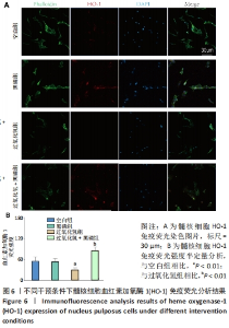
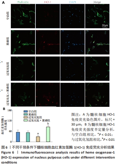
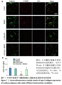
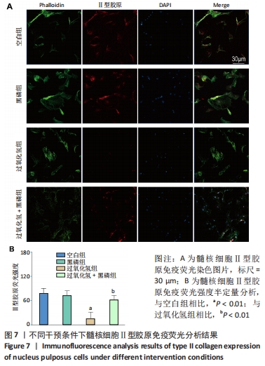
2.2.7 各组髓核细胞免疫荧光染色结果 通过细胞免疫荧光染色进一步验证黑磷量子点在髓核细胞氧化应激的条件下对Nrf2通路下游Ⅱ期解毒酶血红素加氧酶1及细胞外基质成分Ⅱ型胶原表达的影响。结果显示,黑磷组血红素加氧酶1、Ⅱ型胶原表达量与空白组相比无明显差异(P > 0.05),过氧化氢组血红素加氧酶1、Ⅱ型胶原表达量较空白组下降(P < 0.01),而黑磷+过氧化氢组血红素加氧酶1、Ⅱ型胶原表达量高于过氧化氢组(P < 0.01),见图6,7。"
"
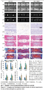
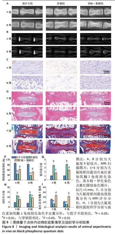
2.3 黑磷量子点动物体内实验结果 2.3.1 实验动物数量分析 30只大鼠全部进入结果分析。 2.3.2 影像学评估结果 如图8A、F所示,穿刺组术后4,8周的椎间盘高度明显低于假手术组(P < 0.01),黑磷+穿刺组后4,8周的椎间盘高度明显高于穿刺组(P < 0.01)。MRI也能可靠地反映椎间盘的再生,T2加权信号较高,表明髓核含水量较高。根据改进的汤姆逊分类法,MRI图像根据T2加权信号强度被分类为Ⅰ-Ⅳ级[35]。术后4,8周,穿刺组T2加权信号显著低于假手术组(P < 0.01),黑磷+穿刺组髓核T2加权信号均高于穿刺组(P < 0.05,P < 0.01),见图8B、G。说明黑磷量子点的加入起到了延缓椎间盘退变的效果。 2.3.3 组织学评价结果 苏木精-伊红和番红固绿染色结果见图8D、E、H所示,不论是在术后4周还是8周,相较于假手术组,穿刺组椎间盘髓核组织胶原含量减少,髓核和纤维环之间的边界模糊;与穿刺组相比,黑磷+穿刺组椎间盘组织的变性和结构损伤均有所改善,髓核和纤维环之间有明确的边界,髓核组织的胶原含量有所提高;术后4周和8周,黑磷+穿刺组的组织学评分显著高于穿刺组(P < 0.05),但低于假手术组(P < 0.05)。 免疫组化染色结果显示,术后4,8周,穿刺组大鼠椎间盘组织内血红素加氧酶1表达量较假手术组降低,黑磷+穿刺组大鼠椎间盘组织内血红素加氧酶1表达量高于穿刺组,见图8C、8I。证明黑磷量子点在体内促进了Nrf2通路的激活,起到了抗氧化应激作用。"
| [1] CIEZA A, CAUSEY K, KAMENOV K, et al. Global estimates of the need for rehabilitation based on the Global Burden of Disease study 2019: a systematic analysis for the Global Burden of Disease Study 2019. Lancet. 2021;396(10267):2006-2017. [2] ADAMS MA, ROUGHLEY PJ. What is intervertebral disc degeneration, and what causes it? Spine (Phila Pa 1976). 2006;31(18):2151-2161. [3] 解志锋,刘清,刘冰,等.腰椎间盘疲劳损伤的生物力学特性[J].中国组织工程研究, 2021,25(3):339-343. [4] LE MAITRE CL, FREEMONT AJ, HOYLAND JA. Accelerated cellular senescence in degenerate intervertebral discs: a possible role in the pathogenesis of intervertebral disc degeneration. Arthritis Res Ther. 2007;9(3):R45. [5] DING F, SHAO ZW, XIONG LM. Cell death in intervertebral disc degeneration. Apoptosis. 2013; 18(7):777-785. [6] DIMOZI A, MAVROGONATOU E, SKLIROU A, et al. Oxidative stress inhibits the proliferation, induces premature senescence and promotes a catabolic phenotype in human nucleus pulposus intervertebral disc cells. Eur Cell Mater. 2015;30:89-102; discussion 103. [7] SONG D, GE J, WANG Y, et al. Tea Polyphenol Attenuates Oxidative Stress-Induced Degeneration of Intervertebral Discs by Regulating the Keap1/Nrf2/ARE Pathway. Oxid Med Cell Longev. 2021; 2021:6684147. [8] LU Y, ZHOU L, HE S, et al. Lycopene alleviates disc degeneration under oxidative stress through the Nrf2 signaling pathway. Mol Cell Probes. 2020;51:101559. [9] VO N, NIEDERNHOFER LJ, NASTO LA, et al. An overview of underlying causes and animal models for the study of age-related degenerative disorders of the spine and synovial joints. J Orthop Res. 2013;31(6):831-837. [10] NAVARRO-YEPES J, BURNS M, ANANDHAN A, et al. Oxidative stress, redox signaling, and autophagy: cell death versus survival. Antioxid Redox Signal. 2014;21(1):66-85. [11] FENG C, YANG M, LAN M, et al. ROS: Crucial Intermediators in the Pathogenesis of Intervertebral Disc Degeneration. Oxid Med Cell Longev. 2017;2017:5601593. [12] MA Q. Role of nrf2 in oxidative stress and toxicity. Annu Rev Pharmacol Toxicol. 2013;53:401-426. [13] ESPINOSA-DIEZ C, MIGUEL V, MENNERICH D, et al. Antioxidant responses and cellular adjustments to oxidative stress. Redox Biol. 2015;6:183-197. [14] MOTOHASHI H, KATSUOKA F, ENGEL JD, et al. Small Maf proteins serve as transcriptional cofactors for keratinocyte differentiation in the Keap1-Nrf2 regulatory pathway. Proc Natl Acad Sci U S A. 2004;101(17):6379-6384. [15] ZHANG X, LIANG S, GAO X, et al. Protective Effect of Chitosan Oligosaccharide against Hydrogen Peroxide-Mediated Oxidative Damage and Cell Apoptosis via Activating Nrf2/ARE Signaling Pathway. Neurotox Res. 2021;39(6):1708-1720. [16] TAO W, SUN W, LIU L, et al. Chitosan Oligosaccharide Attenuates Nonalcoholic Fatty Liver Disease Induced by High Fat Diet through Reducing Lipid Accumulation, Inflammation and Oxidative Stress in C57BL/6 Mice. Mar Drugs. 2019;17(11):645. [17] SALOMONE F, GODOS J, ZELBER-SAGI S. Natural antioxidants for non-alcoholic fatty liver disease: molecular targets and clinical perspectives. Liver Int. 2016;36(1):5-20. [18] ZHANG Y, AHMAD KA, KHAN FU, et al. Chitosan oligosaccharides prevent doxorubicin-induced oxidative stress and cardiac apoptosis through activating p38 and JNK MAPK mediated Nrf2/ARE pathway. Chem Biol Interact. 2019;305:54-65. [19] LI W, KHOR TO, XU C, et al. Activation of Nrf2-antioxidant signaling attenuates NFkappaB-inflammatory response and elicits apoptosis. Biochem Pharmacol. 2008;76(11):1485-1489. [20] MA Q, KINNEER K, YE J, et al. Inhibition of nuclear factor kappaB by phenolic antioxidants: interplay between antioxidant signaling and inflammatory cytokine expression. Mol Pharmacol. 2003;64(2):211-219. [21] ZHANG GZ, LIU MQ, CHEN HW, et al. NF-κB signalling pathways in nucleus pulposus cell function and intervertebral disc degeneration. Cell Prolif. 2021;54(7):e13057. [22] FU HD, LI ZB, XIE HH, et al. Different-sized black phosphorus nanosheets with good cytocompatibility and high photothermal performance. Rsc Adv. 2017;7(24):14618-14624. [23] BRENT JR, GANGULI AK, KUMAR V, et al. On the stability of surfactant-stabilised few-layer black phosphorus in aqueous media. Rsc Adv. 2016;6(90):86955-86958. [24] HOU J, WANG H, GE Z, et al. Treating Acute Kidney Injury with Antioxidative Black Phosphorus Nanosheets. Nano Lett. 2020;20(2):1447-1454. [25] CHENG L, CHEN Z, CAI Z, et al. Bioinspired Functional Black Phosphorus Electrospun Fibers Achieving Recruitment and Biomineralization for Staged Bone Regeneration. Small. 2020;16(50):e2005433. [26] RAUCCI MG, FASOLINO I, CAPORALI M, et al. Exfoliated Black Phosphorus Promotes in Vitro Bone Regeneration and Suppresses Osteosarcoma Progression through Cancer-Related Inflammation Inhibition. ACS Appl Mater Interfaces. 2019;11(9):9333-9342. [27] WANG Z, LIU Z, SU C, et al. Biodegradable Black Phosphorus-based Nanomaterials in Biomedicine: Theranostic Applications. Curr Med Chem. 2019;26(10):1788-1805. [28] BIAN J, CAI F, CHEN H, et al. Modulation of Local Overactive Inflammation via Injectable Hydrogel Microspheres. Nano Lett. 2021;21(6): 2690-2698. [29] XU YC, GU Y, CAI F, et al. Metabolism Balance Regulation via Antagonist-Functionalized Injectable Microsphere for Nucleus Pulposus Regeneration. Adv Funct Mater. 2020;30(52): 14. [30] 叶旭文,顾勇,陈亮.负载姜黄素可注射微球延缓椎间盘的退变[J].中国组织工程研究, 2023,27(12):1884-1891. [31] MASUDA K, AOTA Y, MUEHLEMAN C, et al. A novel rabbit model of mild, reproducible disc degeneration by an anulus needle puncture: correlation between the degree of disc injury and radiological and histological appearances of disc degeneration. Spine (Phila Pa 1976). 2005;30(1):5-14. [32] CHEN H, LIU Z, WEI B, et al. Redox responsive nanoparticle encapsulating black phosphorus quantum dots for cancer theranostics. Bioact Mater. 2021;6(3):655-665. [33] GÓMEZ-PÉREZ J, BARNA B, TÓTH IY, et al. Quantitative Tracking of the Oxidation of Black Phosphorus in the Few-Layer Regime. ACS Omega. 2018;3(10):12482-12488. [34] BELLEZZA I, GIAMBANCO I, MINELLI A, et al. Nrf2-Keap1 signaling in oxidative and reductive stress. Biochim Biophys Acta Mol Cell Res. 2018;1865(5):721-733. [35] HAN B, ZHU K, LI FC, et al. A simple disc degeneration model induced by percutaneous needle puncture in the rat tail. Spine (Phila Pa 1976). 2008;33(18):1925-1934. [36] HUA W, LI S, LUO R, et al. Icariin protects human nucleus pulposus cells from hydrogen peroxide-induced mitochondria-mediated apoptosis by activating nuclear factor erythroid 2-related factor 2. Biochim Biophys Acta Mol Basis Dis. 2020;1866(1):165575. [37] Tang Z, Hu B, Zang F, et al. Nrf2 drives oxidative stress-induced autophagy in nucleus pulposus cells via a Keap1/Nrf2/p62 feedback loop to protect intervertebral disc from degeneration. Cell Death Dis. 2019;10(7):510. [38] HE R, CUI M, LIN H, et al. Melatonin resists oxidative stress-induced apoptosis in nucleus pulposus cells. Life Sci. 2018;199:122-130. [39] KIM KW, CHUNG HN, HA KY, et al. Senescence mechanisms of nucleus pulposus chondrocytes in human intervertebral discs. Spine J. 2009;9(8):658-666. [40] FINKEL T. Signal transduction by reactive oxygen species. J Cell Biol. 2011;194(1):7-15. [41] THURAKKAL S, ZHANG X. Recent Advances in Chemical Functionalization of 2D Black Phosphorous Nanosheets. Adv Sci (Weinh). 2020;7(2):1902359. [42] WANG H, YANG X, SHAO W, et al. Ultrathin Black Phosphorus Nanosheets for Efficient Singlet Oxygen Generation. J Am Chem Soc. 2015;137(35):11376-11382. [43] DING F, SHAO ZW, YANG SH, et al. Role of mitochondrial pathway in compression-induced apoptosis of nucleus pulposus cells. Apoptosis. 2012;17(6):579-590. [44] NASTO LA, ROBINSON AR, NGO K, et al. Mitochondrial-derived reactive oxygen species (ROS) play a causal role in aging-related intervertebral disc degeneration. J Orthop Res. 2013;31(7):1150-1157. [45] BUENDIA I, MICHALSKA P, NAVARRO E, et al. Nrf2-ARE pathway: An emerging target against oxidative stress and neuroinflammation in neurodegenerative diseases. Pharmacol Ther. 2016;157: 84-104. [46] WANG P, ZHANG S, LIU W, et al. Selenium Attenuates TBHP-Induced Apoptosis of Nucleus Pulposus Cells by Suppressing Mitochondrial Fission through Activating Nuclear Factor Erythroid 2-Related Factor 2. Oxid Med Cell Longev. 2022;2022:7531788. [47] LI Y, CHEN L, GAO Y, et al. Oxidative Stress and Intervertebral Disc Degeneration: Pathophysiology, Signaling Pathway, and Therapy. Oxid Med Cell Longev. 2022;2022:1984742. [48] FENG C, ZHANG Y, YANG M, et al. Oxygen-Sensing Nox4 Generates Genotoxic ROS to Induce Premature Senescence of Nucleus Pulposus Cells through MAPK and NF-κB Pathways. Oxid Med Cell Longev. 2017;2017:7426458. [49] 杨林,石军,郭中华,等.基于高分子材料的椎间盘组织工程:研究重点和热点[J].中国组织工程研究,2021,25(16):2589-2596. |
| [1] | Yang Yifeng, Ye Nan, Wang Lin, Guo Shuaicheng, Huang Jian. Signaling pathway of dexmedetomidine against ischemia-reperfusion injury [J]. Chinese Journal of Tissue Engineering Research, 2024, 28(9): 1464-1469. |
| [2] | Wang Weiqing, Zhou Yue. Chronic inflammation regulates adipose tissue fibrosis [J]. Chinese Journal of Tissue Engineering Research, 2024, 28(8): 1307-1312. |
| [3] | Wang Ji, Zhang Min, Li Wenbo, Yang Zhongya, Zhang Long. Effect of aerobic exercise on glycolipid metabolism, skeletal muscle inflammation and autophagy in type 2 diabetic rats [J]. Chinese Journal of Tissue Engineering Research, 2024, 28(8): 1200-1205. |
| [4] | Liu Xin, Hu Man, Zhao Wenjie, Zhang Yu, Meng Bo, Yang Sheng, Peng Qing, Zhang Liang, Wang Jingcheng. Cadmium promotes senescence of annulus fibrosus cells via activation of PI3K/Akt signaling pathway [J]. Chinese Journal of Tissue Engineering Research, 2024, 28(8): 1217-1222. |
| [5] | Mu Bingtao, Yu Jingwen, Liu Chunyun, Guo Minfang, Meng Tao, Yang Pengwei, Wei Wenyue, Song Lijuan, Yu Jiezhong, Ma Cungen. Immunomodulatory effect of astragaloside IV on T cells of experimental autoimmune encephalomyelitis mice [J]. Chinese Journal of Tissue Engineering Research, 2024, 28(7): 1057-1062. |
| [6] | Zhang Kefan, Shi Hui. Research status and application prospect of cytokine therapy for osteoarthritis [J]. Chinese Journal of Tissue Engineering Research, 2024, 28(6): 961-967. |
| [7] | Zhang Ya, Mu Qiuju, Wang Zilin, Liu Hongjie, Zhu Lili. Hydrogel loaded with platelet-rich plasma promotes wound healing in diabetic rats [J]. Chinese Journal of Tissue Engineering Research, 2024, 28(5): 690-696. |
| [8] | Fu Qiangchang, Zheng Liming, Jiang Lifeng. High tibial osteotomy promotes cartilage regeneration in the treatment of knee osteoarthritis [J]. Chinese Journal of Tissue Engineering Research, 2024, 28(32): 5243-5248. |
| [9] | Li Yang, Ma Fei, Leng Yebo, Xu Shicai, He Baoqiang, Zhou Jiajun, Liao Yehui, Tang Qiang, Tang Chao, Wang Qing, Zhong Dejun. Correlation between intervertebral disc degeneration and hyperuricemia [J]. Chinese Journal of Tissue Engineering Research, 2024, 28(32): 5091-5096. |
| [10] | Zhang Yunxin, Zhang Cunxin, Wang Qian, Xu Xinliang, Lyu Chaoliang, Ni Yong. Syringin inhibits intervertebral disc degeneration in rats [J]. Chinese Journal of Tissue Engineering Research, 2024, 28(32): 5104-5109. |
| [11] | Ren Weiliang, Jiao Yongwei, Zhang Jian, Yang Liying, Yang Qi. Modulatory effect of resveratrol on oxidative stress and inflammatory factors in the joint fluid of rats with knee osteoarthritis [J]. Chinese Journal of Tissue Engineering Research, 2024, 28(32): 5154-5158. |
| [12] | Hou Zengtao, Dong Zhiwei, Zhang Jinfeng, Yang Xiaohui, Fan Xiao. Platelet-rich fibrin regulates apoptosis to promote cartilage repair in rats with knee osteoarthritis [J]. Chinese Journal of Tissue Engineering Research, 2024, 28(32): 5167-5171. |
| [13] | Cao Sheng, Kong Lingwei, Xu Kun, Sun Zhijie. Effect of gelatin methacryloyl hydrogel loaded with salvianolic acid B on intervertebral disc degeneration [J]. Chinese Journal of Tissue Engineering Research, 2024, 28(3): 380-386. |
| [14] | Zhang Tiandong, Peng Qingping, Liu Huan, Feng Jianguo, Yi Qian, Huang Wenhua. Semen cuscutae in the treatment of osteoarthritis: network pharmacology analysis and experimental validation [J]. Chinese Journal of Tissue Engineering Research, 2024, 28(28): 4516-4521. |
| [15] | Guan Jinqi, Sun Pingping, Bian Jing, Yan Xue, Zhang Weimin. Gastrodin intervention attenuates inflammatory injury in ischemic stroke rats [J]. Chinese Journal of Tissue Engineering Research, 2024, 28(28): 4535-4540. |
| Viewed | ||||||
|
Full text |
|
|||||
|
Abstract |
|
|||||