[1] LI Y, YANG L, WANG L, et al. Burden of hypertension in China: A nationally representative survey of 174,621 adults.Int J Cardiol. 2017;227:516-523.
[2] OLIVEROS E, PATEL H, KYUNG S, et al. Hypertension in older adults: Assessment, management, and challenges. Clin Cardiol. 2020;43(2):99-107.
[3] MILLS KT, STEFANESCU A, HE J. The global epidemiology of hypertension. Nature Reviews Nephrology. 2020;16(4):223-237.
[4] Touyz RM, Rios FJ, Alves-Lopes R, et al. Oxidative stress: a unifying paradigm in hypertension. Can J Cardiol. 2020;36(5):659-670.
[5] TOUYZ RM, ANAGNOSTOPOULOU A, CAMARGO L, et al. Vascular biology of superoxide-generating NADPH oxidase 5 (Nox5)- implications in hypertension and cardiovascular disease. Antioxid Redox Signal. 2019;30(7):1027-1040.
[6] MA LQ, YU Y, CHEN H, et al. Sweroside Alleviated Aconitine-Induced Cardiac Toxicity in H9c2 Cardiomyoblast Cell Line. Front Pharmacol. 2018;9:1138.
[7] YANG G, JANG JH, KIM SW,et al. Sweroside Prevents Non-Alcoholic Steatohepatitis by Suppressing Activation of the NLRP3 Inflammasome. Int J Mol Sci. 2020;21(8):2790.
[8] WEI S, CHEN G, HE W, et al. Inhibitory effects of secoiridoids from the roots of Gentiana straminea on stimulus-induced superoxide generation, phosphorylation and translocation of cytosolic compounds to plasma membrane in human neutrophils. Phytother Res. 2012;26(2):168-173.
[9] LI J, ZHAO C, ZHU Q, et al. Sweroside Protects Against Myocardial Ischemia-Reperfusion Injury by Inhibiting Oxidative Stress and Pyroptosis Partially via Modulation of the Keap1/Nrf2 Axis. Front Cardiovasc Med. 2021;8:650368.
[10] DHARANEESWARAN H, ABID MR, YUAN L, et al. FOXO1-mediated activation of Akt plays a critical role in vascular homeostasis. Circ Res. 2014;115(2):238-251.
[11] JIAN D, WANG Y, JIAN L, et al. METTL14 aggravates endothelial inflammation and atherosclerosis by increasing FOXO1 N6-methyladeosine modifications. Theranostics. 2020;10(20):8939-8956.
[12] GRAVES DT, MILOVANOVA TN. Mucosal Immunity and the FOXO1 Transcription Factors. Front Immunol. 2019;10:2530.
[13] LIU X, CAO K, LV W, et al. Punicalagin attenuates endothelial dysfunction by activating FoxO1, a pivotal regulating switch of mitochondrial biogenesis. Free Radic Biol Med. 2019;135:251-260.
[14] WANG R, DONG Z, LAN X, et al. Sweroside Alleviated LPS-Induced Inflammation via SIRT1 Mediating NF-κB and FOXO1 Signaling Pathways in RAW264.7 Cells. Molecules. 2019;24(5):872.
[15] NATALIN HM, GARCIA AF, RAMALHO LN, et al. Resveratrol improves vasoprotective effects of captopril on aortic remodeling and fibrosis triggered by renovascular hypertension. Cardiovasc Pathol. 2016;25(2):116-119.
[16] TIAN Z, LIANG M. Renal metabolism and hypertension. Nat Commun. 2021;12(1):963.
[17] DAIBER A, CHLOPICKI S. Revisiting pharmacology of oxidative stress and endothelial dysfunction in cardiovascular disease: Evidence for redox-based therapies. Free Radic Biol Med. 2020;157:15-37.
[18] MIDAOUI AE, FANTUS IG, BOUGHROUS AA, et al. Beneficial Effects of Alpha-Lipoic Acid on Hypertension, Visceral Obesity, UCP-1 Expression and Oxidative Stress in Zucker Diabetic Fatty Rats. Antioxidants. 2019;8(12):648.
[19] GRIENDLING KK, CAMARGO LL, RIOS FJ, et al. Oxidative stress and hypertension. Circ Res. 2021;128(7):993-1020.
[20] NASH KM, SCHIEFER IT, SHAH ZA. Development of a reactive oxygen species-sensitive nitric oxide synthase inhibitor for the treatment of ischemic stroke.Free Radic Biol Med. 2018;115:395-404.
[21] CHEN L, JIANG M. Hypotensive Effect of Magnolin on Spontaneously Hypertensive Rats by Reversing Nitric Oxide Synthase Uncoupling. Revista Brasileira de Farmacognosia. 2022;32(1):65-73.
[22] PICKERING RJ. Oxidative Stress and Inflammation in Cardiovascular Diseases. Antioxidants (Basel). 2021;10(2):171.
[23] MARZIANO C, GENET G, HIRSCHI KK. Vascular endothelial cell specification in health and disease. Angiogenesis. 2021;24(2):213-236.
[24] WILSON C, ZHANG X, BUCKLEY C, et al. Increased Vascular Contractility in Hypertension Results From Impaired Endothelial Calcium Signaling. Hypertension. 2019;74(5):1200-1214.
[25] SILVA I, FIGUEIREDO R, RIOS D. Effect of Different Classes of Antihypertensive Drugs on Endothelial Function and Inflammation. Int J Mol Sci. 2019;20(14):3458.
[26] LIAO L, GONG L, ZHOU M, et al. Leonurine Ameliorates Oxidative Stress and Insufficient Angiogenesis by Regulating the PI3K/Akt-eNOS Signaling Pathway in H2O2-Induced HUVECs.Oxid Med Cell Longev. 2021;2021:9919466.
[27] ZHUANG T, LIU J, CHEN X, et al. Endothelial Foxp1 Suppresses Atherosclerosis via Modulation of Nlrp3 Inflammasome Activation. Circ Res. 2019;125(6):590-605.
[28] HUANG S, SHAO T, LIU H, et al. SIRT6 mediates MRTF-A deacetylation in vascular endothelial cells to antagonize oxLDL-induced ICAM-1 transcription. Cell Death Discov. 2022;8(1):96.
[29] WANG S, SHI M, LI J, et al. Endothelial cell–derived exosomal circHIPK3 promotes the proliferation of vascular smooth muscle cells induced by high glucose via the miR-106a-5p/Foxo1/Vcam1 pathway. Aging (Albany NY). 2021;13(23):25241-25255.
[30] CHEN L, YIN Y, LIU G. Metformin alleviates bevacizumab-induced vascular endothelial injury by up-regulating GDF15 and activating the PI3K/AKT/FOXO/PPARγ signaling pathway. Ann Transl Med. 2021;9(20):1547.
[31] LIU S, LAI L, ZUO Q, et al. PKA turnover by the REGγ-proteasome modulates FoxO1 cellular activity and VEGF-induced angiogenesis. J Mol Cell Cardiol. 2014;72:28-38.
[32] WANG J, HU JQ, SONG YJ, et al. 2′-Fucosyllactose Ameliorates Oxidative Stress Damage in d-Galactose-Induced Aging Mice by Regulating Gut Microbiota and AMPK/SIRT1/FOXO1 Pathway. Foods. 2022;11(2):151.
[33] IANNI A, KUMARI P, TARIGHI S, et al. An Insight into Giant Cell Arteritis Pathogenesis: Evidence for Oxidative Stress and SIRT1 Downregulation. Antioxidants (Basel). 2021;10(6):885.
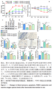
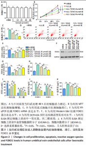
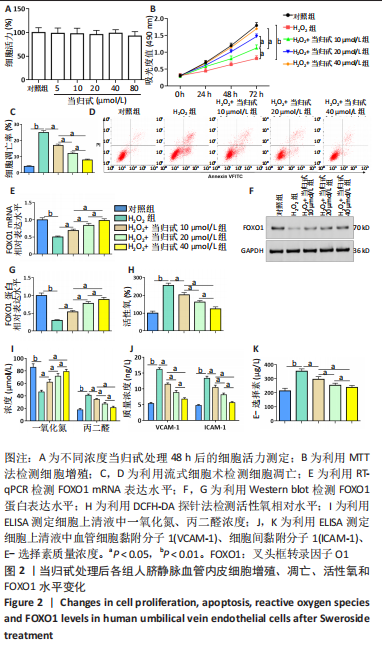
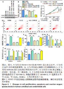
|