Chinese Journal of Tissue Engineering Research ›› 2021, Vol. 25 ›› Issue (22): 3534-3539.doi: 10.3969/j.issn.2095-4344.3189
Previous Articles Next Articles
Structure analysis of platelet-rich fibrin derived from two centrifugation procedures
Bi Qingwei1, Liu Chengpu1, Li Yan1, Zhao Wenwen1, Han Mei2
- 1Heilongjiang Stomatological Disease Center, Harbin 150001, Heilongjiang Province, China; 2Heilongjiang Nursing College, Harbin 150002, Heilongjiang Province, China
-
Received:2020-07-23Revised:2020-07-25Accepted:2020-09-15Online:2021-08-08Published:2021-01-20 -
Contact:Li Yan, Chief Physician, Heilongjiang Stomatological Disease Center, Harbin 150001, Heilongjiang Province, China -
About author:Bi Qingwei, Master, Associate chief physician, Heilongjiang Stomatological Disease Center, Harbin 150001, Heilongjiang Province, China -
Supported by:Scientific research project of Heilongjiang Health Commission, No. 2017552 (to BQW); the Scientific Research Project of Heilongjiang Health Commission, No. 2018033 (to ZWW)
CLC Number:
Cite this article
Bi Qingwei, Liu Chengpu, Li Yan, Zhao Wenwen, Han Mei. Structure analysis of platelet-rich fibrin derived from two centrifugation procedures[J]. Chinese Journal of Tissue Engineering Research, 2021, 25(22): 3534-3539.
share this article
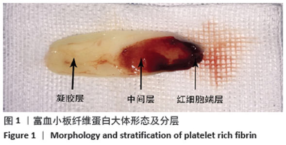
2.1 两种纤维蛋白的大体形态 离心后,离心管上部分凝胶为富血小板纤维蛋白。将A-PRF和L-PRF凝胶分为3部分,包括:黄色部分为纤维蛋白凝胶,占据富血小板纤维蛋白大部分空间(凝胶层);血凝块集中区为红细胞接触部分,即红细胞沉积层(红细胞端层);中间部分为凝胶层与红细胞沉积层两者之间的部分(中间层),见图1;制作完成的2种富血小板纤维蛋白凝胶颜色、形态接近。"

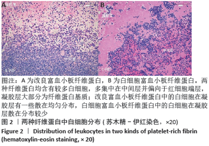
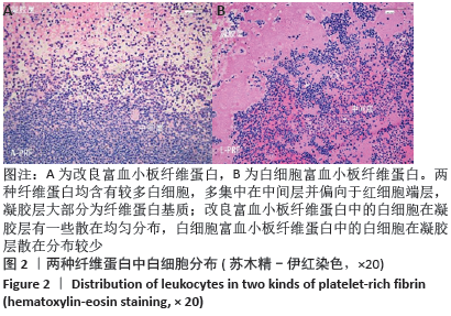
2.2 两种纤维蛋白苏木精-伊红染色结果 光学显微镜下可以观察到,A-PRF和L-PRF均含有较多白细胞,多集中在中间层并偏向于红细胞端层,凝胶层大部分为纤维蛋白基质;A-PRF中的白细胞在凝胶层有一些散在均匀分布,L-PRF中的白细胞在凝胶层散在分布较少(图2)。"

2.3 两种纤维蛋白免疫组化染色结果 免疫组化染色结果显示,两种纤维蛋白在中间层区域含有大量白细胞,包括单核细胞(CD68阳性细胞)和中性粒细胞(CD15阳性细胞),其中A-PRF含有的中性粒细胞分布更广泛、数量更多;L-PRF在中间层含有中性粒细胞数量较多,在凝胶层较少;二者含有的单核细胞数量无明显差别,分布主要集中在中间层,见图3。"

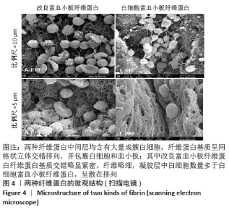
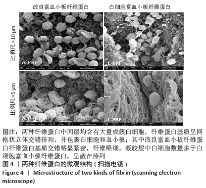
显微镜下随机选择中间层近凝胶层3个视野,按照百分比例分配原则,取3次计数平均数计入结果,A-PRF组、L-PRF组中性粒细胞(CD15阳性细胞)占的比例分别为(55.30±12.15)%和(34.30±11.55)%,两组比较差异有显著性意义(P < 0.05);A-PRF组、L-PRF组单核细胞(CD68阳性细胞)占的比例分别为(29.20±8.15)%和(26.50±8.72)%,两组比较差异无显著性意义 (P > 0.05)。 2.4 两种纤维蛋白扫描电镜观察结果 扫描电镜下观察可见,两种纤维蛋白中间层均含有大量成簇白细胞,白细胞集中区域表面纤维蛋白较少,白细胞数量向凝胶层方向开始减少,纤维蛋白基质呈网格状立体交错排列,并包裹白细胞和血小板;其中A-PRF纤维蛋白基质交错略显紧密,纤维略细,凝胶层中白细胞数量多于L-PRF,呈散在排列,并且血小板活化程度高于L-PRF,见图4。 "
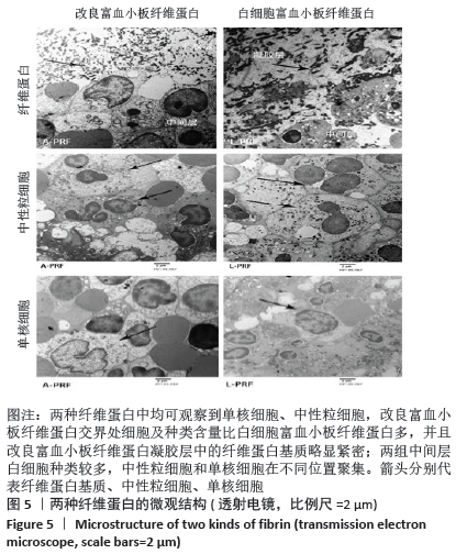
2.5 两种纤维蛋白透射电镜观察结果 透射电镜下可见,两种纤维蛋白中均可观察到单核细胞、中性粒细胞,A-PRF交界处细胞及种类含量比L-PRF多,并且A-PRF凝胶层中的纤维蛋白基质略显紧密;两组中间层白细胞种类较多,中性粒细胞和单核细胞在不同位置聚集,见图5。 2.6 生物相容性 实验制备的两种纤维蛋白来自于人体,具有良好的生物相容性。"
| [1] ANFOSSI G, TROVATI M, MULARONI E, et al. Influence of propranolol on platelet aggregation and thromboxane B2 production from platelet-rich plasma and whole blood. Prostaglandins Leukot Essent Fatty Acids. 1989;36:1-7. [2] FIJNHEER R, PIETERSZ RN, DE KORTE D, et al. Platelet activation during preparation of platelet concentrates:A comparison of the platelet-rich plasma and the buffy coat methods. Transfusion. 1990;30:634-638. [3] MARX RE. Platelet-rich plasma: Evidence to support its use. Oral Maxillofac Surg. 2004;62:489-496. [4] TOFFLER M. Guided bone regeneration (GBR) using cortical bone pins in combination with leukocyte- and platelet-rich fibrin (L-PRF). Compend Contin Educ Dent. 2014;35:192-198. [5] LEKOVIC V, MILINKOVIC I, ALEKSIC Z, et al. Platelet-rich fibrin and bovine porous bone mineral vs. platelet-rich fibrin in the treatment of intrabony periodontal defects. Periodontal Res. 2012;47:409-417. [6] SHIVASHANKAR VY, JOHNS DA, VIDYANATH S, et al. Combination of platelet rich fibrin, hydroxyapatite and PRF membrane in the management of large inflammatory periapical lesion. Conserv Dent. 2013;16:261-264. [7] KOBAYASHI E, FLÜCKIGER L, FUJIOKA-KOBAYASHI M, et al. Comparative release of growth factors from PRP, PRF, and advanced-PRF. Clin Oral Investig. 2016;20(9):2353-2360. [8] DOHAN EHRENFEST DM, ANDIA I, ZUMSTEIN MA, et al. Classification of platelet concentrates (Platelet-Rich Plasma-PRP, Platelet-Rich Fibrin-PRF) for topical and infiltrative use in orthopedic and sports medicine: current consensus, clinical implications and perspectives. Muscles Ligaments Tendons J. 2014;4(1):3-9. [9] SIMONPIERI A, DEL CORSO M, SAMMARTINO G, et al. The relevance of Choukrounan platelet-rich fibrin and metronidazole during complex maxillary rehabilitations using bone allograft. Part I: A new grafting protocol. Implant Dent. 2009;18:102-111. [10] MARTINEZ CE, SMITH PC, PALMA ALVARADO VA. The influence of platelet-derived products on angiogenesis and tissue repair: a concise update. Front Physiol. 2015;6:290. [11] EREN G, KANTARCI A, SCULEAN A, et al. Vascularization after treatment of gingival recession defects with platelet-rich fibrin or connective tissue graft. Clin Oral Investig. 2016;20:2045-2053. [12] CASTRO AB, MESCHI N, TEMMERMAN A, et al. Regenerative potential of leucocyte- and platelet-rich fibrin. Part A: intra-bony defects, furcation defects and periodontal plastic surgery. A systematic review and meta-analysis. Clin Periodontol. 2017;44:67-82. [13] PANDA S, DORAISWAMY J, MALAIAPPAN S, et al. Additive effect of autologous platelet concentrates in treatment of intrabony defects: a systematic review and meta-analysis. J Investig Clin Dent. 2016;7:13-26. [14] SUTTAPREYASRI S, LEEPONG N. Influence of platelet-rich fibrin on alveolar ridge preservation. Craniofac Surg. 2013;24:1088e94. [15] JEONG SM, LEE CU, SON JS, et al. Simultaneous sinus lift and implantation using platelet-rich fibrin as sole grafting material. Craniomaxillofac Surg. 2014;42:990e4. [16] 王灿莉,焦志立,孙勇,等.两种富血小板纤维蛋白对人牙龈成纤维细胞增殖活性影响的比较[J].中国组织工程研究,2019,23(7): 1007-1012. [17] GHANAATI S,BOOMS P,ORLOWSKA A,et al. Advanced platelet-rich fibrin:a new concept for cell-based tissueengineering by means of inflammatorycells. J Oral Implant. 2014;40(6):679-689. [18] DOHAN DM, CHOUKROUN J, DISS A, et al. Platelet-rich fibrin (PRF): a second-generation platelet concentrate. Part III: leucocyte activation: a new feature for platelet concentrates? Oral Surg Oral Med Oral Pathol Oral Radiol Endod. 2006;101(3):e51-e55. [19] CHOUKROUN J, ADDA F, SCHOEFFLER C, et al. Une opportuniteeit paro-implantologie: le PRF. Implantodontie. 2000;42: 55-62. [20] 裴延平,林松杉.富血小板纤维蛋白的临床应用[J].临床口腔医学杂志,2012,28(7):441-443. [21] MASUKI H, OKUDERA T, WATANEBE T, et al. Growth factor and pro-inflammatory cytokine contents in platelet-rich plasma (PRP), plasma rich in growth factors (PRGF), advanced platelet-rich fibrin (A-PRF), and concentrated growth factors(CGF). Int J Implant Dent. 2016;2:19. [22] 李艳秋,周延民,孙晓琳,等.富血小板纤维蛋白体外释放TGF-G和PDGF-AB影响因素的探讨[J].现代口腔医学杂志,2012,26(6): 404-407. [23] DOHAN EHRENFEST DM, DISS A, ODIN G, et al. In vitroeffects of Choukrounro PRF(platelet-rich fibrin) on human gingival fibroblasts, dermal prekeratinocytes,preadipocytes, and maxillofacial osteoblasts in primary cultures. Oral Surg Oral Med Oral Pathol Oral Radiol Endod. 2009;108(3):341-352. [24] DAVID M, JOSEPH C, ANTOINE D, et al. Platelet-rich fibrin (PRF): A second-generation platelet concentrate. Part I: Technological concepts and evolution. Oral Surg Oral Med Oral Pathol Oral Radiol Endod. 2006;101(3):e37-44. [25] DOHAN EHRENFEST DM, DOGLIOLI GM, DEL CM, et al. Choukroun’s platelet-rich fibrin (PRF) stimulates in vitro proliferation and differentiation of human oral bone mesenchymal stem cell in a dose-dependent way. Arch Oral Biol.2010;55(3):185. [26] LIAO HT, CHEN CT, CHEN CH, et al. Combination of guided osteogenesis with autologous platelet-rich fibrin glue and mesenchymal stem cell for mandibular reconstruction. J Trauma. 2011;70(1):228. [27] KNAFL D, THALHAMMER F, VOSSEN MG. In-vitro release pharmacokinetics of amikacin, teicoplanin and polyhexanide in a platelet rich fibrin-layer (PRF)-a laboratory evaluation of a modern, autologous wound treatment. PLoS One. 2017;12(7):e0181090. [28] ASAKA T, OHGA N, YAMAZAKI Y, et al. Platelet-rich fibrin may reduce the risk of delayed recovery in tooth-extracted patients undergoing oral bisphosphonate therapy: a trial study. Clin Oral Investig. 2016;11:1-8. [29] PARK JH, KIM JW, KIM SJ. Does the Addition of Bone Morphogenetic Protein 2 to Platelet-Rich Fibrin Improve Healing After Treatment for Medication-Related Osteonecrosis of the Jaw? J Oral Maxillofac Surg. 2017;75(6):1176-1184. [30] PATHAK H, MOHANTY S, URS AB. Treatment of Oral Mucosal Lesions by Scalpel Excision and Platelet-Rich Fibrin Membrane Grafting: A Review of 26 Sites. J Oral Maxillofac Surg. 2015;73(9):1865-1874. [31] ÖNCÜ E. The Use of Platelet-Rich Fibrin Versus Subepithelial Connective Tissue Graft in Treatment of Multiple Gingival Recessions: A Randomized Clinical Trial. Int J Periodontics Restorative Dent. 2017; 37(2):265-271. [32] DEL CORSO M, VERVELLE A, SIMONPIERI A, et al. Current knowledge and perspectives for the use of platelet-rich plasma(PRP) and platelet-rich fibrin (PRF) in oral and maxillofacial surgery part 1: Periodontal and dentoalveolar surgery. Curr Pharm Biotechnol. 2012;13(7):1207-1230. [33] SIMONPIERI A, DEL CORSO M, VERVELLE A, et al. Current knowledge and perspectives for the use of platelet-rich plasma(PRP) and platelet-rich fibrin (PRF) in oral and maxillofacial surgery part 2: Bone graft, implant and reconstructive surgery. Curr Pharm Biotechnol. 2012;13(7):1231-1256. [34] ZUMSTEIN MA, BERGER S, SCHOBER M, et al. Leukocyte- and platelet-rich fibrin (L-PRF) for long-term delivery of growth factorin rotator cuff repair: review, preliminary results and future directions. Curr Pharm Biotechnol. 2012;13(7):1196-1206. [35] ZUMSTEIN MA, BIELECKI T, DOHAN EHRENFEST DM. The Future of Platelet Concentrates in Sports Medicine: Platelet-Rich Plasma,Platelet-Rich Fibrin, and the Impact of Scaffolds and Cells on the Long-term Delivery of Growth Factors. Oper Tech Sports Med. 2011;19(3): 190-197. [36] MAJID E, MOHAMAD RM, AMIR HN, et al. Platelet-Rich Fibrin: An Autologous Fibrin Matrix in Surgical Procedures: A Case Report and Review of Literature. Iran J Otorhinolaryngol. 2012;24(69): 197-202. [37] DOHAN DM, CHOUKROUN J, DISS A, et al. Platelet-rich fibrin (PRF): a second-generation platelet concentrate. Part II: platelet-related biologic features. Oral Surg Oral Med Oral Pathol Oral Radiol Endod. 2006;101(3):e45-50. [38] CHOUKROUN J, DISS A, SIMONPIERI A, et al. Platelet-rich fibrin (PRF):a second-generationplatelet concentrate. Part V:histologic evaluations of PRFeffects on bone allograft maturation in sinus lift. Oral Surg Oral Med Oral Pathol Oral Radiol Endod. 2006;101(3):299-303. [39] DOHAN EHRENFEST DM, PINTO NR, Pereda A, et al. The impact of the centrifuge characteristics and centrifugation protocols on the cells, growth factors, and fibrin architecture of a leukocyte- and platelet-rich fibrin (L-PRF) clot and membrane. Platelets. 2017;24:1-14. [40] FUJIOKA-KOBAYASHI M, MIRON RJ, HERNANDEZ M, et al. Optimized Platelet-Rich Fibrin With the Low-Speed Concept: Growth Factor Release, Biocompatibility, and Cellular Response. J Periodontol. 2017; 88(1):112-121. [41] MARCHETTI E, MANCINI L, BERNARDI S, et al. Evaluation of Different Autologous Platelet Concentrate Biomaterials: Morphological and Biological Comparisons and Considerations. Materials. 2020:13(10): 2282. [42] DOHAN EHRENFEST DM, DEL CORSO M, DISS A, et al. Three-dimensional architecture and cell composition ofa Choukroun’s platelet-rich fibrin clot and membrane. J Periodontol. 2010;81(4): 546-555. [43] DOHAN EHRENFEST DM, DE PEPPO GM, DOGLIOLI P, et al. Slow release of growth factors and thrombospondin-1 inChoukroun’s platelet-rich fibrin (PRF): a gold standard to achieve for all surgical platelet concentrates technologies. Growth Factors. 2009;27(1):63-69. |
| [1] | Zhang Tongtong, Wang Zhonghua, Wen Jie, Song Yuxin, Liu Lin. Application of three-dimensional printing model in surgical resection and reconstruction of cervical tumor [J]. Chinese Journal of Tissue Engineering Research, 2021, 25(9): 1335-1339. |
| [2] | Gu Xia, Zhao Min, Wang Pingyi, Li Yimei, Li Wenhua. Relationship between hypoxia inducible factor 1 alpha and hypoxia signaling pathway [J]. Chinese Journal of Tissue Engineering Research, 2021, 25(8): 1284-1289. |
| [3] | Geng Qiudong, Ge Haiya, Wang Heming, Li Nan. Role and mechanism of Guilu Erxianjiao in treatment of osteoarthritis based on network pharmacology [J]. Chinese Journal of Tissue Engineering Research, 2021, 25(8): 1229-1236. |
| [4] | Wang Zhengdong, Huang Na, Chen Jingxian, Zheng Zuobing, Hu Xinyu, Li Mei, Su Xiao, Su Xuesen, Yan Nan. Inhibitory effects of sodium butyrate on microglial activation and expression of inflammatory factors induced by fluorosis [J]. Chinese Journal of Tissue Engineering Research, 2021, 25(7): 1075-1080. |
| [5] | Xie Wenjia, Xia Tianjiao, Zhou Qingyun, Liu Yujia, Gu Xiaoping. Role of microglia-mediated neuronal injury in neurodegenerative diseases [J]. Chinese Journal of Tissue Engineering Research, 2021, 25(7): 1109-1115. |
| [6] | Zeng Yanhua, Hao Yanlei. In vitro culture and purification of Schwann cells: a systematic review [J]. Chinese Journal of Tissue Engineering Research, 2021, 25(7): 1135-1141. |
| [7] | Shi Yangyang, Qin Yingfei, Wu Fuling, He Xiao, Zhang Xuejing. Pretreatment of placental mesenchymal stem cells to prevent bronchiolitis in mice [J]. Chinese Journal of Tissue Engineering Research, 2021, 25(7): 991-995. |
| [8] | He Xiangzhong, Chen Haiyun, Liu Jun, Lü Yang, Pan Jianke, Yang Wenbin, He Jingwen, Huang Junhan. Platelet-rich plasma combined with microfracture versus microfracture in the treatment of knee cartilage lesions: a meta-analysis [J]. Chinese Journal of Tissue Engineering Research, 2021, 25(6): 964-969. |
| [9] | Yang Yang, Yao Yu, Shen Xiaotian, Liu Jiajia, Xue Jianhua. Expression and significance of interleukin-21 in intervertebral disc degeneration [J]. Chinese Journal of Tissue Engineering Research, 2021, 25(5): 690-694. |
| [10] | Ma Binxiang, He Wanqing, Zhou Guangchao, Guan Yonglin. Triptolide improves motor dysfunction in rats following spinal cord injury [J]. Chinese Journal of Tissue Engineering Research, 2021, 25(5): 701-706. |
| [11] | Song Shan, Hu Fangyuan, Qiao Jun, Wang Jia, Zhang Shengxiao, Li Xiaofeng. An insight into biomarkers of osteoarthritis synovium based on bioinformatics [J]. Chinese Journal of Tissue Engineering Research, 2021, 25(5): 785-790. |
| [12] | Xu Dongzi, Zhang Ting, Ouyang Zhaolian. The global competitive situation of cardiac tissue engineering based on patent analysis [J]. Chinese Journal of Tissue Engineering Research, 2021, 25(5): 807-812. |
| [13] | Zhao Xiang, Wei Cuilan, Zhang Yeting. Neurogenesis and neuroinflammation under exercise: alteration and regulation [J]. Chinese Journal of Tissue Engineering Research, 2021, 25(5): 813-820. |
| [14] | Li Wenjing, Li Haobo, Liu Congna, Cheng Dongmei, Chen Huizhen, Zhang Zhiyong. Comparison of different bioactive scaffolds in the treatment of regenerative pulp of young permanent teeth [J]. Chinese Journal of Tissue Engineering Research, 2021, 25(4): 499-503. |
| [15] | Chen Junyi, Wang Ning, Peng Chengfei, Zhu Lunjing, Duan Jiangtao, Wang Ye, Bei Chaoyong. Decalcified bone matrix and lentivirus-mediated silencing of P75 neurotrophin receptor transfected bone marrow mesenchymal stem cells to construct tissue-engineered bone [J]. Chinese Journal of Tissue Engineering Research, 2021, 25(4): 510-515. |
| Viewed | ||||||
|
Full text |
|
|||||
|
Abstract |
|
|||||