[1] GENANT HK, ENGELKE K, BOLOGNESE M, et al. Effects of Romosozumab Compared with Teriparatide on Bone Density and Mass at the Spine and Hip in Postmenopausal Women with Low Bone Mass.J Bone Miner Res.2017;32(1):181-187.
[2] LINDSAY R, KREGE J, MARIN F, et al. Teriparatide for osteoporosis: importance of the full course. Osteoporosis Int.2016; 27(8):2395-2410.
[3] KE H, RICHARDS W, LI X, et al. Sclerostin and Dickkopf-1 as therapeutic targets in bone disease.Endoer Rev.2012;33:747-783.
[4] HAY E, BOUAZIZ W, FUNCKBRENTANO T, et al. Sclerostin and Bone Aging: A Mini-Review.Gerontology.2016;62(6):618-623.
[5] MINISOLA S, CIPRIANI C, OCCHIUTO M, et al. New anabolic therapies for osteoporosis.Intern Emerg Med.2017;12:915-921.
[6] WALDORFF EI, ZHANG N, RYABY JT. Pulsed electromagnetic field applications: A corporate perspective.J Orthop Translat. 2017;9:60-68.
[7] ANTONINO C, SAVERIO L, FEDERICA B, et al. Pulsed electromagnetic fields modulate bone metabolism via RANKL/OPG and Wnt/β-catenin pathways in women with postmenopausal osteoporosis: A pilot study.Bone. 2018; 116: 42-46.
[8] 邱裕友,唐翠松,胡剑,等.催产素干预兔骨质疏松模型骨微结构变化规律的实验研究[J].同济大学学报(医学版), 2015,36(5):23-28.
[9] LI X, OMINSKY M, WARMINGTON K, et al. Sclerostin antibody treatment increases bone formation, bone mass, and bone strength in a rat model of postmenopausal osteoporosis.J Bone Miner Res.2009; 24:578-588.
[10] OMINSKY M, VLASSEROS F, JOLETTE J, et al. Two doses of sclerostin antibody in cynomolgus monkeys increases bone formation, bone mineral density, and bone strength.J Bone Miner Res.2010;25:948-959.
[11] XU XJ, SHEN L, YANG YP, et al. Serum sclerostin levels associated with lumbar spine bone mineral density and bone turnover markers in patients with postmenopansal osteoporosis.Chin Med J,2013;126(13): 2480-2484.
[12] SPATZ JM, WEIN MN, GOOI JH, et al. The Wnt inhibitor sclerostin is up-regulated by mechanical unloading in osteocytes in vitro.J Biol Chem.2015;290(27):16744-16758.
[13] MCCOLM J, HU L, WOMACK T, et al. Single-and multiple-dose randomized studies of blosozumab,a monoclonal antibody again stsclerostin in healthy postmenopausal women.J Bone Miner Res. 2014;29:935-943.
[14] MARKOV M. Pulsed electromagnetic field therapy history, state of the art and future. Environmentalist.2007; 27:465-475.
[15] 覃裕,邱冰,朱思刚,等.唑来膦酸盐联合脉冲电磁场治疗绝经后骨质疏松症的临床疗效分析[J].中国骨质疏松杂志, 2015;21(8):945-948.
[16] JUN Z, YUAN L, HAITAO X, et al. Effects of combined treatment with Ibandronate and Pulsed Electromagnetic Field on ovariectomy-induced osteoporosisin rats.Bioelectromagnetics.2017; 38:31-40.
[17] IKEDA KS, TAKESHITA S. The role of osteoclast differentiation and function in skeletal homeostasis. J Biochem.2016;159(1):1-8.
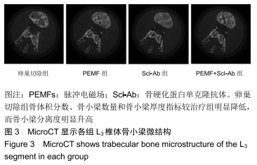
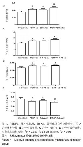
|