[1] PONDÉ N, AFTIMOS P, PICCART M. Antibody-Drug Conjugates in Breast Cancer: a Comprehensive Review.Curr Treat Options Oncol.2019;20(5):37.
[2] 赵洁,于綦悦,侯峰,等.为制作数字化内镜黏膜下剥离术标本评估系统对切片制作流程的改进[J].中华病理学杂志,2019,48(2): 147-149.
[3] 郑波,王鹏姣,薛丽燕,等.复合型环保试剂配合超声快速组织处理仪对肿瘤活检标本HE制片影响因素的分析[J].诊断病理学杂志, 2018,25(9):640-643.
[4] 刘柱新,钟学军,梁英杰.环保脱蜡剂在快速HE染色技术中的应用[J].诊断病理学杂志,2017,24(9):717-718.
[5] 程凯,李颖,何燕,等.新型超声组织处理仪及环保试剂在甲状腺穿刺液基细胞块中的应用[J].诊断病理学杂志,2018,25(5): 384-385.
[6] 朱卫东.新型病理环保系列试剂在病理常规技术中的应用[J].临床与实验病理学杂,2016,32(6):713-714.
[7] TAN PH, ELLIS IO. Myoepithelial and epithelial-myoepithelial, mesenchymal and fibroepithelialbreast lesions: updates from the WHO Classification of Tumours of the Breast2012.J Clin Pathol.2013;66(6):465-470.
[8] 《乳腺癌HER2检测指南(2014版)》编写组.乳腺癌HER2检测指南(2014版)[J].中华病理学杂志,2014,43(4):262-267.
[9] 《乳腺癌HER2检测指南(2019版)》编写组.乳腺癌HER2检测指南(2019版)[J].中华病理学杂志,2019,48(3):169-175.
[10] 陈志强,王莹,米贤军,等.Van-clear替代二甲苯对乳腺癌HER2蛋白表达的影响[J].诊断病理学杂志,2017,24(12):918-921.
[11] 陈志强,王莹,叶才果,等.环保透明脱蜡液Van-Clear与传统试剂在组织处理后HE染色及FISH法检测乳腺癌HER2基因中的比较[J].华中科技大学学报(医学版),2017,46(4):453-458.
[12] 张虹,王双,王颖,等.运用2013版美国临床肿瘤学会/美国病理医师学院乳腺癌HER2检测指南对1501例浸润性乳腺癌的HER2状态进行再评估[J].中华病理学杂志,2015,44(1):42-47.
[13] 许燕,柏乾明,杨飞,等.应用201 3版ASCO/CAP HER2检测指南判读1780例免疫组织化学HER2不确定浸润性乳腺癌的荧光原位杂交结果[J].中华病理学杂志,2016,45(8):545-549.
[14] PRESS MF, VILLALOBOS I, SANTIAGO A, et al.Assessing the New American Society of Clinical Oncology/College of American Pathologists Guidelines for HER2 Testing by Fluorescence In Situ Hybridization: Experience of an Academic Consultation Practice.Arch Pathol Lab Med. 2016. [Epub ahead of print]
[15] SHAH MV, WIKTOR AE, MEYER RG, et al.Change in Pattern of HER2 Fluorescent in Situ Hybridization (FISH) Results in Breast Cancers Submitted for FISH Testing: Experience of a Reference Laboratory Using US Food and Drug Administration Criteria and American Society of Clinical Oncology and College of American Pathologists Guidelines.J Clin Oncol. 2016;34(29): 3502-3510.
[16] 刘月平,步宏,杨文涛,等.2019版中国乳腺癌HER2检测指南更新解读[J].中华病理学杂志,2019,48(3):182-185.
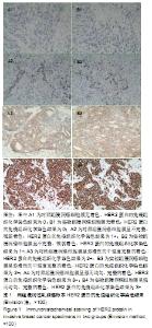
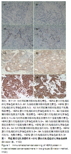
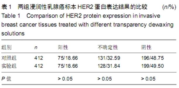
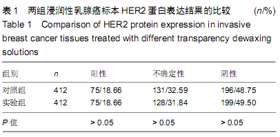
|