Chinese Journal of Tissue Engineering Research ›› 2019, Vol. 23 ›› Issue (22): 3549-3555.doi: 10.3969/j.issn.2095-4344.1281
Previous Articles Next Articles
Protective effect of glucose-dependent incretin analogue DAIa2GIP on knee cartilage injury of rats
- Department of Orthopedics, Second Hospital of Shanxi Medical University, Shanxi Provincial Key Laboratory of Bone and Soft Tissue Injury Repair, Taiyuan 030001, Shanxi Province, China
-
Received:2019-03-14 -
Contact:Wang Yuze, MD, Associate chief physician, Department of Orthopedics, Second Hospital of Shanxi Medical University, Shanxi Provincial Key Laboratory of Bone and Soft Tissue Injury Repair, Taiyuan 030001, Shanxi Province, China -
About author:Yang Xuan, Master candidate, Attending physician, Department of Orthopedics, Second Hospital of Shanxi Medical University, Shanxi Provincial Key Laboratory of Bone and Soft Tissue Injury Repair, Taiyuan 030001, Shanxi Province, China -
Supported by:the National Natural Science Foundation of China, No. 31200728 (to WYZ); the Doctoral Startup Foundation of the Second Hospital of Shanxi Medical University, No. 20120408 (to WYZ)
CLC Number:
Cite this article
Yang Xuan, Han Pengfei, Zhou Xin, Lu Jiangong, Wang Shichuan, Wang Yuze. Protective effect of glucose-dependent incretin analogue DAIa2GIP on knee cartilage injury of rats[J]. Chinese Journal of Tissue Engineering Research, 2019, 23(22): 3549-3555.
share this article
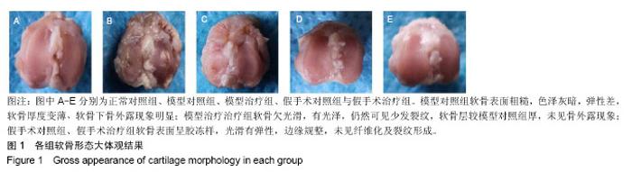
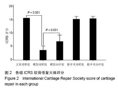
2.1 各组软骨形态大体观结果 见图1。 正常对照组软骨面光滑,有弹性,边缘整齐;模型对照组软骨表面粗糙,色泽灰暗,弹性差,软骨厚度变薄、软骨下骨外露现象明显,ICRS评分为3.80±0.79,明显低于正常对照组(P < 0.001);模型治疗组软骨欠光滑,有光泽,仍然可见少发裂纹,软骨层较模型对照组厚,未见骨外露现象,ICRS评分为7.10±1.20,明显高于模型对照组(P < 0.001);假手术对照组、假手术治疗组软骨表面呈胶冻样,光滑有弹性,边缘规整,未见纤维化及裂纹形成,ICRS评分分别为15.60±0.52,15.80±0.42,此两组评分与正常对照组比较无明显差异(P > 0.05),见图2。"

"

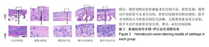
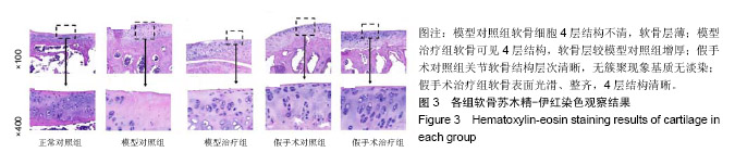
2.2 各组软骨苏木精-伊红染色观察结果 见图3。 正常对照组软骨表面光滑,4层结构清晰,软骨细胞排列整齐,基质无淡染;模型对照组典型的软骨细胞4层结构不清,细胞大小不一,软骨层薄;模型治疗组软骨可见4层结构,软骨层较模型对照组增厚,细胞大小基本一致;假手术对照组关节软骨结构层次清晰,无簇聚现象,软骨细胞大小均匀,基质无淡染;假手术治疗组软骨表面光滑、整齐,4层结构清晰,中层细胞呈柱状排列。"

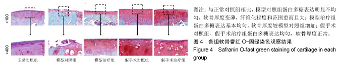
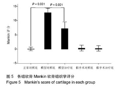
2.3 各组软骨番红O-固绿染色观察结果 见图4。 正常对照组蛋白多糖表达均匀,软骨厚度适中,Mankin评分为0.40±0.52;与正常对照组相比,模型对照组蛋白多糖表达明显不均匀,软骨厚度变薄,纤维化程度和范围重而且大,Mankin评分为12.90±0.74,评分低于正常对照组(P < 0.001);模型治疗组蛋白多糖表达基本均匀,软骨厚度较模型对照组增加,Mankin评分为7.30±1.16,评分低于模型对照组(P < 0.001);假手术对照组、假手术治疗组蛋白多糖表达均匀,软骨厚度正常,Mankin评分分别为0.60±0.70,0.80±0.789,此两组评分与正常对照组比较无明显差异(P > 0.05),见图5。"
"
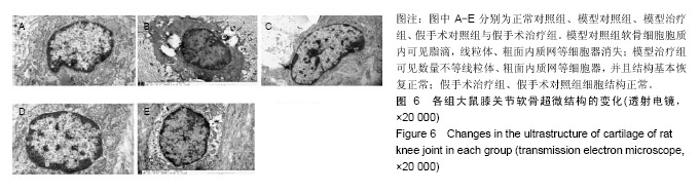
2.4 各组软骨细胞透射电镜观察结果 见图6。 正常对照组细胞核呈卵圆形,染色质分布均匀,细胞内可见大量粗面内质网、线粒体等细胞器,胶原纤维排列呈束状;模型对照组细胞胞浆丢失严重,胞质内见数个脂滴、电子密度增加,胞质内多数细胞器溶解、消失,偶见线粒体、内质网、高尔基体,胶原纤维排列紊乱;模型治疗组染色质分布尚均匀,线粒体较少且线粒体嵴消失,粗面内质网肿胀出现空泡化,可见数量不等的线粒体、内质网、游离核糖体、高尔基体,部分胶原纤维成束装排列;假手术模型组、假手术治疗组细胞核呈椭圆形,染色质分布均匀,细胞内可见大量粗面内质网、线粒体、游离核糖体、高尔基体等细胞器。"

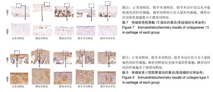
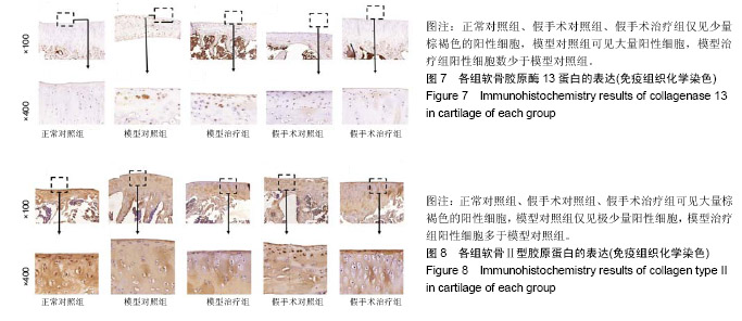
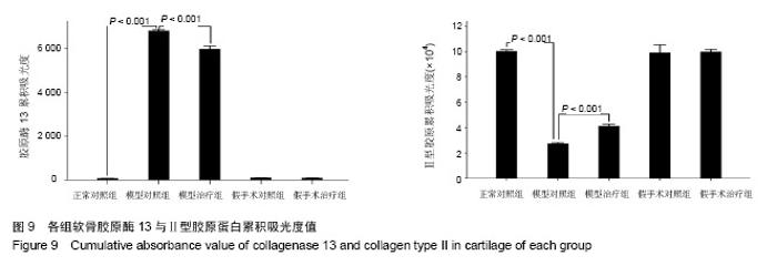
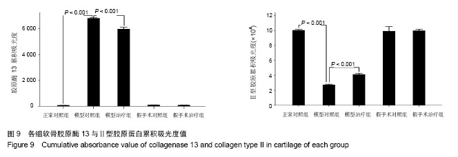
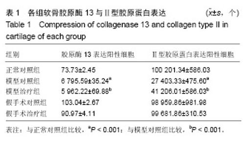
2.5 各组胶原酶13、Ⅱ型胶原蛋白的表达 见图7,8。 正常对照组、假手术对照组、假手术治疗组胶原酶13蛋白呈低表达,Ⅱ型胶原蛋白呈高表达,2组间两指标比较差异无显著性意义(P > 0.05);与正常对照组比较,模型对照组胶原酶13蛋白呈高表达(P < 0.001),Ⅱ型胶原蛋白呈低表达(P < 0.001);与模型对照组比较,模型治疗组胶原酶13蛋白表达显著降低(P < 0.001),Ⅱ型胶原蛋白表达明显增高(P < 0.001),见表1。各组胶原酶13、Ⅱ型胶原蛋白累积吸光度值,见图9。"
"
"

| [1] Yuan GH,Masukohongo K,Kato T,et al.Immunologic intervention in the pathogenesis of osteoarthritis.Arthritis Rheumatol. 2010; 48(3):602-611. [2] 韩廷成,周临东,董松林.中医外治法治疗膝骨关节炎的现状与分析[J].现代中西医结合杂志, 2012,21(5):566-568. [3] Gaudin-Audrain C,Irwin N,Mansur S,et al.Glucose-dependent insulinotropic polypeptide receptor deficiency leads to modifications of trabecular bone volume and quality in mice. Bone. 2013;53(1):221-230. [4] Shimazu-Kuwahara S,Kanemaru Y,Harada N,et al. Glucose- dependent insulinotropic polypeptide deficiency reduced fat accumulation and insulin resistance, but deteriorated bone loss in ovariectomized mice.J Diabetes Investig.2018. doi: 10.1111/jdi.12978. [Epub ahead of print] [5] Kieffer TJ,McIntosh CH,Pederson RA.Degradation of glucose-dependent insulinotropic polypeptide and truncated glucagon-like peptide 1 in vitro and in vivo by dipeptidyl peptidase iv. Endocrinology. 1995;136(8):3585-3596. [6] Usdin TB,Mezey E,Button DC,et al.Gastric inhibitory polypeptide receptor, a member of the secretin-vasoactive intestinal peptide receptor family, is widely distributed in peripheral organs and the brain.Endocrinology.1993;133(6):2861-2870. [7] Joo E,Harada N,Yamane S,et al.Inhibition of gastric inhibitory polypeptide receptor signaling in adipose tissue reduces insulin resistance and hepatic steatosis in high-fat diet-fed mice. Diabetes. 2017;66(4):868-879. [8] Nasteska D,Harada N,Suzuki K,et al.Chronic reduction of gip secretion alleviates obesity and insulin resistance under high-fat diet conditions.Diabetes.2014;63(7):2332-2343. [9] Torekov SS,Harsløf T,Rejnmark L,et al. A functional amino acid substitution in the glucose-dependent insulinotropic polypeptide receptor (GIPR) gene is associated with lower bone mineral density and increased fracture risk.J Clin Endocrinol Metab. 2014;99(4):E729-733. [10] Holst JJ,Windelov JA,Boer GA,et al.Searching for the physiological role of glucose-dependent insulinotropic polypeptide.J Diabetes Investig.2016;7 Suppl 1:8-12. [11] Mieczkowska A,Irwin N,Flatt PR,et al.Glucose-dependent insulinotropic polypeptide (gip) receptor deletion leads to reduced bone strength and quality.Bone.2013;56(2):337-342. [12] Mai X,Lu Z,Lei Z,et al.Phosphorylation of osteopontin in osteoarthritis degenerative cartilage and its effect on matrix metalloprotease 13.Rheumatol Int.2013;33(5):1313-1319. [13] Wayne JS,Mcdowell CL,Shields KJ,et al.In vivo response of polylactic acid-alginate scaffolds and bone marrow-derived cells for cartilage tissue engineering.Tissue Eng Part A. 2005; 11(5-6):953-963. [14] Mankin HJ,Dorfman H,Lippiello L,et al.Biochemical and metabolic abnormalities in articular cartilage from osteo-arthritic human hips. Ii. Correlation of morphology with biochemical and metabolic data.B J Bone Joint Surg Am.1971; 53(3):523-537. [15] 姜旭,吴成爱,王莹,等.膝关节骨性关节炎中wisp-1调控机制的研究[J].中国骨质疏松杂志, 2015,21(5):537-540. [16] Xu M,Zhang L,Zhao L,et al. Phosphorylation of osteopontin in osteoarthritis degenerative cartilage and its effect on matrix metalloprotease 13.Rheumatol Int.2013;33(5):1313-1319. [17] Li Y,Lin T,Duan Y,et al.Upregulation of mmp-13 and timp-1 expression in response to mechanical strain in mc3t3-e1 osteoblastic cells.Bmc Res Notes.2010;3(1):309. [18] Mieczkowska A,Bouvard B,Chappard D,et al.Glucose- dependent insulinotropic polypeptide (gip) directly affects collagen fibril diameter and collagen cross-linking in osteoblast cultures. Bone. 2015;74:29-36. [19] Mabilleau G,Mieczkowska A,Irwin N,et al.Beneficial effects of a n-terminally modified gip agonist on tissue-level bone material properties.Bone.2014;63:61-68. [20] 余洁,许岭翎.肠促胰素与骨代谢[J].中华糖尿病杂志, 2017,9(3): 200-202. [21] 吴松,石俊俊,卫定禄,等.Dala2gip对淀粉样前体蛋白/早老素1转基因小鼠膝关节软骨损伤保护效应的研究[J].中华实验外科杂志, 2017,34(11):1861-1864. [22] 邓莉,王海波.膝骨性关节炎动物模型的研究进展[J].广东医学, 2018,56(12):1895-1897 [23] Duffy AM,Hölscher C.The incretin analogue d-ala 2 gip reduces plaque load, astrogliosis and oxidative stress in an app/ps1 mouse model of alzheimer’s disease.Neuroscience.2013; 228: 294-300. [24] 李保驰,王维山,董金波,等. MMP3、MMP13在骨性关节炎患者滑膜中的表达及意义[J].中国骨质疏松杂志, 2014,20(6):593-596. [25] 包刚,刘永,刘亚丽,等.MMP-13在骨性关节炎滑膜组织中的表达分析[J].中国实验诊断学, 2016,20(9):1548-1549. [26] Hughes A,Oxford AE,Tawara K,et al.Endoplasmic reticulum stress and unfolded protein response in cartilage pathophysiology; contributing factors to apoptosis and osteoarthriti. Int J Mol Sci. 2017;18(3).pii: E665.doi:10.3390/ijms18030665. [27] Li NG,Shi ZH,Tang YP,et al. New hope for the treatment of osteoarthritis through selective inhibition of MMP-13.Curr Med Chem.2011;18(7):977-1001. [28] Phull AR,Eo SH,Abbas Q,et al. Applications of chondrocyte- based cartilage engineering: An overview. BioMed Res Int. 2016;2016(2):1879837. [29] Bollag R,Zhong Q, Ding K, et al. Glucose-dependent insulinotropic peptide is an integrative hormone with osteotropic effects. Mol Cell Endocrinol.2001;177(1-2):35-41. [30] Zhong Q,Ding KH,Mulloy AL,et al.Glucose-dependent insulinotropic peptide stimulates proliferation and tgf-beta release from mg-63 cells.Peptides.2003;24(4):611-616. [31] Howard S,Anastassiades T.Differential effects of bone associated factors on newly synthesized anionic glycoconjugates by articular chondrocyte cultures from adult and immature bovines.J Rheumatol. 1993;20(12):2083-2094. |
| [1] | Huang Dengcheng, Wang Zhike, Cao Xuewei. Comparison of the short-term efficacy of extracorporeal shock wave therapy for middle-aged and elderly knee osteoarthritis: a meta-analysis [J]. Chinese Journal of Tissue Engineering Research, 2021, 25(9): 1471-1476. |
| [2] | Peng Zhihao, Feng Zongquan, Zou Yonggen, Niu Guoqing, Wu Feng. Relationship of lower limb force line and the progression of lateral compartment arthritis after unicompartmental knee arthroplasty with mobile bearing [J]. Chinese Journal of Tissue Engineering Research, 2021, 25(9): 1368-1374. |
| [3] | Liu Xiangxiang, Huang Yunmei, Chen Wenlie, Lin Ruhui, Lu Xiaodong, Li Zuanfang, Xu Yaye, Huang Meiya, Li Xihai. Ultrastructural changes of the white zone cells of the meniscus in a rat model of early osteoarthritis [J]. Chinese Journal of Tissue Engineering Research, 2021, 25(8): 1237-1242. |
| [4] | Liu Xin, Yan Feihua, Hong Kunhao. Delaying cartilage degeneration by regulating the expression of aquaporins in rats with knee osteoarthritis [J]. Chinese Journal of Tissue Engineering Research, 2021, 25(5): 668-673. |
| [5] | Ma Zetao, Zeng Hui, Wang Deli, Weng Jian, Feng Song. MicroRNA-138-5p regulates chondrocyte proliferation and autophagy [J]. Chinese Journal of Tissue Engineering Research, 2021, 25(5): 674-678. |
| [6] | Xie Chongxin, Zhang Lei. Comparison of knee degeneration after anterior cruciate ligament reconstruction with or without remnant preservation [J]. Chinese Journal of Tissue Engineering Research, 2021, 25(5): 735-740. |
| [7] | Cao Xuhan, Bai Zixing, Sun Chengyi, Yang Yanjun, Sun Weidong. Mechanism of “Ruxiang-Moyao” herbal pair in the treatment of knee osteoarthritis based on network pharmacology [J]. Chinese Journal of Tissue Engineering Research, 2021, 25(5): 746-753. |
| [8] | Li Yonghua, Feng Qiang, Tan Renting, Huang Shifu, Qiu Jinlong, Yin Heng. Molecular mechanism of Eucommia ulmoides active ingredients treating synovitis of knee osteoarthritis: an analysis based on network pharmacology [J]. Chinese Journal of Tissue Engineering Research, 2021, 25(5): 765-771. |
| [9] | Song Shan, Hu Fangyuan, Qiao Jun, Wang Jia, Zhang Shengxiao, Li Xiaofeng. An insight into biomarkers of osteoarthritis synovium based on bioinformatics [J]. Chinese Journal of Tissue Engineering Research, 2021, 25(5): 785-790. |
| [10] | Deng Zhenhan, Huang Yong, Xiao Lulu, Chen Yulin, Zhu Weimin, Lu Wei, Wang Daping. Role and application of bone morphogenetic proteins in articular cartilage regeneration [J]. Chinese Journal of Tissue Engineering Research, 2021, 25(5): 798-806. |
| [11] | Lü Jiaxing, Bai Leipeng, Yang Zhaoxin, Miao Yuesong, Jin Yu, Li Zhehong, Sun Guangpu, Xu Ying, Zhang Qingzhu. Evaluation of internal fixation with proximal femoral nail antirotation in elderly knee osteoarthritis patients with femoral intertrochanteric fractures [J]. Chinese Journal of Tissue Engineering Research, 2021, 25(3): 391-396. |
| [12] | Zheng Li, Li Dadi, Hu Weifan, Tang Jinlong, Zhao Fengchao. Risk assessment of contralateral knee arthroplasty after unilateral total knee arthroplasty [J]. Chinese Journal of Tissue Engineering Research, 2021, 25(3): 374-379. |
| [13] | Liu Jinfu, Zeng Ping, Nong Jiao, Fan Siqi, Feng Chengqin, Huang Jiaxing. Integrative analysis of biomarkers and therapeutic targets in synovium of patients with osteoarthritis by multiple microarrays [J]. Chinese Journal of Tissue Engineering Research, 2021, 25(23): 3690-3696. |
| [14] | Chen Feng, Zhang Xiaoyun, Chen Yueping, Liao Jianzhao, Li Jiajun, Song Shilei, Lai Yu. Molecular mechanism of anhydroicaritin in the treatment of osteoarthritis: an analysis based on network pharmacology and bioinformatics [J]. Chinese Journal of Tissue Engineering Research, 2021, 25(23): 3704-3710. |
| [15] | Yang Wei, Chen Zehua, Yi Zhiyong, Huang Xudong, Han Qingmin, Zhang Ronghua. Effectiveness of intra-articular injection of hyaluronic acid versus placebo in the treatment of early and mid-stage knee osteoarthritis: a Meta-analysis based on randomized, double-blind, controlled, clinical trials [J]. Chinese Journal of Tissue Engineering Research, 2021, 25(23): 3760-3766. |
| Viewed | ||||||
|
Full text |
|
|||||
|
Abstract |
|
|||||