Chinese Journal of Tissue Engineering Research ›› 2019, Vol. 23 ›› Issue (21): 3432-3438.doi: 10.3969/j.issn.2095-4344.1746
Previous Articles Next Articles
Biological characteristics of menstrual blood-derived mesenchymal stem cells and their clinical application in tissue repair and reconstruction
Chang Qiyuan1, Tan Jichun1, 2
- 1Second Department of Reproduction, Reproductive Center, Shengjing Hospital of China Medical University, Shenyang 110022, Liaoning Province, China; 2Key Laboratory for Reproductive Disorder and Fertility Remodeling of Liaoning Province, Shenyang 110022, Liaoning Province, China
-
Revised:2019-02-18Online:2019-07-28Published:2019-07-28 -
Contact:Tan Jichun, MD, Doctoral supervisor, Second Department of Reproduction, Reproductive Center, Shengjing Hospital of China Medical University, Shenyang 110022, Liaoning Province, China; Key Laboratory for Reproductive Disorder and Fertility Remodeling of Liaoning Province, Shenyang 110022, Liaoning Province, China -
About author:Chang Qiyuan, Master candidate, Second Department of Reproduction, Reproductive Center, Shengjing Hospital of China Medical University, Shenyang 110022, Liaoning Province, China -
Supported by:the National Key Research &Developmental Program of China, No. 2018YFC1002100 (to TJC); Liaoning Provincial Key R&D Guidance Plan, No. 2018020222 (to TJC); Major Special Construction Plan for Discipline Construction Project of China Medical University, No. 3110118033 (to TJC)
CLC Number:
Cite this article
Chang Qiyuan, Tan Jichun. Biological characteristics of menstrual blood-derived mesenchymal stem cells and their clinical application in tissue repair and reconstruction[J]. Chinese Journal of Tissue Engineering Research, 2019, 23(21): 3432-3438.
share this article
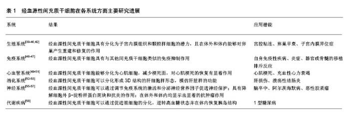
2.1 间充质干细胞和子宫内膜干细胞 以干细胞疗法为首的再生医学近年来一直是治疗多种慢性疾病的研究热点。然而应用胚胎干细胞会产生伦理问题,而诱导多能干细胞需要病毒转染有风险,两者均有移植后导致畸胎瘤等肿瘤的风险。唯有间充质干细胞在应用方面比较安全,其低免疫原性使其在异体移植干细胞时可以产生免疫抑制的作用,最适合用于自体或异体细胞移植替代疗法[1],且能够在体外分化为成骨细胞、脂肪细胞和成软骨细胞[11]。根据国际细胞治疗学会委员会的定义[11],间充质干细胞应在标准培养条件下能够贴附于塑料壁,表达表面分子CD105,CD73和CD90。迄今为止,研究者们已从多种人体组织中发现并分离出间充质干细胞,包括骨髓、牙髓、脐带、脂肪、胎盘等[12-16]。 2004年Chan等[2]从子宫组织中分离了子宫内膜干细胞,其不仅具有成纤维样细胞的形态、较强的克隆增殖能力以及多向分化潜能[3-7],而且能表达CD73,CD90,CD105,CD29,CD44,MSI1和NOTCH1[3,6,17]。通过刮宫获得的子宫内膜组织,可以利用CD146和血小板衍生生长因子受体b双阳性表达的亚群筛选得到子宫内膜间充质干细胞[18]。 2.2 经血源性间充质干细胞 在2007年Meng等[10]从经血中分离出具有间充质干细胞表型和多向分化潜能的子宫内膜再生细胞。之后2008年Patel等[19]分离出的同种细胞能够表达胚胎干细胞的表面标记,具有分化潜能。随着干细胞分离和鉴定技术的提高,2013年Gang等[20]改善了分离子宫内膜间充质干细胞的方法,提高了分离效率。从经血中分离得到子宫内膜间充质干细胞都称为经血源性间充质干细胞。 2.2.1 经血源性间充质干细胞的生物学特性 经血源性间充质干细胞可以通过密度梯度离心法从经血中分离出来[9],在体外利用间充质干细胞贴壁的特性进行培养。经血源性间充质干细胞的增殖能力很强,在体外的倍增时间仅为数小时,且在传代次数达到20-30代的情况下仍然能够保持细胞核型不变[20-21]。有研究发现,在年龄为30-60的女性供者中,年龄越小,其贡献的经血源性间充质干细胞的增殖克隆能力越强[22]。 除了增殖能力外,经血源性间充质干细胞还拥有非常强的多向分化潜能。已有大量研究证明经血源性间充质干细胞能够在不同细胞培养条件下分化为外胚层、中胚层和内胚层三个胚胎层的多种细胞系,并表达胚胎干细胞标记物Oct-4[10,19]。经血源性间充质干细胞经结缔组织生长因子诱导后可分化为成纤维样细胞[23],也能分化成为胰腺样细胞、许旺细胞、子宫内膜样上皮细胞、脂肪细胞、神经细胞、成骨细胞等[8,24-28]。Li等[29]通过慢病毒转导OCT4,SOX2和KLF4对经血源性间充质干细胞进行重编程,得到的经血源性间充质干细胞来源的诱导多能干细胞在形态学、多能性标记和基因表达等方面均显示出与人胚胎干细胞相同的特征。 经血源性间充质干细胞也具有间充质干细胞的免疫调节功能。已有研究表明,来源于脐带、脂肪组织和骨髓的间充质干细胞都具有不同程度的抑制外周血中B,T淋巴细胞和自然杀伤细胞的能力[30]。而间充质干细胞对其他免疫活性细胞也有免疫抑制作用。间充质干细胞能通过影响树突状细胞的募集,成熟和功能,产生免疫抑制。进一步研究发现,通过释放细胞因子如干扰素γ和肿瘤坏死因子α。间充质干细胞还能够抑制适应性和先天性免疫系统,不管是体外或者体内都能抑制免疫反应,尤其在暴露于促炎症因子时,其免疫抑制功能更强[31]。对于发现时间较短的经血源性间充质干细胞来说,其在免疫调节方面的作用还不是完全清楚。目前有一些研究表明,经血源性间充质干细胞具有低免疫原性和免疫抑制的作用[32-33]。从应用的角度来看,经血源性间充质干细胞在免疫调节方面的作用将是临床实验的关注点,值得对其进行更深层次的研究。 2.2.2 经血源性间充质干细胞的旁分泌作用 绝大多数细胞可以通过释放细胞外囊泡,细胞因子等物质产生旁分泌作用。已有研究表明,骨髓来源的间充质干细胞可以分泌多种细胞因子,通过组织间隙作用于周围细胞,促进受损组织血管内皮细胞的修复,发挥重要的旁分泌作用[34]。同时,除了对于血管的影响,骨髓来源的间充质干细胞的旁分泌作用还广泛参与免疫调节、细胞增殖、凋亡、血管再生等病理生理作用[34]。有研究发现,体外培养的早期传代经血源性间充质干细胞,能产生高水平的NT3,NT4,脑源性神经生长因子和神经生长因子等,发挥对神经保护的旁分泌作用[35]。Wang等[36]将体外培养的经血源性间充质干细胞的条件培养基通过尾静脉注射到顺铂建立的卵巢早衰小鼠模型后,观察发现条件培养基组能够部分地通过分泌成纤维细胞生长因子2对受损的卵巢发挥保护作用。Chen等[37]利用Transwell共培养,观察发现经血源性间充质干细胞通过分泌单核细胞趋化蛋白1、白细胞介素6、白细胞介素8、肝细胞生长因子、生长相关癌基因和骨保护素来抑制LX-2细胞(永生化肝星状细胞系)的增殖,进而抑制肝脏纤维化。随着对旁分泌作用的进一步理解,关于经血源性间充质干细胞通过外泌体产生的旁分泌作用也产生了许多研究。Chen等[38]进一步用细胞因子阵列分析经血源性间充质干细胞来源的外泌体,发现其表达细胞间黏附分子1、促血管生成素2、Axl受体酪氨酸激酶、胰岛素样生长因子结合蛋白6、白细胞介素6、白细胞介素8和骨保护素等细胞因子。通过在尾静脉注射和细胞培养基内添加,外泌体能够分别迁移至损伤部位和AML12细胞(小鼠肝细胞系),显着改善模型小鼠的肝功能,提高存活率,减少了受损肝脏中肝脏单核细胞的数量和活性凋亡蛋白caspase-3的数量[38]。 经血源性间充质干细胞来源丰富,一名生育期的健康女性每年可以贡献约12次月经血,为用于治疗和实验的细胞储备提供了充足的细胞来源。与骨髓间充质干细胞相比,其获取过程为非侵入性的无创过程,对于供者来说更容易接受并且没有了伦理道德的限制。经血源性间充质干细胞在获得方法、分离培养、免疫调节以及组织修复方面的优点使经血源性间充质干细胞能够替代其他间充质干细胞作为细胞疗法中一类绝佳的低免疫反应细胞来源。而经血源性间充质干细胞分泌的营养因子和功能性物质所产生的旁分泌作用值得进一步研究其作用机制和靶向调控,以更好地将经血源性间充质干细胞应用在再生医疗和生物医药方面。 2.3 经血源性间充质干细胞的应用 2.3.1 经血源性间充质干细胞在生殖系统方面的应用 经血源性间充质干细胞凭借多向分化潜能和相似的组织特异性的优势,可以作为治疗妇科疾病,特别是某些难以修复的生殖系统组织损伤的干细胞疗法的绝佳细胞来源。已经有实验证实,经血源性间充质干细胞在雌二醇诱导下能够体外分化为子宫内膜上皮和基质细胞[39]。在非肥胖糖尿病/重症联合免疫缺陷雌性小鼠皮下植入经血源性间充质干细胞后可以重建子宫内膜组织[39]。2组研究的子宫内膜损伤小鼠模型在植入经血源性间充质干细胞后,子宫内膜的厚度和微血管密度增加,提示子宫内膜的恢复。宫腔粘连是一类妇产科常见的、影响生育功能的疾病,可见宫腔内结缔组织增生,纤维化瘢痕形成,主要发生于子宫内膜基底层损伤后,术后极易复发。目前,已有临床研究用经血源性间充质干细胞治疗中、重度宫腔粘连并获得初步疗效。Tan等[40]利用自体经血源性间充质干细胞移植到评级为重度的宫腔粘连患者子宫腔内,经2个周期的治疗后子宫内膜达到胚胎植入的最低标准,后通过辅助生殖技术成功令患者妊娠。研究者进一步应用富血小板血浆培养经血源性间充质干细胞,发现其能够显著提高治疗宫腔粘连的疗效[41]。 将经血源性间充质干细胞注射到卵巢早衰小鼠模型后,经血源性间充质干细胞能够存活并表达卵巢颗粒细胞的特异性蛋白质,提示经血源性间充质干细胞可以分化成卵巢颗粒细胞[42]。Lai等[43]在应用经血源性间充质干细胞治疗后,卵巢质量、雌二醇水平和正常卵泡数量均明显增加。用绿色荧光蛋白标记经血源性间充质干细胞后能在卵巢基质中检测到绿色荧光。同时,雌二醇水平的增加也可以促进卵泡的发育。 目前,有研究人员推测经血源性间充质干细胞在子宫内膜异位症的形成中也可能起十分重要的作用。血管生成和细胞迁移是形成子宫内膜异位病变的重要过程。Yerlikaya等[44]研究发现,子宫内膜异位灶中血管内皮生长因子受体和血小板衍生生长因子B亚单位的表达高于在位子宫内膜,而在位内膜的血管因子表达又高于健康受试者。Proestling等[45]发现子宫内膜异位病灶中OCT4,SOX15和TWIST1的表达与在位子宫内膜和无子宫内膜异位症患者相比有所增加,提示这些干性标志物的上调有助于异位子宫内膜病灶的建立。根据经血源性间充质干细胞的生物学特性,可以提出假设,经血源性间充质干细胞迁移到异位病灶并分泌促进血管生成的因子,从而形成子宫内膜组织。从这个角度出发,经血源性间充质干细胞可能是治疗子宫内膜异位症的关键,经血源性间充质干细胞在子宫内膜异位症的具体机制中能起到何种作用仍有待进一步研究。 在生殖系统方面,上述几个实验可以证明经血源性间充质干细胞具有分化为子宫内膜组织和颗粒样细胞的潜力,且在体外和体内能够对卵巢产生重建和修复的作用。同时,经血源性间充质干细胞的定向趋化特性使其在生殖系统疾病的靶向治疗方面拥有广阔的发展前景。 2.3.2 经血源性间充质干细胞在免疫系统方面的应用 Nikoo等[46]研究发现,当共培养外周血单核细胞和经血源性间充质干细胞时,经血源性间充质干细胞在免疫调节方面发挥双重作用。当经血源性间充质干细胞和外周血单核细胞之间的比例在较高的比例(1∶1至1∶2)下,经血源性间充质干细胞会抑制外周血单核细胞的增殖能力;而在较低的比例(1∶32至1∶64)时,经血源性间充质干细胞能够促进外周血单核细胞增殖。这种剂量依赖性效应还需要进一步的研究来确定其分子机制。Mahmood等[47]在2014年的研究中发现,与经血源性间充质干细胞共培养后,单核细胞向未成熟树突状细胞的最佳表型分化受到抑制。在培养基中能检测到更高水平的白细胞介素6和白细胞介素10。但是将经血源性间充质干细胞添加到未成熟树突状细胞培养物中却不能阻止未成熟树突状细胞成熟,只有从分化过程开始将经血源性间充质干细胞与单核细胞共培养才可以有效地阻碍完全成熟树突状细胞的产生。经血源性间充质干细胞同时还具有低免疫原性,是用于临床试验的基础。将经血源性间充质干细胞注入人体后,只会有较低程度的免疫反应,避免了免疫损伤。 在免疫系统方面,经血源性间充质干细胞具有与其他间充质干细胞类似的免疫抑制作用。经血源性间充质干细胞的免疫学特性鼓励研究人员使用经血源性间充质干细胞作为干细胞疗法治疗卵巢早衰、糖尿病、帕金森病等疾病[36,48]。未来,经血源性间充质干细胞或还可用于抑制自身免疫或有害的过度免疫反应,治疗自身免疫性疾病、炎症、器官或骨髓的移植排斥反应。 2.3.3 经血源性间充质干细胞在心血管系统方面的应用 Hida等[49]将经血源性间充质干细胞移植到心肌梗死模型裸鼠的梗死区域,发现经血源性间充质干细胞能够分化为心肌细胞,且心肌梗死面积明显减少,表明经血源性间充质干细胞对心肌梗死的恢复有显著作用。Zhi等[50]将经血源性间充质干细胞注入大鼠心肌缺血模型的梗死区后,观察到移植组的心功能明显改善,心血管密度增高,且经血源性间充质干细胞的移植能够促进心肌细胞再生。同时,经血源性间充质干细胞和心肌细胞共培养实验提示经血源性间充质干细胞主要通过旁分泌细胞因子进而促进现有心肌细胞的增殖。在临床试验方面,Ichim等[51]通过静脉注射经血源性间充质干细胞到充血性心力衰竭患者体内,观察发现患者的射血分数从30%增加到40%。 经血源性间充质干细胞应用于心血管系统方面的临床试验文章较少,可能有2个原因。首先,目前对于经血源性间充质干细胞机制的研究还不是很彻底;其次,关于经血源性间充质干细胞的临床应用有太多的不确定性,比如经血源性间充质干细胞的致瘤性。由此看来,经血源性间充质干细胞的旁分泌产物比经血源性间充质干细胞更加安全有效,且无栓塞的风险,更适合应用于临床。 2.3.4 经血源性间充质干细胞在消化系统方面的应用 经血源性间充质干细胞在消化系统方面的研究较少。将经血源性间充质干细胞在体外与肝脏组织匀浆共培养,或是在添加向肝诱导分化的培养基中培养时,可以分化形成3D结构的肝细胞样形态,模仿肝脏样的功能[52]。当经血源性间充质干细胞与受损的肝组织匀浆共培养时,研究人员检测发现白蛋白和甲胎蛋白的表达增高,表明经血源性间充质干细胞有修复肝损伤的临床潜力[52]。在用经血源性间充质干细胞对溃疡性结肠炎进行治疗时发现,经血源性间充质干细胞治疗组对溃疡性结肠可以产生明显的疗效但未能完全达到健康对照者的状态[53]。 2.3.5 经血源性间充质干细胞在神经系统方面的应用 脑卒中是一种危险的神经系统疾病,具有极高的致死率、致残率,对人类生命和生活质量构成严重威胁。目前临床上最好的脑卒中治疗剂是组织纤溶酶原激活剂。但由于治疗药物给药窗口期短,患者通常不能及时地接受治疗。Borlongan等[54]将氧葡萄糖剥夺暴露的原代大鼠神经元与经血源性间充质干细胞共培养或暴露于经血源性间充质干细胞的培养基,结果显示细胞死亡显著减少。之后,他们将经血源性间充质干细胞移植到脑卒中大鼠中,证明了经血源性间充质干细胞的神经保护和神经发生作用。在进一步研究中,Rodrigues等[55]发现经血源性间充质干细胞可以通过调节免疫系统的激活和分泌神经营养因子促进神经保护。阿尔茨海默病是较为常见的痴呆类型,Zhao等[56]将经血源性间充质干细胞移植入APP/PS1小鼠脑内后显著改善小鼠的空间学习和记忆。研究结果表明经血源性间充质干细胞具有降解细胞外β-淀粉样蛋白斑块和抗炎的作用。恶性胶质瘤是一种高度侵袭性的原发性颅内肿瘤,Wang等[57]研究发现,经血源性间充质干细胞显示出人类恶性胶质瘤的趋向性。用过表达TRAIL分泌型三聚体的腺病毒感染经血源性间充质干细胞在体外和体内均显示出显着的抗肿瘤作用。 在神经系统方面,已在脑卒中、阿尔茨海默病等疾病中证实了经血源性间充质干细胞可以通过旁分泌作用发挥对神经的保护功能,同时其作为细胞递送载体同样具有广泛的治疗潜能。 2.3.6 经血源性间充质干细胞在代谢疾病方面的应用 1型糖尿病是一种慢性代谢性疾病,是由于胰岛β细胞被自身免疫选择性破坏所致。2014年,Wu等[58]用经血源性间充质干细胞治疗链脲佐菌素诱导的1型糖尿病小鼠。通过体质量和血糖的指标,研究结果证实了经血源性间充质干细胞对糖尿病的治疗作用。进一步的研究结果表明,经血源性间充质干细胞可以逆转高血糖状态并在体内恢复胰岛结构。需要注意的是,这个研究的治疗作用机制并不是通过经血源性间充质干细胞自身分化替代受损的B细胞,而是通过促进祖细胞的分化,与细胞的旁分泌有关。 在代谢疾病方面,由于糖尿病的发病机制十分复杂,再未研究彻底之前尚不能将干细胞治疗纳入糖尿病的临床应用。经血源性间充质干细胞在转化用于临床应用之前还有很长的一段路要走。 经血源性间充质干细胞在各系统方面主要研究进展见表1。"

| [1] Mutlu L, Hufnagel D, Taylor HS. The endometrium as a source of mesenchymal stem cells for regenerative medicine. Biol Reprod. 2015;92(6):138.[2] Chan RW, Schwab KE, Gargett CE. Clonogenicity of human endometrial epithelial and stromal cells. Biol Reprod. 2004; 70(6):1738-1750. [3] Schwab KE, Hutchinson P, Gargett CE. Identification of surface markers for prospective isolation of human endometrial stromal colony-forming cells. Hum Reprod. 2008; 23(4):934-943.[4] Dimitrov R, Timeva T, Kyurkchiev D, et al. Characterization of clonogenic stromal cells isolated from human endometrium. Reproduction. 2008;135(4):551-558. [5] Gargett CE, Schwab KE, Zillwood RM, et al. Isolation and culture of epithelial progenitors and mesenchymal stem cells from human endometrium. Biol Reprod. 2009;80(6): 1136-1145. [6] Schüring AN, Schulte N, Kelsch R, et al. Characterization of endometrial mesenchymal stem-like cells obtained by endometrial biopsy during routine diagnostics. Fertil Steril. 2011;95(1):423-426. [7] Spitzer TL, Rojas A, Zelenko Z, et al. Perivascular human endometrial mesenchymal stem cells express pathways relevant to self-renewal, lineage specification, and functional phenotype. Biol Reprod. 2012;86(2):58. [8] Gargett CE, Schwab KE, Deane JA. Endometrial stem/ progenitor cells: the first 10 years. Hum Reprod Update. 2016; 22(2):137-163. [9] 谭季春,李雅璇,王秋实,等.利用月经血建立的经血源性基质干细胞系[J].中国组织工程研究,2015,19(50):8155-8160.[10] Meng X, Ichim TE, Zhong J, et al. Endometrial regenerative cells: a novel stem cell population. J Transl Med. 2007;5:57.[11] Dominici M, Le Blanc K, Mueller I,et al. Minimal criteria for defining multipotent mesenchymal stromal cells. The International Society for Cellular Therapy position statement. Cytotherapy. 2006;8(4):315-317.[12] Crane JL, Cao X. Bone marrow mesenchymal stem cells and TGF-β signaling in bone remodeling. J Clin Invest. 2014; 124(2):466-472.[13] Gronthos S, Mankani M, Brahim J, et al. Postnatal human dental pulp stem cells (DPSCs) in vitro and in vivo. Proc Natl Acad Sci U S A. 2000;97(25):13625-13630.[14] Li T, Xia M, Gao Y, et al. Human umbilical cord mesenchymal stem cells: an overview of their potential in cell-based therapy. Expert Opin Biol Ther. 2015;15(9):1293-1306. [15] Gimble JM, Katz AJ, Bunnell BA. Adipose-derived stem cells for regenerative medicine. Circ Res. 2007;100(9):1249-1260.[16] Makhoul G, Chiu RC, Cecere R. Placental mesenchymal stem cells: a unique source for cellular cardiomyoplasty. Ann Thorac Surg. 2013;95(5):1827-1833. [17] Schwab KE, Gargett CE. Co-expression of two perivascular cell markers isolates mesenchymal stem-like cells from human endometrium. Hum Reprod. 2007;22(11):2903-2911.[18] Xu Y, Zhu H, Zhao D, et al. Endometrial stem cells: clinical application and pathological roles. Int J Clin Exp Med. 2015; 8(12):22039-22044. [19] Patel AN, Park E, Kuzman M, et al. Multipotent menstrual blood stromal stem cells: isolation, characterization, and differentiation. Cell Transplant. 2008;17(3):303-311.[20] Gang EJ, Bosnakovski D, Figueiredo CA, et al. SSEA-4 identifies mesenchymal stem cells from bone marrow. Blood. 2007;109(4):1743-1751. [21] Rossignoli F, Caselli A, Grisendi G, et al. Isolation, characterization, and transduction of endometrial decidual tissue multipotent mesenchymal stromal/stem cells from menstrual blood. Biomed Res Int. 2013;2013:901821. [22] Mehrabani D, Nazarabadi RB, Kasraeian M, et al. Growth Kinetics, Characterization, and Plasticity of Human Menstrual Blood Stem Cells. Iran J Med Sci. 2016;41(2):132-139.[23] Su K, Edwards SL, Tan KS, et al. Induction of endometrial mesenchymal stem cells into tissue-forming cells suitable for fascial repair. Acta Biomater. 2014;10(12):5012-5020. [24] Bayat N, Ebrahimi-Barough S, Ardakan MM, et al. Differentiation of Human Endometrial Stem Cells into Schwann Cells in Fibrin Hydrogel as 3D Culture. Mol Neurobiol. 2016;53(10):7170-7176. [25] 陈波.人早孕蜕膜组织子宫内膜间质干细胞向子宫内膜上皮细胞的分化[J].中国组织工程研究,2016,20(14):2098-2103.[26] Ai J, Shahverdi AR, Barough SE, et al. Derivation of Adipocytes from Human Endometrial Stem Cells (EnSCs). J Reprod Infertil. 2012;13(3):151-157.[27] Noureddini M, Verdi J, Mortazavi-Tabatabaei SA, et al. Human endometrial stem cell neurogenesis in response to NGF and bFGF. Cell Biol Int. 2012;36(10):961-966.[28] Rossignoli F, Caselli A, Grisendi G, et al. Isolation, characterization, and transduction of endometrial decidual tissue multipotent mesenchymal stromal/stem cells from menstrual blood. Biomed Res Int. 2013;2013:901821. [29] Li Y, Li X, Zhao H, et al. Efficient induction of pluripotent stem cells from menstrual blood. Stem Cells Dev. 2013;22(7): 1147-1158. [30] Ribeiro A, Laranjeira P, Mendes S, et al. Mesenchymal stem cells from umbilical cord matrix, adipose tissue and bone marrow exhibit different capability to suppress peripheral blood B, natural killer and T cells. Stem Cell Res Ther. 2013; 4(5):125. [31] Wang Y, Chen X, Cao W, et al. Plasticity of mesenchymal stem cells in immunomodulation: pathological and therapeutic implications. Nat Immunol. 2014;15(11):1009-1016. [32] Nikoo S, Ebtekar M, Jeddi-Tehrani M, et al. Effect of menstrual blood-derived stromal stem cells on proliferative capacity of peripheral blood mononuclear cells in allogeneic mixed lymphocyte reaction. J Obstet Gynaecol Res. 2012; 38(5):804-809. [33] Bozorgmehr M, Moazzeni SM, Salehnia M, et al. Menstrual blood-derived stromal stem cells inhibit optimal generation and maturation of human monocyte-derived dendritic cells. Immunol Lett. 2014;162(2 Pt B):239-246. [34] 王劲,陈燕,王琳.骨髓间充质干细胞的旁分泌作用研究进展[J].中国小儿急救医学, 2016,23(1):57-61.[35] Liu Y, Yang F, Liang S, et al. N-Cadherin Upregulation Promotes the Neurogenic Differentiation of Menstrual Blood-Derived Endometrial Stem Cells. Stem Cells Int. 2018;2018:3250379.[36] Wang Z, Wang Y, Yang T, et al. Erratum to: Study of the reparative effects of menstrual-derived stem cells on premature ovarian failure in mice. Stem Cell Res Ther. 2017;8(1):49.[37] Chen L, Zhang C, Chen L, et al. Human Menstrual Blood-Derived Stem Cells Ameliorate Liver Fibrosis in Mice by Targeting Hepatic Stellate Cells via Paracrine Mediators. Stem Cells Transl Med. 2017;6(1):272-284. [38] Chen L, Xiang B, Wang X, et al. Exosomes derived from human menstrual blood-derived stem cells alleviate fulminant hepatic failure. Stem Cell Res Ther. 2017;8(1):9. [39] Zhang Y, Lin X, Dai Y, et al. Endometrial stem cells repair injured endometrium and induce angiogenesis via AKT and ERK pathways. Reproduction. 2016;152(5):389-402. [40] Tan J, Li P, Wang Q, et al. Autologous menstrual blood-derived stromal cells transplantation for severe Asherman's syndrome. Hum Reprod. 2016;31(12): 2723-2729. [41] Zhang S, Li P, Yuan Z, et al. Effects of platelet-rich plasma on the activity of human menstrual blood-derived stromal cells in vitro. Stem Cell Res Ther. 2018;9(1):48. [42] Liu T, Huang Y, Zhang J, et al. Transplantation of human menstrual blood stem cells to treat premature ovarian failure in mouse model. Stem Cells Dev. 2014;23(13):1548-1557.[43] Lai D, Wang F, Yao X, et al. Human endometrial mesenchymal stem cells restore ovarian function through improving the renewal of germline stem cells in a mouse model of premature ovarian failure. J Transl Med. 2015;13: 155.[44] Yerlikaya G, Balendran S, Pröstling K, et al. Comprehensive study of angiogenic factors in women with endometriosis compared to women without endometriosis. Eur J Obstet Gynecol Reprod Biol. 2016;204:88-98. [45] Proestling K, Birner P, Balendran S, et al. Enhanced expression of the stemness-related factors OCT4, SOX15 and TWIST1 in ectopic endometrium of endometriosis patients. Reprod Biol Endocrinol. 2016;14(1):81.[46] Nikoo S, Ebtekar M, Jeddi-Tehrani M, et al. Effect of menstrual blood-derived stromal stem cells on proliferative capacity of peripheral blood mononuclear cells in allogeneic mixed lymphocyte reaction. J Obstet Gynaecol Res. 2012; 38(5):804-809. [47] Bozorgmehr M, Moazzeni SM, Salehnia M, et al. Menstrual blood-derived stromal stem cells inhibit optimal generation and maturation of human monocyte-derived dendritic cells. Immunol Lett. 2014;162(2 Pt B):239-246. [48] Wu X, Luo Y, Chen J, et al. Transplantation of human menstrual blood progenitor cells improves hyperglycemia by promoting endogenous progenitor differentiation in type 1 diabetic mice. Stem Cells Dev. 2014;23(11):1245-1257. [49] Hida N, Nishiyama N, Miyoshi S, et al. Novel cardiac precursor-like cells from human menstrual blood-derived mesenchymal cells. Stem Cells. 2008;26(7):1695-1704.[50] Jiang Z, Hu X, Yu H, et al. Human endometrial stem cells confer enhanced myocardial salvage and regeneration by paracrine mechanisms. J Cell Mol Med. 2013;17(10): 1247-1260. [51] Ichim TE, Solano F, Lara F, et al. Combination stem cell therapy for heart failure. Int Arch Med. 2010;3(1):5. [52] Bornstein R, Macias MI, de la Torre P, et al. Human decidua-derived mesenchymal stromal cells differentiate into hepatic-like cells and form functional three-dimensional structures. Cytotherapy. 2012;14(10):1182-1192. [53] Lv Y, Xu X, Zhang B, et al. Endometrial regenerative cells as a novel cell therapy attenuate experimental colitis in mice. J Transl Med. 2014;12:344. [54] Borlongan CV, Kaneko Y, Maki M, et al. Menstrual blood cells display stem cell-like phenotypic markers and exert neuroprotection following transplantation in experimental stroke. Stem Cells Dev. 2010;19(4):439-452. [55] Rodrigues MC, Dmitriev D, Rodrigues A Jr, et al. Menstrual blood transplantation for ischemic stroke: Therapeutic mechanisms and practical issues. Interv Med Appl Sci. 2012; 4(2):59-68.[56] Zhao Y, Chen X, Wu Y, et al. Transplantation of Human Menstrual Blood-Derived Mesenchymal Stem Cells Alleviates Alzheimer's Disease-Like Pathology in APP/PS1 Transgenic Mice. Front Mol Neurosci. 2018;11:140. [57] Wang XJ, Xiang BY, Ding YH, et al. Human menstrual blood-derived mesenchymal stem cells as a cellular vehicle for malignant glioma gene therapy. Oncotarget. 2017;8(35): 58309-58321. [58] Wu X, Luo Y, Chen J, et al. Transplantation of human menstrual blood progenitor cells improves hyperglycemia by promoting endogenous progenitor differentiation in type 1 diabetic mice. Stem Cells Dev. 2014;23(11):1245-1257. |
| [1] | Pu Rui, Chen Ziyang, Yuan Lingyan. Characteristics and effects of exosomes from different cell sources in cardioprotection [J]. Chinese Journal of Tissue Engineering Research, 2021, 25(在线): 1-. |
| [2] | Lin Qingfan, Xie Yixin, Chen Wanqing, Ye Zhenzhong, Chen Youfang. Human placenta-derived mesenchymal stem cell conditioned medium can upregulate BeWo cell viability and zonula occludens expression under hypoxia [J]. Chinese Journal of Tissue Engineering Research, 2021, 25(在线): 4970-4975. |
| [3] | Zhang Xiumei, Zhai Yunkai, Zhao Jie, Zhao Meng. Research hotspots of organoid models in recent 10 years: a search in domestic and foreign databases [J]. Chinese Journal of Tissue Engineering Research, 2021, 25(8): 1249-1255. |
| [4] | Liu Cong, Liu Su. Molecular mechanism of miR-17-5p regulation of hypoxia inducible factor-1α mediated adipocyte differentiation and angiogenesis [J]. Chinese Journal of Tissue Engineering Research, 2021, 25(7): 1069-1074. |
| [5] | Wang Zhengdong, Huang Na, Chen Jingxian, Zheng Zuobing, Hu Xinyu, Li Mei, Su Xiao, Su Xuesen, Yan Nan. Inhibitory effects of sodium butyrate on microglial activation and expression of inflammatory factors induced by fluorosis [J]. Chinese Journal of Tissue Engineering Research, 2021, 25(7): 1075-1080. |
| [6] | Wang Xianyao, Guan Yalin, Liu Zhongshan. Strategies for improving the therapeutic efficacy of mesenchymal stem cells in the treatment of nonhealing wounds [J]. Chinese Journal of Tissue Engineering Research, 2021, 25(7): 1081-1087. |
| [7] | Liao Chengcheng, An Jiaxing, Tan Zhangxue, Wang Qian, Liu Jianguo. Therapeutic target and application prospects of oral squamous cell carcinoma stem cells [J]. Chinese Journal of Tissue Engineering Research, 2021, 25(7): 1096-1103. |
| [8] | Xie Wenjia, Xia Tianjiao, Zhou Qingyun, Liu Yujia, Gu Xiaoping. Role of microglia-mediated neuronal injury in neurodegenerative diseases [J]. Chinese Journal of Tissue Engineering Research, 2021, 25(7): 1109-1115. |
| [9] | Li Shanshan, Guo Xiaoxiao, You Ran, Yang Xiufen, Zhao Lu, Chen Xi, Wang Yanling. Photoreceptor cell replacement therapy for retinal degeneration diseases [J]. Chinese Journal of Tissue Engineering Research, 2021, 25(7): 1116-1121. |
| [10] | Jiao Hui, Zhang Yining, Song Yuqing, Lin Yu, Wang Xiuli. Advances in research and application of breast cancer organoids [J]. Chinese Journal of Tissue Engineering Research, 2021, 25(7): 1122-1128. |
| [11] | Wang Shiqi, Zhang Jinsheng. Effects of Chinese medicine on proliferation, differentiation and aging of bone marrow mesenchymal stem cells regulating ischemia-hypoxia microenvironment [J]. Chinese Journal of Tissue Engineering Research, 2021, 25(7): 1129-1134. |
| [12] | Zeng Yanhua, Hao Yanlei. In vitro culture and purification of Schwann cells: a systematic review [J]. Chinese Journal of Tissue Engineering Research, 2021, 25(7): 1135-1141. |
| [13] | Kong Desheng, He Jingjing, Feng Baofeng, Guo Ruiyun, Asiamah Ernest Amponsah, Lü Fei, Zhang Shuhan, Zhang Xiaolin, Ma Jun, Cui Huixian. Efficacy of mesenchymal stem cells in the spinal cord injury of large animal models: a meta-analysis [J]. Chinese Journal of Tissue Engineering Research, 2021, 25(7): 1142-1148. |
| [14] | Hou Jingying, Yu Menglei, Guo Tianzhu, Long Huibao, Wu Hao. Hypoxia preconditioning promotes bone marrow mesenchymal stem cells survival and vascularization through the activation of HIF-1α/MALAT1/VEGFA pathway [J]. Chinese Journal of Tissue Engineering Research, 2021, 25(7): 985-990. |
| [15] | Shi Yangyang, Qin Yingfei, Wu Fuling, He Xiao, Zhang Xuejing. Pretreatment of placental mesenchymal stem cells to prevent bronchiolitis in mice [J]. Chinese Journal of Tissue Engineering Research, 2021, 25(7): 991-995. |
| Viewed | ||||||
|
Full text |
|
|||||
|
Abstract |
|
|||||