Chinese Journal of Tissue Engineering Research ›› 2019, Vol. 23 ›› Issue (19): 3031-3036.doi: 10.3969/j.issn.2095-4344.1249
Previous Articles Next Articles
Metformin affects changes of cartilage and subchondral bone in mouse models of early osteoarthritis
Feng Xiaofeng, Zhang Rongkai, Qi Weizhong, Pan Jianying, Li Junyan, Bai Xiaochun, Cai Daozhang
- (the Third Affiliated Hospital of Southern Medical University, Guangzhou 510630, Guangdong Province, China)
-
Received:2019-01-31Online:2019-07-08Published:2019-07-08 -
Contact:Cai Daozhang, MD, Chief physician, the Third Affiliated Hospital of Southern Medical University, Guangzhou 510630, Guangdong Province, China -
About author:Feng Xiaofeng, Master candidate, the Third Affiliated Hospital of Southern Medical University, Guangzhou 510630, Guangdong Province, China -
Supported by:the National Natural Science Foundation of China, No. 81371990 (to CDZ)
CLC Number:
Cite this article
Feng Xiaofeng, Zhang Rongkai, Qi Weizhong, Pan Jianying, Li Junyan, Bai Xiaochun, Cai Daozhang . Metformin affects changes of cartilage and subchondral bone in mouse models of early osteoarthritis[J]. Chinese Journal of Tissue Engineering Research, 2019, 23(19): 3031-3036.
share this article
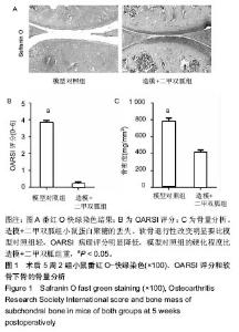
2.1 实验动物数量分析 实验选用小鼠16只,分为2组,实验过程无脱失,全部进入结果分析。 2.2 组织形态学分析 术后5周时,番红O快绿染色结果显示:模型对照组小鼠关节软骨面的蛋白聚糖丢失、软骨发生了退行性改变,部分区域出现软骨剥脱;而造模+二甲双胍组小鼠的关节面改变相比于模型对照组来说,蛋白聚糖的丢失、软骨的退行性改变明显要轻,关节面也比较光滑、完整;模型对照组OARSI病理评分明显高于造模+二甲双胍组(P < 0.05,见图1A,B),从软骨下骨的硬化程度来看,模型对照组的硬化程度也比造模+二甲双胍组的硬化程度重(P < 0.05,见图1C)。"

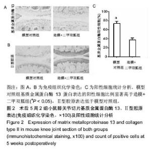
2.3 小鼠膝关节切片基质金属蛋白酶13及Ⅱ型胶原的表达 术后5周时,模型对照组基质金属蛋白酶13蛋白表达的细胞比例显著高于造模+二甲双胍组,并且这些阳性细胞主要集中在关节软骨面(图2A,B,P < 0.05);经过二甲双胍处理的造模+二甲双胍组Ⅱ型胶原的免疫组织化学染色表达的强度显著高于模型对照组(图2C)。"

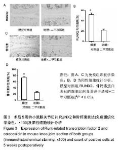
2.4 小鼠膝关节软骨下骨成骨蛋白表达的变化 术后5周时,模型对照组RUNX2 (图3A,B,P < 0.05)、骨钙素(图3C,D,P < 0.05)蛋白表达的细胞比例显著高于造模+二甲双胍组。从模型对照组的结果也可以看出,这些阳性细胞表达的部位不仅仅在关节软骨表达,在软骨下骨的区域同样存在高表达的情况。"


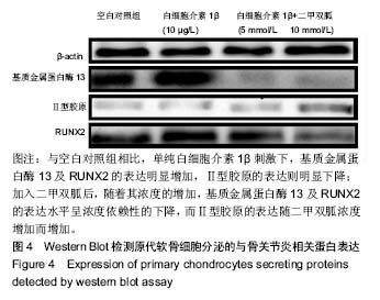
2.5 新生小鼠原代软骨细胞的体外实验结果 单纯的白细胞介素1β(10 μg/L)刺激下,基质金属蛋白酶13及RUNX2的表达水平与模型对照组相比明显增加,而Ⅱ型胶原的表达水平则明显下降;在加入二甲双胍后,随着其浓度的增加,基质金属蛋白酶13及RUNX2的表达水平呈浓度依赖性的下降,而Ⅱ型胶原的表达水平则随着二甲双胍的浓度增加呈浓度依赖性的增加,见图4。"
| [1] Glyn-Jones S,Palmer AJ,Agricola R,et al.Osteoarthritis. Lancet.2015; 386(9991):376-387.[2] Dieppe PA,Lohmander LS.Pathogenesis and management of pain in osteoarthritis.Lancet.2005; 365(9463):965-973.[3] Hootman JM,Helmick CG.Projections of US prevalence of arthritis and associated activity limitations. Arthritis Rheum.2006;54(1):226-229.[4] Woolf AD,Pfleger B.Burden of major musculoskeletal conditions. Bull World Health Organ.2003; 81: 646–656.[5] Murray CJ,Vos T,Lozano R,et al.Disability-adjusted life years (DALYs) for 291 diseases and injuries in 21 regions,1990–2010: a systematic analysis for the Global Burden of Disease Study 2010.Lancet 2012; 380: 2197–223.[6] Coxib and traditional NSAID Trialists’ (CNT) Collaboration, Bhala N,Emberson J,Merhi A,et al. Vascular and upper gastrointestinal effects of non-steroidal anti-inflammatory drugs: meta-analyses of individual participant data from randomised trials.Lancet.2013; 382:769–779.[7] Pap T, Korb-Pap A. Cartilage damage in osteoarthritis and rheumatoid arthritis—two unequal siblings. Nat Rev Rheumatol. 2015;11(10):606-15.[8] Wojdasiewicz P, Poniatowski ?A, Szukiewicz D. The role of inflammatory and anti-inflammatory cytokines in the pathogenesis of osteoarthritis, Mediators Inflamm. 2014 (2014) 561459.[9] Blasioli DJ, Kaplan DL.The roles of catabolic factors in the development of osteoarthritis, Tissue Eng. Tissue Eng Part B Rev. 2014;20(4):355-363. [10] Wang M, Shen J, Jin H, et al.Recent progress in understanding molecular mechanisms of cartilage degeneration during osteoarthritis. Ann N Y Acad Sci. 2011;1240:61-69. [11] 范梅琳,刘云峰,章毅,等;二甲双胍对脂多糖诱导的THP-1细胞相关炎症因子及凋亡的影响[J].中华内分泌代谢杂志,2013, 29(9): 801-805.[12] Sag D,Carling D,Stout RD,et al.Adenosine 5'-monophosphate-activated protein kinase promotes macrophage polarization to an anti-inflammatory functional phenotype.J Immunol.2008; 181(12):8633-8641.[13] Glasson SS,Chambers MG,Van Den Berg WB,et al.The OARSI histopathology initiative e recommendations for histological assessments of osteoarthritis in the mouse. Osteoarthritis Cartilage 2010;18 Suppl 3 S17-23.[14] Zhen G, Wen C, Jia X,et al. Inhibition of TGF-beta signaling in mesenchymal stem cells of subchondral bone attenuates osteoarthritis. Nat Med.2013;19: 70412.[15] Poulet B,de Souza R,Kent AV,et al.Intermittent applied mechanical loading induces subchondral bone thickening that may be intensified locally by contiguous articular cartilage lesions. Osteoarthritis Cartilage.2015; 23:9408. [16] Botter SM, van Osch GJ, Clockaerts S,et al.Osteoarthritis induction leads to early and temporal subchondral plate porosity in the tibial plateau of mice: an in vivo microfocal computed tomography study.Arthritis Rheum.2011; 63: 26909. [17] Chen LY, Wang Y, Terkeltaub R,et al. Activation of AMPK-SIRT3 Signaling is Chondroprotective by Preserving Mitochondrial DNA Integrity and Function. Osteoarthritis Cartilage. 2018 Nov;26(11):1539-1550.[18] Sebastian Alers, Antje S. Löffler, Sebastian Wesselborg et al. Role of AMPK-mTOR-Ulk1/2 in the Regulation of Autophagy: Cross Talk, Shortcuts, and Feedbacks. Mol. Cell. Biol. 2012, 32(1):2.[19] Sahu BD, Kuncha M, Sindhura GJ, et al. Hesperidin attenuates cisplatin-induced acute renal injury by decreasing oxidative stress, inflammation and DNA damage. Phytomedicine. 2013;20(5):453-460.[20] Liu XX, Yu DD, Chen MJ, et al.Hesperidin ameliorates lipopolysaccharide- induced acute lung injury in mice by inhibiting HMGB1 release. Int Immunopharmacol. 2015;25(2):370-376..[21] Guilak F.Biomechanical factors in osteoarthritis. Best Pract Res Clin Rheumatol. 2011;25(6):815-823.[22] Scholtes S, Krämer E, Weisser M, et al.Global chondrocyte gene expression after a single anabolic loading period: Time evolution and reinducibility of mechano-responses, J Cell Physiol 233 (2018) 699-711.[23] O'Conor CJ, Leddy HA, Benefield HC,et al.TRPV4-mediated mechanotransduction regulates the metabolic response of chondrocytes to dynamic loading, Proc Natl Acad Sci U S A 111 (2014) 1316-1321.[24] Praxenthaler H, Krämer E, Weisser M, et al.Extracellular matrix content and WNT/β-catenin levels of cartilage determine the chondrocyte response to compressive load. Biochim Biophys Acta Mol Basis Dis. 2018;1864(3): 851-859.[25] Bay-Jensen AC,Hoegh-Madsen S,Dam E,et al.Which elements are involved in reversible and irreversible cartilage degradation in osteoarthritis? Rheumatol Int 2010;30: 435-442.[26] Reimann I,Mankin HJ,Trahan C.Quantitative histologic analyses of articular-cartilage and subchondral bone from osteoarthritic and normal human hips.Acta Orthop Scand.1977; 48: 63-73.[27] Vela-Anero Á, Hermida-Gómez T, Gato-Calvo L, et al.Long-term effects of hydrogen sulfide on the anabolic-catabolic balance of articular cartilage in vitro.Nitric Oxide.2017; 70:42-50.[28] Bauge C, Girard N, Leclercq S,et al. Regulatory mechanism of transforming growth factor beta receptor type II degradation by interleukin-1 in primary chondrocytes. Biochim Biophys Acta.2012; 1823:983-986.[29] Song YM, Lee YH, Kim JW,et al.Metformin alleviates hepatosteatosis by restoring SIRT1-mediated autophagy induction via an AMP-activated protein kinase-independent pathway, Autophagy, 2015 January 11:1, 46-59.[30] Lacourt M,Gao C,Li A,et al.Relationship between cartilage and subchondral bone lesions in repetitive impact trauma-induced equine osteoarthritis. Osteoarthritis Cartilage. 2012; 20:572-583.[31] Sharma AR, Jagga S, Lee SS, Nam JS.Interplay between cartilage and subchondral bone contributing to pathogenesis of osteoarthritis. Int J Mol Sci.2013;14:19805-19830.[32] Yuan XL, Meng HY, Wang YC,et al.Bonecartilage interface crosstalk in osteoarthritis: Potential pathways and future therapeutic strategies. Osteoarthritis Cartilage.2014; 22:1077-1089. |
| [1] | Huang Dengcheng, Wang Zhike, Cao Xuewei. Comparison of the short-term efficacy of extracorporeal shock wave therapy for middle-aged and elderly knee osteoarthritis: a meta-analysis [J]. Chinese Journal of Tissue Engineering Research, 2021, 25(9): 1471-1476. |
| [2] | Peng Zhihao, Feng Zongquan, Zou Yonggen, Niu Guoqing, Wu Feng. Relationship of lower limb force line and the progression of lateral compartment arthritis after unicompartmental knee arthroplasty with mobile bearing [J]. Chinese Journal of Tissue Engineering Research, 2021, 25(9): 1368-1374. |
| [3] | Liu Xiangxiang, Huang Yunmei, Chen Wenlie, Lin Ruhui, Lu Xiaodong, Li Zuanfang, Xu Yaye, Huang Meiya, Li Xihai. Ultrastructural changes of the white zone cells of the meniscus in a rat model of early osteoarthritis [J]. Chinese Journal of Tissue Engineering Research, 2021, 25(8): 1237-1242. |
| [4] | Liu Xin, Yan Feihua, Hong Kunhao. Delaying cartilage degeneration by regulating the expression of aquaporins in rats with knee osteoarthritis [J]. Chinese Journal of Tissue Engineering Research, 2021, 25(5): 668-673. |
| [5] | Ma Zetao, Zeng Hui, Wang Deli, Weng Jian, Feng Song. MicroRNA-138-5p regulates chondrocyte proliferation and autophagy [J]. Chinese Journal of Tissue Engineering Research, 2021, 25(5): 674-678. |
| [6] | Cao Xuhan, Bai Zixing, Sun Chengyi, Yang Yanjun, Sun Weidong. Mechanism of “Ruxiang-Moyao” herbal pair in the treatment of knee osteoarthritis based on network pharmacology [J]. Chinese Journal of Tissue Engineering Research, 2021, 25(5): 746-753. |
| [7] | Li Yonghua, Feng Qiang, Tan Renting, Huang Shifu, Qiu Jinlong, Yin Heng. Molecular mechanism of Eucommia ulmoides active ingredients treating synovitis of knee osteoarthritis: an analysis based on network pharmacology [J]. Chinese Journal of Tissue Engineering Research, 2021, 25(5): 765-771. |
| [8] | Song Shan, Hu Fangyuan, Qiao Jun, Wang Jia, Zhang Shengxiao, Li Xiaofeng. An insight into biomarkers of osteoarthritis synovium based on bioinformatics [J]. Chinese Journal of Tissue Engineering Research, 2021, 25(5): 785-790. |
| [9] | Deng Zhenhan, Huang Yong, Xiao Lulu, Chen Yulin, Zhu Weimin, Lu Wei, Wang Daping. Role and application of bone morphogenetic proteins in articular cartilage regeneration [J]. Chinese Journal of Tissue Engineering Research, 2021, 25(5): 798-806. |
| [10] | Lü Jiaxing, Bai Leipeng, Yang Zhaoxin, Miao Yuesong, Jin Yu, Li Zhehong, Sun Guangpu, Xu Ying, Zhang Qingzhu. Evaluation of internal fixation with proximal femoral nail antirotation in elderly knee osteoarthritis patients with femoral intertrochanteric fractures [J]. Chinese Journal of Tissue Engineering Research, 2021, 25(3): 391-396. |
| [11] | Zheng Li, Li Dadi, Hu Weifan, Tang Jinlong, Zhao Fengchao. Risk assessment of contralateral knee arthroplasty after unilateral total knee arthroplasty [J]. Chinese Journal of Tissue Engineering Research, 2021, 25(3): 374-379. |
| [12] | Luo Anyu, Liu Hanlin, Xie Xiaofei, Huang Chen. Effect of antioxidant mixture on structural degeneration of an osteoarthritis rat model [J]. Chinese Journal of Tissue Engineering Research, 2021, 25(23): 3625-3629. |
| [13] | Gao Kun, Chen Dayu, Zhang Yong, Liu Weidong, Sun Shufen, Lai Wenqiang, Ma Dujun, Wu Yihong, Lin Zhanpeng, Jiang Yinglu, Yu Weiji. Achyranthes bidentata alcohol extract inhibits extracellular matrix degradation of the cartilage by regulating synovial fibroblast exosomes [J]. Chinese Journal of Tissue Engineering Research, 2021, 25(23): 3636-3640. |
| [14] | Liu Jinfu, Zeng Ping, Nong Jiao, Fan Siqi, Feng Chengqin, Huang Jiaxing. Integrative analysis of biomarkers and therapeutic targets in synovium of patients with osteoarthritis by multiple microarrays [J]. Chinese Journal of Tissue Engineering Research, 2021, 25(23): 3690-3696. |
| [15] | Chen Feng, Zhang Xiaoyun, Chen Yueping, Liao Jianzhao, Li Jiajun, Song Shilei, Lai Yu. Molecular mechanism of anhydroicaritin in the treatment of osteoarthritis: an analysis based on network pharmacology and bioinformatics [J]. Chinese Journal of Tissue Engineering Research, 2021, 25(23): 3704-3710. |
| Viewed | ||||||
|
Full text |
|
|||||
|
Abstract |
|
|||||