Chinese Journal of Tissue Engineering Research ›› 2019, Vol. 23 ›› Issue (13): 1982-1988.doi: 10.3969/j.issn.2095-4344.1678
Previous Articles Next Articles
Preincubation with low-concentration hydrogen peroxide enhances anti-oxidative stress ability of bone marrow mesenchymal stem cells
Yuan Dajiang, Peng Wuxun, Zhang Fei, Wang Zhenwen, Zheng Yinggang
- Guizhou Medical University, Guiyang 550004, Guizhou Province, China
-
Revised:2018-12-19Online:2019-05-08Published:2019-05-08 -
Contact:Peng Wuxun, MD, Professor, Chief physician, Guizhou Medical University, Guiyang 550004, Guizhou Province, China -
About author:Yuan Dajiang, Master candidate, Guizhou Medical University, Guiyang 550004, Guizhou Province, China -
Supported by:Guizhou Provincial Department of Technology & Guizhou Medical University Joint Fund Project, No. LH[2017]7197 (to PWX); Guizhou Province Health and Welfare Commission Fund Project, No. gzwjkj 2016-1-001 (to PWX); Guiyang Science and Technology Bureau Fund Project, No. GY2016-3 (to PWX)
CLC Number:
Cite this article
Yuan Dajiang, Peng Wuxun, Zhang Fei, Wang Zhenwen, Zheng Yinggang. Preincubation with low-concentration hydrogen peroxide enhances anti-oxidative stress ability of bone marrow mesenchymal stem cells[J]. Chinese Journal of Tissue Engineering Research, 2019, 23(13): 1982-1988.
share this article
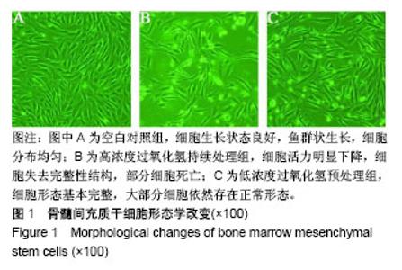
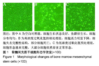
2.1 细胞形态学变化 倒置显微镜下观察细胞形态,空白对照组细胞生长状态良好,鱼群状生长,细胞分布均匀,见图1A;高浓度过氧化氢持续处理组细胞活力明显下降,细胞形态发生显著变化,失去完整性结构,部分细胞死亡,漂浮在正常细胞层上面,见图1B;低浓度过氧化氢预处理组细胞形态基本完整,大部分细胞依然存在正常形态,见图1C。"
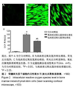
2.2 骨髓间充质干细胞内活性氧水平 骨髓间充质干细胞经500 µmol/L过氧化氢处理24 h后,能够结合更多的DCFH-DA探针,在525 nm处有更高的吸光强度,反应出强烈绿色荧光,与空白对照组相比差异有显著性意义,提示经高浓度过氧化氢处理后,骨髓间充质干细胞内处于一个活性氧积累大于清除的代谢失衡状态。低浓度过氧化氢预处理组荧光反应与高浓度过氧化氢持续处理组相比明显降低,氧化应激现象得到明显改善,差异有显著性意义(P < 0.01),见图2。"

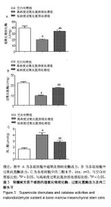
2.3 骨髓间充质干细胞内超氧化物歧化酶、过氧化氢酶活力及丙二醛水平 如图3所示,高浓度过氧化氢持续处理组细胞中超氧化物歧化酶、过氧化氢酶活性分别为(20.06±1.53) U/mg,(9.63±0.55) U/mg,明显低于空白对照组(39.60±3.13) U/mg,(22.73±1.23) U/mg (P < 0.01);低浓度过氧化氢预处理组细胞中超氧化物歧化酶、过氧化氢酶活力升高,分别为(34.11±1.72) U/mg,(17.59± 0.68) U/mg,高于高浓度过氧化氢持续处理组(P < 0.05),说明低浓度过氧化氢预处理可显著升高超氧化物歧化酶、过氧化氢酶活力。丙二醛是脂质过氧化的主要生成物之一,在生物体内可引起蛋白质、核酸等大分子的聚合反应,也可影响线粒体内关键酶活性,故而丙二醛是研究氧化损伤常用的重要指标之一。高浓度过氧化氢持续处理组细胞中丙二醛水平为(4.03±0.41) μmol/g,明显高于空白对照组(1.68±0.21) μmol/g (P < 0.01),经低浓度过氧化氢预处理后,细胞中丙二醛水平明显降低,为(2.83±0.28) μmol/g,与高浓度过氧化氢持续处理组相比差异有显著性意义(P < 0.05),说明低浓度过氧化氢预处理可明显降低丙二醛产生。"

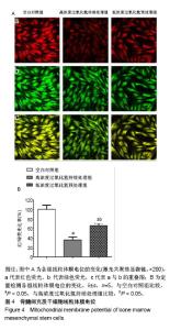
2.4 骨髓间充质干细胞线粒体膜电位分析 线粒体功能紊乱被认为是氧化应激损伤的主要机制之一,因此实验选用线粒体膜电位这一检测指标来间接反映线粒体的功能。正常细胞表达高红、高绿荧光。然而,当细胞暴露于高浓度过氧化氢(500 µmol/L)24 h时,Δψm快速去极化,绿色荧光增加和伴随红色荧光消失,见图4A。高浓度过氧化氢持续处理组红绿比为(36.34±5.62)%,低浓度过氧化氢预处理组红绿比为(66.52±4.71)%,见图4B,低浓度过氧化氢预处理组红绿比明显升高,提示预处理可能通过抑制线粒体损伤途径来实现抗细胞凋亡作用。"

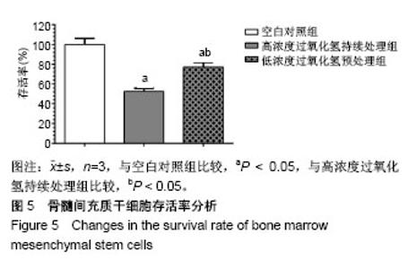
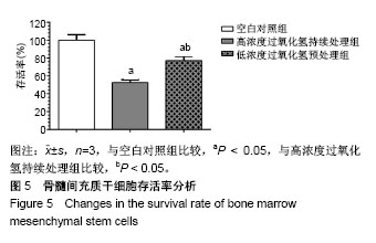
2.5 骨髓间充质干细胞存活率分析 经500 µmol/L过氧化氢处理24 h,高浓度过氧化氢持续处理组细胞在450 nm处的吸光度明显降低,细胞增殖活力只有空白对照组的(52.41±3.26)%,差异有显著性意义(P < 0.01)。低浓度过氧化氢预处理组吸光度值与高浓度过氧化氢持续处理组相比明显升高,为(77.08±4.88)%,细胞生长抑制现象明显改善,两组相比差异有显著性意义(P < 0.01),见图5。"
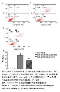
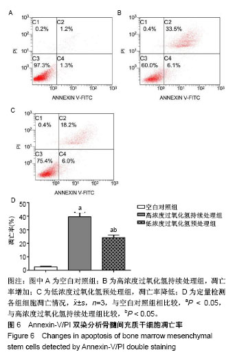
2.6 Annexin-V/PI双染分析细胞凋亡率 500 µmol/L过氧化氢处理24 h后,细胞凋亡率(39.67±3.30)%与空白对照组相比明显增加,差异有显著性意义(P < 0.01),而低浓度过氧化氢预处理可明显减少细胞凋亡率(24.26±2.20)%,低浓度过氧化氢预处理组与高浓度过氧化氢持续处理组相比,差异有显著性意义(P < 0.01),见图6。"
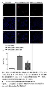
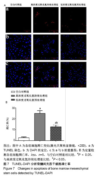
2.7 TUNEL-DAPI双染分析细胞凋亡率 采用TUNEL-DAPI双染法检测细胞核固缩和DNA断裂情况来反映低浓度过氧化氢预处理减少骨髓间充质干细胞凋亡的程度。空白对照组几乎不产生红色荧光,经500 µmol/L过氧化氢处理的骨髓间充质干细胞呈现较多红色荧光,细胞中出现DNA的碎片,凋亡率为(25.72±2.33)%,低浓度过氧化氢预处理组红色荧光明显减少,凋亡率为(12.67± 0.72)%。结果表明低浓度过氧化氢预处理能减少DNA碎片、凋亡小体的产生,有效抑制细胞凋亡,见图7。"
| [1] Hu W, Jing P, Wang L, et al. The positive effects of Ginsenoside Rg1 upon the hematopoietic microenvironment in a D-Galactose-induced aged rat model. BMC Complement Altern Med. 2015;15:119.[2] Thanan R, Techasen A, Hou B, et al. Development and characterization of a hydrogen peroxide-resistant cholangiocyte cell line: A novel model of oxidative stress-related cholangiocarcinoma genesis. Biochem Biophys Res Commun. 2015;464(1):182-188.[3] Ni S, Wang D, Qiu X, et al. Bone marrow mesenchymal stem cells protect against bleomycin-induced pulmonary fibrosis in rat by activating Nrf2 signaling. Int J Clin Exp Pathol. 2015;8(7): 7752-7761.[4] Ashour RH, Saad MA, Sobh MA, et al. Comparative study of allogenic and xenogeneic mesenchymal stem cells on cisplatin-induced acute kidney injury in Sprague-Dawley rats. Stem Cell Res Ther. 2016;7(1):126.[5] Fan CD, Sun JY, Fu XT, et al. Astaxanthin Attenuates Homocysteine-Induced Cardiotoxicity in Vitro and in Vivo by Inhibiting Mitochondrial Dysfunction and Oxidative Damage. Front Physiol. 2017;8:1041.[6] Liu D, Zhang H, Gu W, et al. Neuroprotective effects of ginsenoside Rb1 on high glucose-induced neurotoxicity in primary cultured rat hippocampal neurons. PLoS One. 2013;8(11):e79399.[7] Li Y, Zhang Y, Wang T, et al. Proteomic identification and characterization of Ctenopharyngodon idella tumor necrosis factor receptor-associated protein 1 (CiTrap1): an anti-apoptosis factor upregulated by grass carp reovirus infection. Fish Shellfish Immunol. 2015;43(2):449-459.[8] Zhang R, Zhang N, Zhang H, et al. Celastrol prevents cadmium-induced neuronal cell death by blocking reactive oxygen species-mediated mammalian target of rapamycin pathway. Br J Pharmacol. 2017;174(1):82-100.[9] Zhou Y, He W, Sun W, et al. Sulfotanshinone IIA Sodium Ameliorates Glucose Peritoneal Dialysis Solution-Induced Human Peritoneal Mesothelial Cell Injury via Suppression of ASK1-P38- mediated Oxidative Stress. Cell Physiol Biochem. 2018;46(6): 2434-2444.[10] He R, Cui M, Lin H, et al. Melatonin resists oxidative stress-induced apoptosis in nucleus pulposus cells. Life Sci. 2018;199:122-130.[11] Zhou Y, Huang S, Shen H, et al. Detection of Glutathione in Oral Squamous Cell Carcinoma Cells With a Fluorescent Probe During the Course of Oxidative Stress and Apoptosis. J Oral Maxillofac Surg. 2017;75(1):223.e1-223.e10.[12] Bebensee DF, Can K, Müller M. Increased Mitochondrial Mass and Cytosolic Redox Imbalance in Hippocampal Astrocytes of a Mouse Model of Rett Syndrome: Subcellular Changes Revealed by Ratiometric Imaging of JC-1 and roGFP1 Fluorescence. Oxid Med Cell Longev. 2017;2017:3064016.[13] Wang LL, Yu QL, Han L, et al. Study on the effect of reactive oxygen species-mediated oxidative stress on the activation of mitochondrial apoptosis and the tenderness of yak meat. Food Chem. 2018;244: 394-402.[14] Xue T, Luo P, Zhu H, et al. Oxidative stress is involved in Dasatinib-induced apoptosis in rat primary hepatocytes. Toxicol Appl Pharmacol. 2012;261(3):280-291.[15] Kapoor R, Kakkar P. Naringenin accords hepatoprotection from streptozotocin induced diabetes in vivo by modulating mitochondrial dysfunction and apoptotic signaling cascade. Toxicol Rep. 2014; 1:569-581.[16] Chan S, Chan GC, Ye J, et al. Thrombopoietin Protects Cardiomyocytes from Iron-Overload Induced Oxidative Stress and Mitochondrial Injury. Cell Physiol Biochem. 2015;36(5):2063-2071.[17] Bing W, Pang X, Qu Q, et al. Simvastatin improves the homing of BMSCs via the PI3K/AKT/miR-9 pathway. J Cell Mol Med. 2016; 20(5):949-961.[18] Ren M, Yang S, Li J, et al. Ginkgo biloba L. extract enhances the effectiveness of syngeneic bone marrow mesenchymal stem cells in lowering blood glucose levels and reversing oxidative stress. Endocrine. 2013;43(2):360-369.[19] Tao SC, Yuan T, Rui BY, et al. Exosomes derived from human platelet-rich plasma prevent apoptosis induced by glucocorticoid-associated endoplasmic reticulum stress in rat osteonecrosis of the femoral head via the Akt/Bad/Bcl-2 signal pathway. Theranostics. 2017;7(3):733-750.[20] Chen R, Liu S, Piao F, et al. 2,5-hexanedione induced apoptosis in mesenchymal stem cells from rat bone marrow via mitochondria-dependent caspase-3 pathway. Ind Health. 2015; 53(3):222-235.[21] Amiri F, Jahanian-Najafabadi A, Roudkenar MH. In vitro augmentation of mesenchymal stem cells viability in stressful microenvironments : In vitro augmentation of mesenchymal stem cells viability. Cell Stress Chaperones. 2015;20(2):237-251.[22] Mahrouf-Yorgov M, Augeul L, Da Silva CC, et al. Mesenchymal stem cells sense mitochondria released from damaged cells as danger signals to activate their rescue properties. Cell Death Differ. 2017;24(7):1224-1238.[23] Wang Y, Ma J, Du Y, et al. Human Amnion-Derived Mesenchymal Stem Cells Protect Human Bone Marrow Mesenchymal Stem Cells against Oxidative Stress-Mediated Dysfunction via ERK1/2 MAPK Signaling. Mol Cells. 2016;39(3):186-194.[24] Kavanagh DP, Suresh S, Newsome PN, et al. Pretreatment of Mesenchymal Stem Cells Manipulates Their Vasculoprotective Potential While Not Altering Their Homing Within the Injured Gut. Stem Cells. 2015;33(9):2785-2797.[25] Liu GY, Jiang XX, Zhu X, et al. ROS activates JNK-mediated autophagy to counteract apoptosis in mouse mesenchymal stem cells in vitro. Acta Pharmacol Sin. 2015;36(12):1473-1479.[26] Yu HH, Xu Q, Chen HP, et al. Stable overexpression of DJ-1 protects H9c2 cells against oxidative stress under a hypoxia condition. Cell Biochem Funct. 2013;31(8):643-651.[27] Román F, Urra C, Porras O, et al. Real-Time H2O2 Measurements in Bone Marrow Mesenchymal Stem Cells (MSCs) Show Increased Antioxidant Capacity in Cells From Osteoporotic Women. J Cell Biochem. 2017;118(3):585-593.[28] Lv C, Hao Y, Han Y, et al. Role and mechanism of microRNA-21 in H2O2-induced apoptosis in bone marrow mesenchymal stem cells. J Clin Neurosci. 2016;27:154-160.[29] Liu H, Yang X, Zhang Y, et al. Fullerol antagonizes dexamethasone-induced oxidative stress and adipogenesis while enhancing osteogenesis in a cloned bone marrow mesenchymal stem cell. J Orthop Res. 2012;30(7):1051-1057. [30] Sun ZB, Wang JW, Xiao H, et al. Icariin may benefit the mesenchymal stem cells of patients with steroid-associated osteonecrosis by ABCB1-promoter demethylation: a preliminary study. Osteoporos Int. 2015;26(1):187-197.[31] Fan L, Zhang C, Yu Z, et al. Transplantation of hypoxia preconditioned bone marrow mesenchymal stem cells enhances angiogenesis and osteogenesis in rabbit femoral head osteonecrosis. Bone. 2015;81:544-553.[32] Li J, Huang Z, Chen L, et al. Restoration of bone defects using modified heterogeneous deproteinized bone seeded with bone marrow mesenchymal stem cells. Am J Transl Res. 2017;9(7): 3200-3211.[33] Kim JY, Lee JS, Han YS, et al. Pretreatment with Lycopene Attenuates Oxidative Stress-Induced Apoptosis in Human Mesenchymal Stem Cells. Biomol Ther (Seoul). 2015;23(6): 517-524.[34] Jeong HJ, Kim DW, Kim MJ, et al. Protective effects of transduced Tat-DJ-1 protein against oxidative stress and ischemic brain injury. Exp Mol Med. 2012;44(10):586-593.[35] Shen ZY, Sun Q, Xia ZY, et al. Overexpression of DJ-1 reduces oxidative stress and attenuates hypoxia/reoxygenation injury in NRK-52E cells exposed to high glucose. Int J Mol Med. 2016;38(3): 729-736. |
| [1] | Pu Rui, Chen Ziyang, Yuan Lingyan. Characteristics and effects of exosomes from different cell sources in cardioprotection [J]. Chinese Journal of Tissue Engineering Research, 2021, 25(在线): 1-. |
| [2] | Lin Qingfan, Xie Yixin, Chen Wanqing, Ye Zhenzhong, Chen Youfang. Human placenta-derived mesenchymal stem cell conditioned medium can upregulate BeWo cell viability and zonula occludens expression under hypoxia [J]. Chinese Journal of Tissue Engineering Research, 2021, 25(在线): 4970-4975. |
| [3] | Zhang Tongtong, Wang Zhonghua, Wen Jie, Song Yuxin, Liu Lin. Application of three-dimensional printing model in surgical resection and reconstruction of cervical tumor [J]. Chinese Journal of Tissue Engineering Research, 2021, 25(9): 1335-1339. |
| [4] | Geng Qiudong, Ge Haiya, Wang Heming, Li Nan. Role and mechanism of Guilu Erxianjiao in treatment of osteoarthritis based on network pharmacology [J]. Chinese Journal of Tissue Engineering Research, 2021, 25(8): 1229-1236. |
| [5] | Wang Xianyao, Guan Yalin, Liu Zhongshan. Strategies for improving the therapeutic efficacy of mesenchymal stem cells in the treatment of nonhealing wounds [J]. Chinese Journal of Tissue Engineering Research, 2021, 25(7): 1081-1087. |
| [6] | Wang Shiqi, Zhang Jinsheng. Effects of Chinese medicine on proliferation, differentiation and aging of bone marrow mesenchymal stem cells regulating ischemia-hypoxia microenvironment [J]. Chinese Journal of Tissue Engineering Research, 2021, 25(7): 1129-1134. |
| [7] | Zeng Yanhua, Hao Yanlei. In vitro culture and purification of Schwann cells: a systematic review [J]. Chinese Journal of Tissue Engineering Research, 2021, 25(7): 1135-1141. |
| [8] | Kong Desheng, He Jingjing, Feng Baofeng, Guo Ruiyun, Asiamah Ernest Amponsah, Lü Fei, Zhang Shuhan, Zhang Xiaolin, Ma Jun, Cui Huixian. Efficacy of mesenchymal stem cells in the spinal cord injury of large animal models: a meta-analysis [J]. Chinese Journal of Tissue Engineering Research, 2021, 25(7): 1142-1148. |
| [9] | Hou Jingying, Yu Menglei, Guo Tianzhu, Long Huibao, Wu Hao. Hypoxia preconditioning promotes bone marrow mesenchymal stem cells survival and vascularization through the activation of HIF-1α/MALAT1/VEGFA pathway [J]. Chinese Journal of Tissue Engineering Research, 2021, 25(7): 985-990. |
| [10] | Shi Yangyang, Qin Yingfei, Wu Fuling, He Xiao, Zhang Xuejing. Pretreatment of placental mesenchymal stem cells to prevent bronchiolitis in mice [J]. Chinese Journal of Tissue Engineering Research, 2021, 25(7): 991-995. |
| [11] | Liang Xueqi, Guo Lijiao, Chen Hejie, Wu Jie, Sun Yaqi, Xing Zhikun, Zou Hailiang, Chen Xueling, Wu Xiangwei. Alveolar echinococcosis protoscolices inhibits the differentiation of bone marrow mesenchymal stem cells into fibroblasts [J]. Chinese Journal of Tissue Engineering Research, 2021, 25(7): 996-1001. |
| [12] | Fan Quanbao, Luo Huina, Wang Bingyun, Chen Shengfeng, Cui Lianxu, Jiang Wenkang, Zhao Mingming, Wang Jingjing, Luo Dongzhang, Chen Zhisheng, Bai Yinshan, Liu Canying, Zhang Hui. Biological characteristics of canine adipose-derived mesenchymal stem cells cultured in hypoxia [J]. Chinese Journal of Tissue Engineering Research, 2021, 25(7): 1002-1007. |
| [13] | Geng Yao, Yin Zhiliang, Li Xingping, Xiao Dongqin, Hou Weiguang. Role of hsa-miRNA-223-3p in regulating osteogenic differentiation of human bone marrow mesenchymal stem cells [J]. Chinese Journal of Tissue Engineering Research, 2021, 25(7): 1008-1013. |
| [14] | Lun Zhigang, Jin Jing, Wang Tianyan, Li Aimin. Effect of peroxiredoxin 6 on proliferation and differentiation of bone marrow mesenchymal stem cells into neural lineage in vitro [J]. Chinese Journal of Tissue Engineering Research, 2021, 25(7): 1014-1018. |
| [15] | Zhu Xuefen, Huang Cheng, Ding Jian, Dai Yongping, Liu Yuanbing, Le Lixiang, Wang Liangliang, Yang Jiandong. Mechanism of bone marrow mesenchymal stem cells differentiation into functional neurons induced by glial cell line derived neurotrophic factor [J]. Chinese Journal of Tissue Engineering Research, 2021, 25(7): 1019-1025. |
| Viewed | ||||||
|
Full text |
|
|||||
|
Abstract |
|
|||||