Chinese Journal of Tissue Engineering Research ›› 2018, Vol. 22 ›› Issue (26): 4202-4207.doi: 10.3969/j.issn.2095-4344.0944
Previous Articles Next Articles
Degradability of poly(trimethylene carbonate) in vitro
Zhang Wei1, Li Hong-yuan2, Yang Li-qun1, Li Miao1, Jin Ying1, Yi Dong-xu1
- 1Liaoning Research Institute of Family Planning, Key Laboratory of Reproductive Health and Medical Genetics, National Health and Family Planning Commission, Shenyang 110031, Liaoning Province, China; 2the Fourth Affiliated Hospital of China Medical University, Shenyang 110031, Liaoning Province, China
-
Received:2018-04-01 -
Contact:Yang Li-qun, M.D., Associate researcher, Liaoning Research Institute of Family Planning, Key Laboratory of Reproductive Health and Medical Genetics, National Health and Family Planning Commission, Shenyang 110031, Liaoning Province, China -
About author:Zhang Wei, Master, Associate researcher, Liaoning Research Institute of Family Planning, Key Laboratory of Reproductive Health and Medical Genetics, National Health and Family Planning Commission, Shenyang 110031, Liaoning Province, China -
Supported by:the National Key Research and Development Program of China, No. 2016YFC1000902; the National Natural Science Foundation of China, No. 51503093; the Natural Science Foundation of Liaoning Province, No. 20170540491; the Doctoral Fund of Liaoning Province, No. 201501116; the Science and Technology Research Project of Shenyang, No. F16-205-1-37
CLC Number:
Cite this article
Zhang Wei, Li Hong-yuan, Yang Li-qun, Li Miao, Jin Ying, Yi Dong-xu. Degradability of poly(trimethylene carbonate) in vitro[J]. Chinese Journal of Tissue Engineering Research, 2018, 22(26): 4202-4207.
share this article
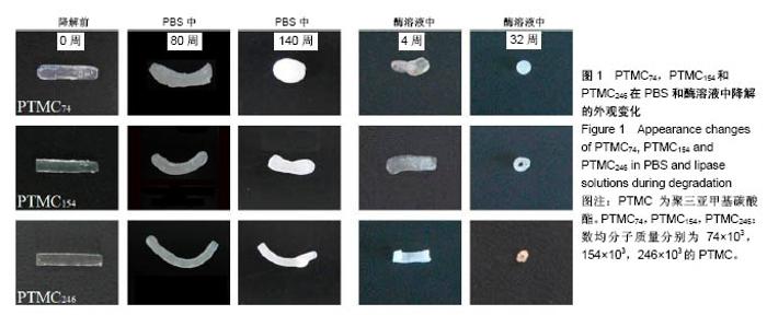
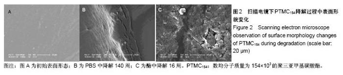
2.1 表面形貌随时间的变化 图1是样条在PBS和酶溶液中降解的外观变化,降解前样条无色并且基本透明,边缘较为工整。随着降解实验的持续,样条颜色因为吸收水分逐渐变的白色不透明。外形上相对分子质量越高样条形状保持越好,相对分子质量较小样条的边缘变得具有流动性,最终变成球形。在酶降解实验中,样品的表面会有逐渐增加的小凹坑。相对分子质量越大的样品,凹坑会越来越大变成裂痕。样品虽然保持基本形状,但外观尺寸在整体缩小,且遍布裂痕和孔洞,直到降解完全。 扫描电镜(SEM)对PTMC154在PBS中降解到140周和在酶溶液中降解到16周时的表面形貌分别进行了观察,结果见图2。从图中可以看出,降解前样条表面除了模具表面造成的压痕和一些杂质外,基本光滑无裂痕无孔洞。在PBS中降解到140周后,表面局部出现了波纹状褶皱和小的裂缝。在脂肪酶溶液中降解到16周后,表面被腐蚀变得碎裂,凹凸不平,伴随着零星的小孔洞以及一些分布不均的光滑条状区域。"
"
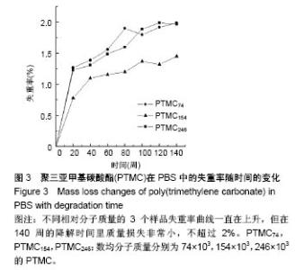
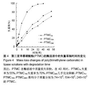
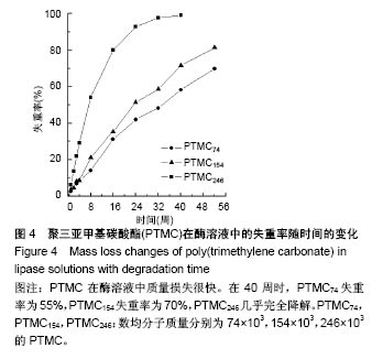
2.2 失重率随时间的变化 图3是PTMC在PBS中降解失重率的变化曲线。实验持续了将近3年,在140周的时候样品最大失重率不超过2%,降解非常缓慢,失重率可忽略不计。在酶降解实验中,高相对分子质量PTMC246的质量损失速率很快,在8周时失重率为55%,40周时几乎完全降解,但降解速率先快后慢(图4)。中和低相对分子质量PTMC的降解也很快,失重率曲线均呈线性。从总体趋势来看,PTMC的相对分子质量越高,在脂肪酶中降解的越快。"

"
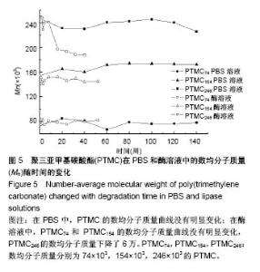
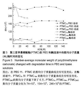
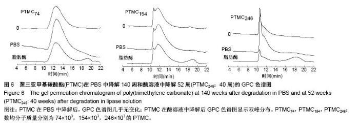
2.3 相对分子质量随时间的变化 图5是PTMC分别在PBS和脂肪酶溶液中的数均相对分子质量的变化曲线。经过对比发现在两种溶液中,除了酶溶液中的PTMC246的数均相对分子质量有较大幅度的下降,PTMC的数均相对分子质量变化趋势不明显。 实验把最后一个降解时间点样品的GPC谱图与原始0点做比较(图6),在PBS中降解PTMC的色谱图基本无变化(去除测定时设备信号响应的强弱),但是酶降解PTMC的色谱图显示了双峰分布,在主峰和溶剂峰之间出现了1个小分子峰,说明GPC检测取样时的样品是两个不同相对分子质量PTMC的共混物。有研究认为只有在降解实验的最后阶段,当表面低聚物是样品的主要构成时,双峰分布才能出现[24]。实际上,PTMC在脂肪酶中降解前期的GPC色谱图中就出现了双峰相对分子质量分布,并且样品相对分子质量越低,双峰出现时间越早。PTMC74和PTMC154的双峰分布出现在1周时间点,PTMC246的出现在8周,后峰面积均随降解时间增大。"
"
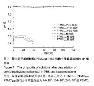
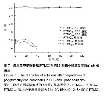
2.4 pH值随时间的变化 为了验证PTMC在降解过程中是否产生酸性产物,实验对降解液的pH值进行了检测。图7显示,在整个降解过程中,降解液的pH值分别恒定保持在近7.4和6.44,结果表明降解中没有形成酸性产物,这一结论与文献报道的一致[25]。"
| [1] Arora P, Arora RS, Cahill D. Essure(®) for management of hydrosalpinx prior to in vitro fertilisation-a systematic review and pooled analysis. BJOG. 2014;121(5):527-536. [2] Barbosa MW, Sotiriadis A, Papatheodorou SI, et al. High miscarriage rate in women treated with Essure® for hydrosalpinx before embryo transfer: a systematic review and meta-analysis. Ultrasound Obstet Gynecol. 2016;48(5): 556-565.[3] Matorras R, Rabanal A, Prieto B, et al. Hysteroscopic hydrosalpinx occlusion with Essure device in IVF patients when salpingectomy or laparoscopy is contraindicated. Eur J Obstet Gynecol Reprod Biol. 2013;169(1):54-59.[4] 欧阳振波,王黎,陈春林,等.输卵管微栓Essure的临床应用进展[J].生殖与避孕,2012,32(3):195-198. [5] Cohen SB, Bouaziz J, Schiff E, et al. In vitro fertilization outcomes after placement of essure microinserts in patients with hydrosalpinges who previously failed in vitro fertilization treatment: a multicenter study. J Minim Invasive Gynecol. 2016;23(6):939-943.[6] 杜鹏,倪才方,邹建伟,等.介入封堵治疗输卵管积水的疗效分析[J].中国介入影像与治疗学,2016,13(3):138-141.[7] 钱朝霞,陈克敏,宋富珍,等.栓塞治疗输卵管积水对体外授精-胚胎移植结局的影响[J].介入放射学杂志,2014,23(4):311-313.[8] 王靖辉,赵亚琼,郭海鸥,等.输卵管栓堵术在输卵管积水预处理中的应用价值[J].实用放射学杂志,2014,30(4):653-655.[9] 刘嵘,钱朝霞,宋富珍,等.介入栓塞术治疗输卵管积水的疗效分析[J].中国临床医学,2014,21(2):202-205.[10] 范玉荣,王凤英,唐帅,等.聚乳酸基形状记忆材料对兔输卵管黏膜的影响[J].生殖与避孕,2009,29(1):12-16.[11] 唐帅,范玉荣,张觇宇,等.聚乳酸基形状记忆输卵管节育器对兔输卵管的堵塞效果研究[J].生殖与避孕,2010,30(4):235-239.[12] 唐宝悌,董军,张其兰,等.家兔聚氨酯铋栓堵输卵管与复通后的病理形态、超微结构研究[J].生殖与避孕,1992,6(12):16-19.[13] 张巍,杨丹,王萍,等.ε-己内酯与DL-丙交酯共聚物的体内降解性能[J].中国组织工程研究,2012,16(38):7131-7134.[14] Karp JM, Shoichet MS, Davies JE. Bone formation on two-dimensional poly (DL-lactide-co-glycolide) (PLGA) films and three-dimensional PLGA tissue engineering scaffolds in vitro. J Biomed Mater Res A. 2003;64(2):388-396.[15] Spaans CJ, Belgraver VW, Rienstra O, et al. Solvent-free fabrication of micro-porous polyurethane amide and polyurethane-urea scaffolds for repair and replacement of the knee-joint meniscus. Biomaterials. 2000;21(23):2453-2460.[16] 杨立群,周群华,张巍,等.聚三亚甲基碳酸酯的研究进展[J].高分子通报,2016,(8):29-43.[17] Song Y, Wennink JW, Kamphuis MM, et al. Effective seeding of smooth muscle cells into tubular poly(trimethylene carbonate) scaffolds for vascular tissue engineering. J Biomed Mater Res A. 2010;95(2):440-446.[18] Narra N, Blanquer SB, Haimi SP, et al. μCT based assessment of mechanical deformation of designed PTMC scaffolds. Clin Hemorheol Microcirc. 2015;60(1):99-108.[19] Papenburg BJ, Schüller-Ravoo S, Bolhuis-Versteeg LA, et al. Designing porosity and topography of poly (1,3-trimethylene carbonate) scaffolds. Acta Biomater. 2009;5(9):3281-3294.[20] Buttafoco L, Boks NP, Engbers-Buijtenhuijs P, et al. Porous hybrid structures based on P (DLLA-co-TMC) and collagen for tissue engineering of small-diameter blood vessels. J Biomed Mater Res B Appl Biomater. 2006;79(2):425-434.[21] Zhang Y, Zhuo RX. Synthesis and drug release behavior of poly (trimethylene carbonate)-poly (ethylene glycol)-poly (trimethylene carbonate) nanoparticles. Biomaterials. 2005; 26(14):2089-2094.[22] Kluin OS, Busscher HJ, Neut D, et al. Poly(trimethylene carbonate) as a carrier for rifampicin and vancomycin to target therapy-recalcitrant staphylococcal biofilms. J Orthop Res. 2016;34(10):1828-1837.[23] Zhang Y, Liang RJ, Xu JJ, et al. Efficient induction of antimicrobial activity with vancomycin nanoparticle-loaded poly (trimethylene carbonate) localized drug delivery system. Int J Nanomedicine. 2017;12:1201-1214.[24] Zhang Z, Kuijer R, Sjoerd K, et al. The in vivo and in vitro degradation behavior of poly(trimethylene carbonate). Biomaterials. 2006;27(9):1741-1748.[25] Yang LQ, Li JX, Zhang W, et al. The degradation of poly (trimethylene carbonate) implants: the role of molecular weight and enzymes. Polymer Degradation Stability. 2015; 122:77-87.[26] Pêgo AP, Van Luyn MJ, Brouwer LA, et al. In vivo behavior of poly (1,3-trimethylene carbonate) and copolymers of 1,3-trimethylene carbonate with D,L-lactide or epsilon-caprolactone: Degradation and tissue response. J Biomed Mater Res A. 2003;67(3):1044-1054.[27] Pêgo AP, Poot AA, Grijpma DW, et al. In Vitro Degradation of Trimethylene Carbonate Based (Co)polymers. Macromol Biosci. 2002;2(9):411-419.[28] Albertsson AC, Eklund M. Influence of molecular structure on the degradation mechanism of degradable polymers: in vitro degradation of poly (trimethylene carbonate), poly (trimethylene carbonate-co-caprolactone), and poly(adipic anhydride). J Appl Polym Sci. 1995;57:87-103.[29] Gopferich A. Mechanisms of polymer degradation and erosion. Biomaterials. 1996;17(2):103-114.[30] Ali SA, Zhong SP, Doherty PJ, et al. Mechanisms of polymer degradation in implantable devices. I. Poly (caprolactone). Biomaterials. 1993;14(9):648-656.[31] Bisht KS, Svirkin YY, Henderson LA, et al. Lipase-catalyzed ring-opening polymerization of trimethylene carbonate. Macromolecules. 1997;30:7735-7742. |
| [1] | Yuan Bo, Wang Zhiwei, Tang Yifan, Zhou Shengyuan, Chen Xiongsheng, Jia Lianshun. Construction of polycaprolactone-tricalcium phosphate with different mixture ratios using three-dimensional printing technology and its osteoinductivity in vitro [J]. Chinese Journal of Tissue Engineering Research, 2019, 23(6): 821-826. |
| [2] | Wei Wei, Liu Yanfei, Zhang Ling, Xiong Na . Self-assembling peptide hydrogel: hemostatic effect and mechanism [J]. Chinese Journal of Tissue Engineering Research, 2019, 23(2): 310-316. |
| [3] | Zhang Minbo, Peng Qifeng, Ma Yaping, Kong Weijun, Liao Wenbo. Physical properties and biocompatibility of 3D printed bone microparticle/poly(lactic-co-glycolic acid) scaffold [J]. Chinese Journal of Tissue Engineering Research, 2019, 23(14): 2215-2222. |
| [4] | Li Zhi, Tan Chunhua, Cai Xianhua, Wang Huasong, Ding Xiaoming, Zhao Yanhong. Fabrication and biocompatibility assessment of the scaffold with biomimetic interconnected macropore structure [J]. Chinese Journal of Tissue Engineering Research, 2019, 23(14): 2223-2227. |
| [5] | Bai Yulong, Gao Yufeng, Zhong Hongbin, Zhao Yantao, Guo Ruizhou, Li Li. Allogeneic and xenogeneic tissue repair materials: how to choose a suitable virus inactivation process [J]. Chinese Journal of Tissue Engineering Research, 2019, 23(14): 2261-2268. |
| [6] | Luo Kai, Yang Yafeng, Ma Teng, Xia Bing, Huang Liangliang, Huang Jinghui, Luo Zhuojing. Effects of perfluorotributylamine/alginate/bioglass biomaterials on viability and osteogenic differentiation of adipose-derived stem cells [J]. Chinese Journal of Tissue Engineering Research, 2019, 23(13): 1995-2001. |
| [7] | Wang Yuanyuan, Song Wenshan, Yu Dejun, Dai Yuankun, Li Bafang. Preparation and evaluation of fish skin acellular dermal matrix for oral guided tissue regeneration [J]. Chinese Journal of Tissue Engineering Research, 2019, 23(10): 1526-1532. |
| [8] | Liu Shaoyang, Zou Hanlin . Biocompatibility of anodic titanium oxide: an experimental research [J]. Chinese Journal of Tissue Engineering Research, 2019, 23(10): 1575-1580. |
| [9] | Cai Yuhui, Hu Kesu, Zhang Yi. Biosafety evaluation of chitosan lactate/hyaluronate sponge [J]. Chinese Journal of Tissue Engineering Research, 2019, 23(10): 1558-1563. |
| [10] | Zhang Lei, Zhou Song, Cheng Bao-chang. Actuality and challenge of biomaterials in annulus fibrosus repair [J]. Chinese Journal of Tissue Engineering Research, 2018, 22(6): 958-963. |
| [11] | Li Zhi-yue, Zhu Zhi-bo, Zhao Qun, Xiang Si-yu, Zhao Peng, Xu Zhe-wei. Biocompatibility of basic fibroblast growth factor-poly(lactic-co-glycolic acid) microspheres/ hydroxyapatite/poly(l-lactic acid) porous materialsBiocompatibility of basic fibroblast growth factor-poly(lactic-co-glycolic acid) microspheres/ hydroxyapatite/poly(l-lactic acid) porous materials [J]. Chinese Journal of Tissue Engineering Research, 2018, 22(6): 914-920. |
| [12] | Meng Ai, Wang Jian, Sui Lei. Effect of mixed acid reflux time on purification and biocompatibility of large-inner-diameter multi-walled carbon nanotubes [J]. Chinese Journal of Tissue Engineering Research, 2018, 22(6): 896-901. |
| [13] | Yang Ji-bin, Zhu Xi-zhong, Xiong Hua-zhang, Li Yu-wan, Jin Ying, Liu Yi. Biocompatibility of human amniotic mesenchymal stem cells with human acellular amniotic membrane scaffold [J]. Chinese Journal of Tissue Engineering Research, 2018, 22(5): 742-747. |
| [14] | Wang Jing, Wang Mengyang, Feng Qiaoqiao, Sun Jijun, Ci Haosu. Perforation of the pulp chamber floor: repair with mineral trioxide aggregate, light-cured glass ionomer cement and light-cured calcium hydroxide [J]. Chinese Journal of Tissue Engineering Research, 2018, 22(34): 5458-5463. |
| [15] | Yan Xia, Zhang Ya-nan, Meng Zeng-dong. Research progress of non-human primate models for bone defect implant materials [J]. Chinese Journal of Tissue Engineering Research, 2018, 22(31): 5021-5026. |
| Viewed | ||||||
|
Full text |
|
|||||
|
Abstract |
|
|||||