Chinese Journal of Tissue Engineering Research ›› 2018, Vol. 22 ›› Issue (13): 1969-1975.doi: 10.3969/j.issn.2095-4344.0430
Long non-coding RNA H19 facilitates bone marrow mesenchymal stem cell survival and vascularization in hypoxic-ischemic conditions in vitro
Hou Jing-ying, Wang Lei, Long Hui-bao, Wu Hao, Wu Quan-hua, Zhong Ting-ting, Zhou Chang-qing, Guo Tian-zhu, Chen Xu-xiang, Wang Tong
- Department of Emergency, Sun Yat-sen Memorial Hospital, Sun Yat-sen University, Guangzhou 510120, Guangdong Province, China
-
Revised:2017-11-23Online:2018-05-08Published:2018-05-08 -
Contact:Wang Tong, M.D., Doctoral supervisor, Professor, Chief physician, Researcher, Department of Emergency, Sun Yat-sen Memorial Hospital, Sun Yat-sen University, Guangzhou 510120, Guangdong Province, China -
About author:Hou Jing-ying, Master, Attending physician, Department of Emergency, Sun Yat-sen Memorial Hospital, Sun Yat-sen University, Guangzhou 510120, Guangdong Province, China -
Supported by:the National Natural Science Foundation of China, No. 81270213, 81670306, 81700242; the Science and Technology Plan Project of Guangdong Province, No. 2014A020211002, 2017A020215176; the Fundamental Research Funds for Culture, Innovation, Interdisciplinary Plan of Sun Yat-sen University, No. 17ykjc18; the Medical Research Foundation of Guangdong Province, No. A2016264, A2017001
CLC Number:
Cite this article
Hou Jing-ying, Wang Lei, Long Hui-bao, Wu Hao, Wu Quan-hua, Zhong Ting-ting, Zhou Chang-qing, Guo Tian-zhu, Chen Xu-xiang, Wang Tong. Long non-coding RNA H19 facilitates bone marrow mesenchymal stem cell survival and vascularization in hypoxic-ischemic conditions in vitro[J]. Chinese Journal of Tissue Engineering Research, 2018, 22(13): 1969-1975.
share this article
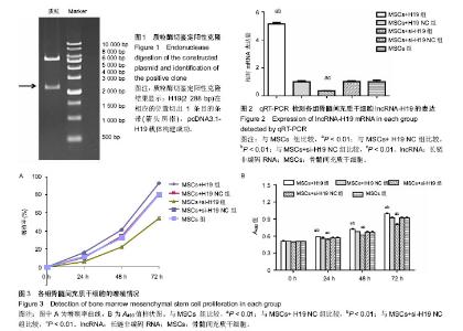
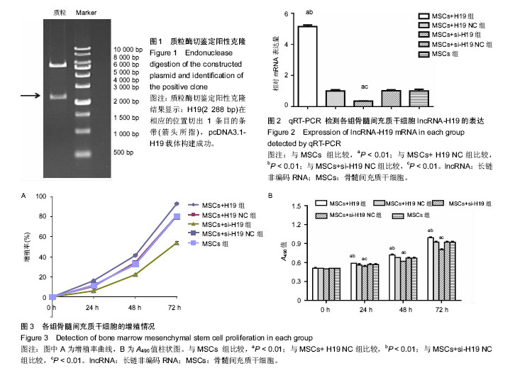
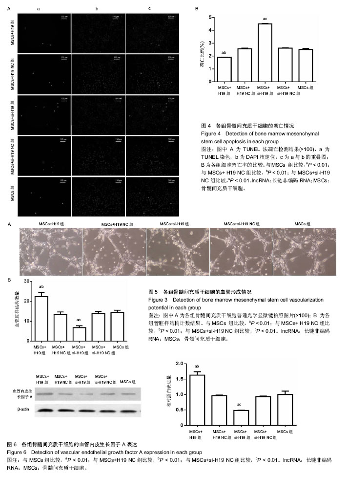
2.1 pcDNA3.1-H19载体酶切鉴定和测序结果 pcDNA3.1-H19酶切鉴定及测序结果均显示,目的基因H19已成功转入pcDNA3.1载体中(图1)。H19(2 288 bp)在相应的位置切出1条目的条带(箭头所指),pcDNA3.1-H19载体构建成功。 2.2 各组lncRNA-H19表达情况 MSCs+H19组的lncRNA-H19表达量较MSCs+H19 NC组和MSCs组明显增高(P < 0.01,图2),而 MSCs+si-H19组较MSCs+si-H19 NC和MSCs组明显降低(P < 0.01,图2),表明骨髓间充质干细胞成功转染了lncRNA-H19和lncRNA-H19siRNA。 2.3 lncRNA-H19对骨髓间充质干细胞增殖的影响 MTS检测结果显示:MSCs+H19组在体外培养24,48,72 h的细胞增殖率均明显高于MSCs+H19 NC组和MSCs组(P < 0.01,图3A),A490值均高于MSCs+H19 NC组和MSCs组(P < 0.01,图3B),MSCs+si-H19组上述不同时间点细胞增殖率明显低于MSCs+si-H19 NC组和MSCs组(P < 0.01,图3A),A490值均低于MSCs+si-H19 NC组和MSCs组(P < 0.01,图3B),表明lncRNA-H19能够促进骨髓间充质干细胞的增殖。 2.4 lncRNA-H19 对骨髓间充质干细胞凋亡的影响 TUNEL检测结果显示:MSCs+H19组、MSCs+H19 NC组、MSCs+si-H19组、MSCs+si-H19 NC组和MSCs组的凋亡比例分别为(1.90±0.01)%,(2.56±0.06)%,(4.50±0.04)%,(2.62± 0.02)%和(2.50±0.09)%,MSCs+H19组的凋亡比例低于MSCs+H19 NC组和MSCs组(P < 0.01,图4),而MSCs+ si-H19组凋亡比例高于 MSCs+si-H19 NC组和MSCs组(P < 0.01,图4)。 2.5 lncRNA-H19对骨髓间充质干细胞血管形成能力的影响 人脐静脉内皮细胞分别与各组骨髓间充质干细胞培养基上清液共培养后,在matrigel中均形成血管腔样结构(图5)。MSCs+H19组、MSCs+H19 NC组、MSCs+si-H19组、MSCs+si-H19 NC组和MSCs组血管腔样结构的数量分别为22.40±2.02,13.40±1.30,6.8±0.98,13.80±1.21和14.4±1.20。与MSCs+H19 NC组和MSCs组相比较,MSCs+H19组血管腔样结构的数量明显增多(P < 0.01);与 MSCs+si-H19 NC组和MSCs组相比较,MSCs+si-H19组血管腔样结构数量显著减少(P < 0.01),以上表明 lncRNA-H19对骨髓间充质干细胞的成管能力具有促进作用。 2.6 各组骨髓间充质干细胞血管内皮生长因子A的表达 与MSCs组及MSCs+H19 NC组相比,MSCs+H19组血管内皮生长因子A表达显著增高,而与MSCs组及MSCs+si-H19 NC组相比,MSCs+si-H19组血管内皮生长因子A表达显著减少,差异有显著性意义(P < 0.01,图6)。"

"
| [1] Wen Z, Zheng S, Zhou C, et al. Repair mechanisms of bone marrow mesenchymal stem cells in myocardial infarction. J Cell Mol Med. 2011;15(5):1032-1043.[2] Faiella W, Atoui R. Therapeutic use of stem cells for cardiovascular disease. Clin Transl Med. 2016;5(1):34.[3] Safari S, Malekvandfard F, Babashah S, et al. Mesenchymal stem cell-derived exosomes: A novel potential therapeutic avenue for cardiac regeneration. Cell Mol Biol (Noisy-le-grand). 2016;62(7): 66-73.[4] Wang T, Tang W, Sun S, et al. Mesenchymal stem cells improve outcomes of cardiopulmonary resuscitation in myocardial infarcted rats. J Mol Cell Cardiol. 2009;46(3):378-384.[5] Wang T, Tang W, Sun S, et al. Improved outcomes of cardiopulmonary resuscitation in rats with myocardial infarction treated with allogenic bone marrow mesenchymal stem cells. Crit Care Med. 2009;37(3):833-839.[6] Xing Y, Hou J, Guo T, et al. microRNA-378 promotes mesenchymal stem cell survival and vascularization under hypoxic-ischemic conditions in vitro. Stem Cell Res Ther. 2014; 5(6):130.[7] 侯婧瑛,汪蕾,钟婷婷,等. apelin干预骨髓间充质干细胞在缺血缺氧条件下的生存和血管再生[J].中国组织工程研究,2017,21(1):6-12.[8] 侯婧瑛,周长青,郑韶欣,等. 用双萤光素酶报告基因技术验证小鼠lncRNA-H19与miR-199a-5p的靶向关系[J]. 中国病理生理杂志,2016, 32(12):2256-2260.[9] Schmitz SU, Grote P, Herrmann BG. Mechanisms of long noncoding RNA function in development and disease. Cell Mol Life Sci. 2016; 73(13):2491-2509.[10] Boon RA, Jaé N, Holdt L, et al. Long Noncoding RNAs: From Clinical Genetics to Therapeutic Targets. J Am Coll Cardiol. 2016;67(10): 1214-1226.[11] Rosa A, Ballarino M. Long noncoding RNA regulation of pluripotency. Stem Cells Int. 2016; 2016:1797692. [12] Ounzain S, Micheletti R, Arnan C, et al. CARMEN, a human super enhancer-associated long noncoding RNA controlling cardiac specification, differentiation and homeostasis. Mol Cell Cardiol. 2015; 89(Pt A):98-112.[13] Hou J, Long H, Zhou C, et al. Long noncoding RNA Braveheart promotes cardiogenic differentiation of mesenchymal stem cells in vitro. Stem Cell Res Ther. 2017;8(1):4.[14] Ratajczak MZ. Igf2-H19, an imprinted tandem gene, is an important regulator of embryonic development, a guardian of proliferation of adult pluripotent stem cells, a regulator of longevity, and a 'passkey' to cancerogenesis. Folia Histochem Cytobiol. 2012;50(2):171-179.[15] Deng Y, Yang Z, Terry T, et al. Prostacyclin-producing human mesenchymal cells target H19 lncRNA to augment endogenous progenitor function in hindlimb ischaemia. Nat Commun. 2016;7: 11276.[16] Voellenkle C, Garcia-Manteiga JM, Pedrotti S, et al. Implication of Long noncoding RNAs in the endothelial cell response to hypoxia revealed by RNA-sequencing. Sci Rep. 2016;6:24141.[17] Hou J, Zhong T, Guo T, et al. Apelin promotes mesenchymal stem cells survival and vascularization under hypoxic-ischemic condition in vitro involving the upregulation of vascular endothelial growth factor. Exp Mol Pathol. 2017;102(2):203-209.[18] 郭天柱,邢越,侯婧瑛, 等. miR-378对缺血缺氧条件下骨髓间充质干细胞增殖及凋亡的影响[J].中国组织工程研究,2014,18(19):2961-2967.[19] Carvalho E, Verma P, Hourigan K, et al. Myocardial infarction: stem cell transplantation for cardiac regeneration. Regen Med. 2015; 10(8):1025-1043.[20] Narita T, Suzuki K. Bone marrow-derived mesenchymal stem cells for the treatment of heart failure. Heart Fail Rev. 2015;20(1):53-68.[21] Hou J, Wang L, Hou J, et al. Peroxisome Proliferator-Activated Receptor Gamma Promotes Mesenchymal Stem Cells to Express Connexin43 via the Inhibition of TGF-β1/Smads Signaling in a Rat Model of Myocardial Infarction. Stem Cell Rev. 2015;11(6):885-899.[22] Karpov AA, Drahova AV, Buslova DV, et al. Modification of mesenchymal stem cells as a way to improve the effectiveness of cell therapy of ischemic myocardial injury. Ross Fiziol Zh Im I M Sechenova. 2015;101(9):985-998.[23] 伍权华,侯婧瑛,郭天柱,等. 吡格列酮联合骨髓间充质干细胞移植治疗改善大鼠心肌梗死后的心功能[J].中国组织工程研究,2015,19(23): 3698-3704.[24] Park JS, Suryaprakash S, Lao YH, et al. Engineering mesenchymal stem cells for regenerative medicine and drug delivery. Methods. 2015;84:3-16.[25] Karantalis V, Suncion-Loescher VY, Bagno L, et al. Synergistic Effects of Combined Cell Therapy for Chronic Ischemic Cardiomyopathy. J Am Coll Cardiol. 2015;66(18):1990-1999.[26] Tanaka Y, Shirasawa B, Takeuchi Y, et al. Autologous preconditioned mesenchymal stem cell sheets improve left ventricular function in a rabbit old myocardial infarction model. Am J Transl Res. 2016;8(5): 2222-2233.[27] Bader AM, Klose K, Bieback K, et al. Hypoxic Preconditioning Increases Survival and Pro-Angiogenic Capacity of Human Cord Blood Mesenchymal Stromal Cells In Vitro. PLoS One. 2015;10(9): e0138477.[28] Ounzain S, Pedrazzini T. The promise of enhancer-associated long noncoding RNAs in cardiac regeneration. Trends Cardiovasc Med. 2015;25(7):592-602.[29] Shen S, Jiang H, Bei Y, et al. Long Non-Coding RNAs in Cardiac Remodeling. Cell Physiol Biochem. 2017;41(5):1830-1837.[30] Hou J, Zhou C, Long H, et al. Long noncoding RNAs: Novel molecules in cardiovascular biology, disease and regeneration. Exp Mol Pathol. 2016;100(3):493-501.[31] Xue M, Zhuo Y, Shan B. MicroRNAs, Long Noncoding RNAs, and Their Functions in Human Disease. Methods Mol Biol. 2017;1617: 1-25.[32] Zhang J, Liu CY, Wan Y, et al. Long non-coding RNA H19 promotes the proliferation of fibroblasts in keloid scarring. Oncol Lett. 2016; 12(4):2835-2839.[33] He P, Zhang Z, Huang G, et al. miR-141 modulates osteoblastic cell proliferation by regulating the target gene of lncRNA H19 and lncRNA H19-derived miR-675. Am J Transl Res. 2016;8(4): 1780-1788.[34] Ravid O, Shoshani O, Sela M, et al. Relative genomic stability of adipose tissue derived mesenchymal stem cells: analysis of ploidy, H19 long non-coding RNA and p53 activity. Stem Cell Res Ther. 2014;5(6):139.[35] Jiang X, Yan Y, Hu M, et al. Increased level of H19 long noncoding RNA promotes invasion, angiogenesis, and stemness of glioblastoma cells. J Neurosurg. 2016;124(1):129-136.[36] Jia P, Cai H, Liu X, et al. Long non-coding RNA H19 regulates glioma angiogenesis and the biological behavior of glioma-associated endothelial cells by inhibiting microRNA-29a. Cancer Lett. 2016; 381(2):359-369. [37] Conigliaro A, Costa V, Lo Dico A, et al. CD90+ liver cancer cells modulate endothelial cell phenotype through the release of exosomes containing H19 lncRNA. Mol Cancer. 2015;14:155.[38] Smadja DM, Levy M, Huang L, et al. Treprostinil indirectly regulates endothelial colony forming cell angiogenic properties by increasing VEGF-A produced by mesenchymal stem cells. Thromb Haemost. 2015;114(4):735-747.[39] Moon HH, Joo MK, Mok H, et al. MSC-based VEGF gene therapy in rat myocardial infarction model using facial amphipathic bile acid-conjugated polyethyleneimine. Biomaterials. 2014;35(5): 1744-1754. |
| [1] | Pu Rui, Chen Ziyang, Yuan Lingyan. Characteristics and effects of exosomes from different cell sources in cardioprotection [J]. Chinese Journal of Tissue Engineering Research, 2021, 25(在线): 1-. |
| [2] | Lin Qingfan, Xie Yixin, Chen Wanqing, Ye Zhenzhong, Chen Youfang. Human placenta-derived mesenchymal stem cell conditioned medium can upregulate BeWo cell viability and zonula occludens expression under hypoxia [J]. Chinese Journal of Tissue Engineering Research, 2021, 25(在线): 4970-4975. |
| [3] | Zhang Tongtong, Wang Zhonghua, Wen Jie, Song Yuxin, Liu Lin. Application of three-dimensional printing model in surgical resection and reconstruction of cervical tumor [J]. Chinese Journal of Tissue Engineering Research, 2021, 25(9): 1335-1339. |
| [4] | Hou Jingying, Yu Menglei, Guo Tianzhu, Long Huibao, Wu Hao. Hypoxia preconditioning promotes bone marrow mesenchymal stem cells survival and vascularization through the activation of HIF-1α/MALAT1/VEGFA pathway [J]. Chinese Journal of Tissue Engineering Research, 2021, 25(7): 985-990. |
| [5] | Shi Yangyang, Qin Yingfei, Wu Fuling, He Xiao, Zhang Xuejing. Pretreatment of placental mesenchymal stem cells to prevent bronchiolitis in mice [J]. Chinese Journal of Tissue Engineering Research, 2021, 25(7): 991-995. |
| [6] | Liang Xueqi, Guo Lijiao, Chen Hejie, Wu Jie, Sun Yaqi, Xing Zhikun, Zou Hailiang, Chen Xueling, Wu Xiangwei. Alveolar echinococcosis protoscolices inhibits the differentiation of bone marrow mesenchymal stem cells into fibroblasts [J]. Chinese Journal of Tissue Engineering Research, 2021, 25(7): 996-1001. |
| [7] | Fan Quanbao, Luo Huina, Wang Bingyun, Chen Shengfeng, Cui Lianxu, Jiang Wenkang, Zhao Mingming, Wang Jingjing, Luo Dongzhang, Chen Zhisheng, Bai Yinshan, Liu Canying, Zhang Hui. Biological characteristics of canine adipose-derived mesenchymal stem cells cultured in hypoxia [J]. Chinese Journal of Tissue Engineering Research, 2021, 25(7): 1002-1007. |
| [8] | Geng Yao, Yin Zhiliang, Li Xingping, Xiao Dongqin, Hou Weiguang. Role of hsa-miRNA-223-3p in regulating osteogenic differentiation of human bone marrow mesenchymal stem cells [J]. Chinese Journal of Tissue Engineering Research, 2021, 25(7): 1008-1013. |
| [9] | Lun Zhigang, Jin Jing, Wang Tianyan, Li Aimin. Effect of peroxiredoxin 6 on proliferation and differentiation of bone marrow mesenchymal stem cells into neural lineage in vitro [J]. Chinese Journal of Tissue Engineering Research, 2021, 25(7): 1014-1018. |
| [10] | Zhu Xuefen, Huang Cheng, Ding Jian, Dai Yongping, Liu Yuanbing, Le Lixiang, Wang Liangliang, Yang Jiandong. Mechanism of bone marrow mesenchymal stem cells differentiation into functional neurons induced by glial cell line derived neurotrophic factor [J]. Chinese Journal of Tissue Engineering Research, 2021, 25(7): 1019-1025. |
| [11] | Duan Liyun, Cao Xiaocang. Human placenta mesenchymal stem cells-derived extracellular vesicles regulate collagen deposition in intestinal mucosa of mice with colitis [J]. Chinese Journal of Tissue Engineering Research, 2021, 25(7): 1026-1031. |
| [12] | Pei Lili, Sun Guicai, Wang Di. Salvianolic acid B inhibits oxidative damage of bone marrow mesenchymal stem cells and promotes differentiation into cardiomyocytes [J]. Chinese Journal of Tissue Engineering Research, 2021, 25(7): 1032-1036. |
| [13] | Wang Xianyao, Guan Yalin, Liu Zhongshan. Strategies for improving the therapeutic efficacy of mesenchymal stem cells in the treatment of nonhealing wounds [J]. Chinese Journal of Tissue Engineering Research, 2021, 25(7): 1081-1087. |
| [14] | Wang Shiqi, Zhang Jinsheng. Effects of Chinese medicine on proliferation, differentiation and aging of bone marrow mesenchymal stem cells regulating ischemia-hypoxia microenvironment [J]. Chinese Journal of Tissue Engineering Research, 2021, 25(7): 1129-1134. |
| [15] | Zeng Yanhua, Hao Yanlei. In vitro culture and purification of Schwann cells: a systematic review [J]. Chinese Journal of Tissue Engineering Research, 2021, 25(7): 1135-1141. |
| Viewed | ||||||
|
Full text |
|
|||||
|
Abstract |
|
|||||