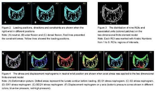
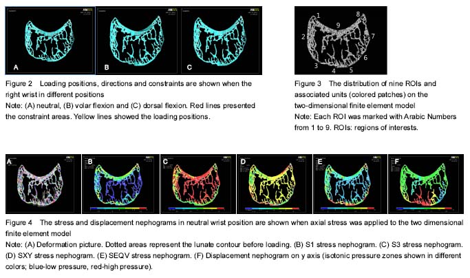
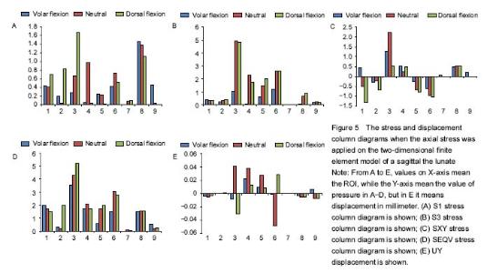
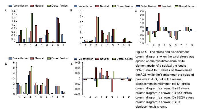
| [1] Kinney JH, Johnson QC, Nichols MC, et al. X-ray micro-tomography X at SSRL. Environ Sci Technol. 1989; 60(7):2471-2474.[2] Neldam CA, Pinholt EM. Synchrotron μCT imaging of bone, titanium implants and bone substitutes-a systematic review of the literature. J Craniomaxillofac Surg. 2014;42(6): 801-805.[3] Ritman EL. Current status of developments and applications of Micro-CT. Annu Rev Biomed Eng. 2011; 13(1): 531-552.[4] Ledoux P, Lamblin D, Wuilbaut A, et al. A finite-element analysis of Kienbock’s disease. J Hand Surg Eur Vol. 2008; 33(3):286-291.[5] Mayfield JK. Mechanism of carpal injuries. Clin Orthop Relat Res. 1980;149(1):45-54.[6] Xiao ZR, Xaiong G, Tao JF, et al. Microstructure study of normal lunates with microCT. Zhonghua Shouwaike Zazhi. 2015;31(6):455-459.[7] Bonzar M, Firrell JC, Hainer M, et al. Kienbock disease and negative ulnar variance. J Bone Joint Surg Am. 1998;80(8): 1154-1157.[8] Watson HK, Guidera PM. Aetiology of Kienbock’s disease. J Hand Surg Br. 1997;22(1):5-7.[9] Low SC, Bain GI, Findlay DM, et al. External and internal bone micro-architecture in normal and Kienbock’s the lunates: a whole-bone micro-computed tomography study. J Orthop Res. 2014;32(6):826-833.[10] Han KJ, Kim JY, Chung NS, et al. Trabecular microstructure of the human the lunate in Kienbock’s disease. J Hand Surg Br. 2012;37(4):336-341. [11] Jaecques SV, Van OH, Muraru L, et al. Individualised, micro CT-based finite element modelling as a tool for biomechanical analysis related to tissue engineering of bone. Biomaterials. 2004;25(9):1683-1696.[12] Majima M, Horii E, Matsuki H, et al. Load transmission through the wrist in the extended position. J Hand Surg Am. 2008;33(2):182-188. [13] Luo YH, Lin YW. The blood supply of lunar bone and its clinical significance. Zhongguo Linchuang Jiepouxue Zazhi. 1990;14(3):151-152.[14] Anderson DD, Daniel TE. A contact-coupled finite element analysis of the radiocarpal joint. Semin Arthroplasty. 1995; 6(1):30-36.[15] Gaisne E, Dap F, Bour C, et al. Arthrodesis of the wrist in manual workers. Apropos of 36 cases. Rev Chir Orthop Reparatrice Appar Mot. 1990;77(8):537-544.[16] Schuind F, Eslami S, Ledoux P. Kienbock’s disease. J Bone Joint Surg Br. 2008;90(2):133-139.[17] Pappas ND, Lee DH. Perilunate injuries. Am J Orthop. 2015; 44(9):E300.[18] Imhof H, Sulzbacher I, Grampp S, et al. Subchondral bone and cartilage disease: a rediscovered functional unit. Invest Radiol. 2000;35(10):581-588.[19] Low SC, Bain GI, Findlay DM, et al. External and internal bone micro-architecture in normal and Kienbock’s the lunates: a whole-bone micro-computed tomography study. J Orthop Res. 2014;32(6):826-833.[20] Dempster DW, Compston JE, Drezner MK, et al. Standardized nomenclature, symbols, and units for bone histomorphometry: a 2012 update of the report of the ASBMR Histomorphometry Nomenclature Committee. J Bone Miner Res. 2013;28(1):2-17.[21] Schiltenwolf M, Martini AK, Eversheim S, et al. Significance of intraosseous pressure for pathogenesis of Kienbock’s disease. Handchir Mikrochir Plast Chir. 1996;28(4): 215-219.[22] Watson HK, Guidera PM. Aetiology of Kienbock’s disease. J Hand Surg Br. 1997;22(1):5-7.[23] Hashizume H, Asahara H, Nishida K, et al. Histopathology of Kienbock’s disease: correlation with magnetic resonance and other imaging techniques. J Hand Surg Br. 1996;21(1): 89-93.[24] AssoulineDY, Chang C, Greenspan A, et al. Pathogenesis and natural history of osteonecrosis. Semin Arthritis Rheum. 2002;32(2):94-124.[25] Klas T, Per A, Karl-Goran T. Lipid extracted bank bone: bone conductive and mechanical properties. Clin Orthop Related Res. 1995;311(1):232-246. |