Chinese Journal of Tissue Engineering Research ›› 2017, Vol. 21 ›› Issue (6): 877-882.doi: 10.3969/j.issn.2095-4344.2017.06.010
Previous Articles Next Articles
Basic fibroblast growth factor-chitosan carrier induces neural stem cells to differentiate into neurons followed by co-culture with myocytes
- 1Department of Neurobiology, Capital Medical University, Beijing 100069, China; 2School of Biological Science and Medical Engineering, Beihang University, Beijing 100191, China
-
Received:2017-01-16Online:2017-02-28Published:2017-03-16 -
Contact:Li Xiao-guang, M.D., Professor, Department of Neurobiology, Capital Medical University, Beijing 100069, China; School of Biological Science and Medical Engineering, Beihang University, Beijing 100191, China -
About author:Zhai Jing-yan, Studying for master’s degree, Department of Neurobiology, Capital Medical University, Beijing 100069, China -
Supported by:Fibroblast Growth Factor 2; Neurons; Tissue Engineering
Funding: the International Cooperation and Communication Project of National Natural Science Foundation of China, No. 31320103903; the National Natural Science Foundation of China, No. 31130022
CLC Number:
Cite this article
Zhai Jing-yan, Duan Hong-mei, Shang Jun-kui, Yang Zhao-yang, Li Xiao-guang. Basic fibroblast growth factor-chitosan carrier induces neural stem cells to differentiate into neurons followed by co-culture with myocytes[J]. Chinese Journal of Tissue Engineering Research, 2017, 21(6): 877-882.
share this article
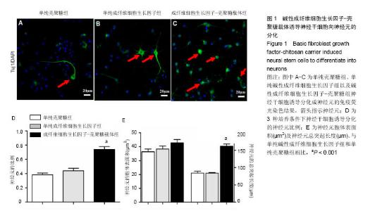
2.1 神经干细胞向神经元的分化 体外培养的神经干细胞具有自我更新和多项分化的潜能,在分化培养条件下,能够分化形成神经元、星形胶质细胞和少突胶质细胞。研究发现,碱性成纤维细胞生长因子-壳聚糖载体组,神经干细胞向神经元分化的比例达74%,大于单纯碱性成纤维细胞生长因子组和单纯壳聚糖组。因此,碱性成纤维细胞生长因子-壳聚糖载体促进神经干细胞向神经元的分化(图1)。"
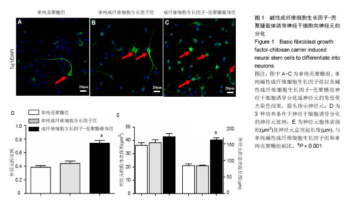
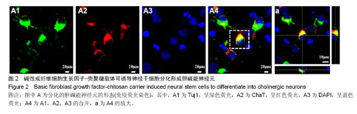
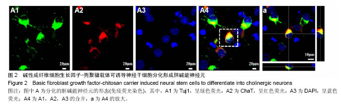
2.2 神经元的形态 3种培养条件下均能诱导分化形成多极神经元、双极神经元和假单极神经元,其中碱性成纤维细胞生长因子-壳聚糖载体组分化形成的神经元有多级、双极和假单极神经元(图1)。 2.3 神经元的突起生长情况 3种分化条件下,分化的神经元的胞体表面积接近,而其中碱性成纤维细胞生长因子-壳聚糖载体组神经元具有更多的突起,且突起长度明显高于其他2组。因此,碱性成纤维细胞生长因子-壳聚糖载体促进神经元的生长(图1)。 2.4 神经干细胞分化形成胆碱能神经元 碱性成纤维细胞生长因子-壳聚糖载体能够高比例诱导神经干细胞向神经元分化。脊髓前角运动神经元为运动传递的最后通路,其为胆碱能神经元。因此,实验进一步探索神经干细胞向胆碱能神经元分化的情况。研究发现神经干细胞可以分化为ChaT阳性的胆碱能神经元(图2)。"

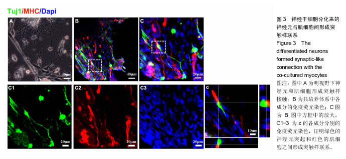
2.5 神经干细胞与肌细胞的共培养 正常人体内,脊髓前角运动神经元能够和骨骼肌纤维形成功能性突触,称为神 经肌肉接头[13-14]。碱性成纤维细胞生长因子-壳聚糖载体可以诱导神经干细胞向胆碱能神经元分化。因此,进一步研究碱性成纤维细胞生长因子-壳聚糖载体培养体系中,神经干细胞和肌细胞是否形成突触样联系。研究发现,神经干细胞分化的神经元能够和肌细胞之间形成突触样联系。因此,碱性成纤维细胞生长因子-壳聚糖载体能够诱导神经干细胞分化为胆碱能神经元,且分化形成的胆碱能神经元能够和肌细胞形成突触样联系(图3)。"
| [1]Pinho AC, Fonseca AC, Serra AC, et al. Peripheral Nerve Regeneration: Current Status and New Strategies Using Polymeric Materials. Adv Healthc Mater. 2016;5(21): 2732-2744. [2]Johnson PJ, Wood MD, Moore AM, et al. Tissue engineered constructs for peripheral nerve surgery. Eur Surg. 2013;45(3). [3]Young W. Spinal cord regeneration. Cell Transplant. 2014; 23(4-5):573-611.[4]Silver J, Miller JH. Regeneration beyond the glial scar. Nat Rev Neurosci. 2004;5(2):146-156.[5]Anderson MA, Burda JE, Ren Y, et al. Astrocyte scar formation aids central nervous system axon regeneration. Nature. 2016;532(7598):195-200. [6]Liu K, Tedeschi A, Park KK, et al. Neuronal intrinsic mechanisms of axon regeneration. Annu Rev Neurosci. 2011;34:131-152.[7]Tam RY, Fuehrmann T, Mitrousis N, et al. Regenerative therapies for central nervous system diseases: a biomaterials approach. Neuropsychopharmacology. 2014; 39(1):169-188. [8]Yang Z, Zhang A, Duan H, et al. NT3-chitosan elicits robust endogenous neurogenesis to enable functional recovery after spinal cord injury. Proc Natl Acad Sci U S A. 2015;112(43): 13354-13359. [9]Duan H, Ge W, Zhang A, et al. Transcriptome analyses reveal molecular mechanisms underlying functional recovery after spinal cord injury. Proc Natl Acad Sci U S A. 2015;112(43): 13360-13365. [10]Palmer TD, Markakis EA, Willhoite AR, et al. Fibroblast growth factor-2 activates a latent neurogenic program in neural stem cells from diverse regions of the adult CNS. J Neurosci. 1999;19(19):8487-8497.[11]王聪,杨朝阳,段红梅,等.碱性成纤维细胞生长因子-壳聚糖载体诱导神经干细胞向神经元分化并形成突触的研究[J].中国康复理论与实验,2015,21(4):406-411.[12]段红梅,王聪,杨朝阳,等.碱性成纤维细胞生长因子-壳聚糖载体诱导神经干细胞向神经元分化的机制[J].中国康复理论与实践, 2016,22(5):528-534.[13]Guo X, Gonzalez M, Stancescu M, et al. Neuromuscular junction formation between human stem cell-derived motoneurons and human skeletal muscle in a defined system. Biomaterials. 2011;32(36):9602-9611.[14]Morimoto Y, Kato-Negishi M, Onoe H, et al. Three-dimensional neuron-muscle constructs with neuromuscular junctions. Biomaterials. 2013;34(37): 9413-9419. [15]Cameron HA, Woolley CS, McEwen BS, et al. Differentiation of newly born neurons and glia in the dentate gyrus of the adult rat. Neuroscience. 1993;56(2):337-344.[16]Margolis RK, Margolis RU. Nervous tissue proteoglycans. Experientia. 1993;49(5):429-446.[17]Arber S. Motor circuits in action: specification, connectivity, and function. Neuron. 2012;74(6):975-989. [18]Lemon RN. Descending pathways in motor control. Annu Rev Neurosci. 2008;31:195-218. [19]Canedo A. Primary motor cortex influences on the descending and ascending systems. Prog Neurobiol. 1997; 51(3):287-335.[20]Lemon RN, Kirkwood PA, Maier MA, et al. Direct and indirect pathways for corticospinal control of upper limb motoneurons in the primate. Prog Brain Res. 2004;143:263-279.[21]ten Donkelaar HJ. Development and regenerative capacity of descending supraspinal pathways in tetrapods: a comparative approach. Adv Anat Embryol Cell Biol. 2000; 154:iii-ix, 1-145.[22]Cheney PD, Fetz EE, Mewes K. Neural mechanisms underlying corticospinal and rubrospinal control of limb movements. Prog Brain Res. 1991;87:213-252.[23]Tripodi M, Arber S. Regulation of motor circuit assembly by spatial and temporal mechanisms. Curr Opin Neurobiol. 2012; 22(4):615-623. [24]Dasen JS. Transcriptional networks in the early development of sensory-motor circuits. Curr Top Dev Biol. 2009;87: 119-148. [25]McCrea DA. Supraspinal and segmental interactions. Can J Physiol Pharmacol. 1996;74(4):513-517.[26]Stein PS. Spinal cord circuits for motor pattern selection in the turtle. Ann N Y Acad Sci. 1989;563:1-10.[27]Pun S, Santos AF, Saxena S, et al. Selective vulnerability and pruning of phasic motoneuron axons in motoneuron disease alleviated by CNTF. Nat Neurosci. 2006;9(3):408-419.[28]Burns AS, Jawaid S, Zhong H, et al. Paralysis elicited by spinal cord injury evokes selective disassembly of neuromuscular synapses with and without terminal sprouting in ankle flexors of the adult rat. J Comp Neurol. 2007;500(1): 116-133.[29]Jablonka S, Dombert B, Asan E, et al. Mechanisms for axon maintenance and plasticity in motoneurons: alterations in motoneuron disease. J Anat. 2014;224(1):3-14. [30]Sendtner M, Dittrich F, Hughes RA, et al. Actions of CNTF and neurotrophins on degenerating motoneurons: preclinical studies and clinical implications. J Neurol Sci. 1994;124 Suppl:77-83.[31]Kang H, Tian L, Mikesh M, et al. Terminal Schwann cells participate in neuromuscular synapse remodeling during reinnervation following nerve injury. J Neurosci. 2014;34(18): 6323-6333.[32]Lee YI, Li Y, Mikesh M, et al. Neuregulin1 displayed on motor axons regulates terminal Schwann cell-mediated synapse elimination at developing neuromuscular junctions. Proc Natl Acad Sci U S A. 2016;113(4):E479-487. [33]De Winter F, Vo T, Stam FJ, et al. The expression of the chemorepellent Semaphorin 3A is selectively induced in terminal Schwann cells of a subset of neuromuscular synapses that display limited anatomical plasticity and enhanced vulnerability in motor neuron disease. Mol Cell Neurosci. 2006;32(1-2):102-117. [34]Ko CP, Chen L. Synaptic remodeling revealed by repeated in vivo observations and electron microscopy of identified frog neuromuscular junctions. J Neurosci. 1996;16(5):1780-1790. |
| [1] | Li Shengkai, Li Tao, Wei Chao, Shi Ming. Comparison of biomechanical properties of calcium phosphate/polymethyl methacrylate composite bone cement and polymethyl methacrylate bone cement [J]. Chinese Journal of Tissue Engineering Research, 2022, 10(16): 2581-2586. |
| [2] | Yang Xue, Wang Baoqun, Jiang Xiaowen, Zou Shengcan, Ming Jinfa, Lin Shasha. Preparation and properties of biodegradable plant polysaccharide hemostatic microspheres [J]. Chinese Journal of Tissue Engineering Research, 2022, 10(16): 2607-2611. |
| [3] | Liu Jiali, Suo Hairui, Yang Han, Wang Ling, Xu Mingen. Influence of lay-down angles on mechanical properties of three-dimensional printed polycaprolactone scaffolds [J]. Chinese Journal of Tissue Engineering Research, 2022, 10(16): 2612-2617. |
| [4] | Liu Xiaojun, Xu Yuyin, Liu Kangbo, Zhou Jing, Han Ying, Xiong Yue, Tian Yuan. Preparation and properties of carboxymethylated cotton linters hemostatic gauze [J]. Chinese Journal of Tissue Engineering Research, 2022, 10(16): 2593-2599. |
| [5] | Huang Bo, Chen Mingxue, Peng Liqing, Luo Xujiang, Li Huo, Wang Hao, Tian Qinyu, Lu Xiaobo, Liu Shuyun, Guo Quanyi . Fabrication and biocompatibility of injectable gelatin-methacryloyl/cartilage-derived matrix particles composite hydrogel scaffold [J]. Chinese Journal of Tissue Engineering Research, 2022, 10(16): 2600-2606. |
| [6] | Luo Di, Liang Xuezhen, Liu Jinbao, Li Jiacheng, Yan Bozhao, Xu Bo, Li Gang. Difference in osteogenesis- and angiogenesis-related protein expression in femoral head samples from patients with femoral head necrosis of different etiologies [J]. Chinese Journal of Tissue Engineering Research, 2022, 26(11): 1641-1647. |
| [7] | Zhu Chunhui, Zhang Yi, Song Huanghe, Liang Wenwei. Protective effect of astaxanthin on tert-butyl hydrogen peroxide-induced chondrocyte damage [J]. Chinese Journal of Tissue Engineering Research, 2022, 26(11): 1648-1655. |
| [8] | Zhao Tianyu, Jin Song, Zhang Di, Liu Xiaoxiao, Ma Jiang, Wang Ju. Baduanjin training for patellar tendinopathy in a randomized controlled trial: improving pain, muscle flexibility and lower limb balance stability [J]. Chinese Journal of Tissue Engineering Research, 2022, 26(11): 1662-1668. |
| [9] | Zhang Lei, Xiu Chunmei, Ni Li, Chen Jianquan. Identification and expression analysis of mouse nucleus pulposus specific markers [J]. Chinese Journal of Tissue Engineering Research, 2022, 26(11): 1669-1674. |
| [10] | Yu Dong, Liu Kan, Shi Zongting, Yang Xiaoxia, Liu Hengping, Zhang Qingfeng. Pathological changes of the cervical intervertebral discs and rules of migration and apoptosis in endplate chondrocytes in a rabbit model of dynamic disequilibrium [J]. Chinese Journal of Tissue Engineering Research, 2022, 26(11): 1675-1679. |
| [11] | Tian Zhuang, Wang Diaodiao, Zhang Chu, Li Hanchen, Zhou Jian, Yao Qi. The mechanism by which bone morphogenetic protein 2 indirectly regulates sclerostin expression in osteocytes [J]. Chinese Journal of Tissue Engineering Research, 2022, 26(11): 1686-1691. |
| [12] | Li Jingyu, Su Yingying, Bai Ding. Morphological characteristics of subchondral bone in a mouse model of early osteoarthritis [J]. Chinese Journal of Tissue Engineering Research, 2022, 26(11): 1692-1698. |
| [13] | Bao Hongyu Lü Dongmei, He Yun, Xia Delin, Chen Junliang. Incidence of osteonecrosis in rats with jaw versus femoral defects following zoledronic acid injection [J]. Chinese Journal of Tissue Engineering Research, 2022, 26(11): 1699-1704. |
| [14] | Tan Qian, Li Bocun, Li Jing, Li Jia, Xiang Hongchun, Cai Guowei. Acupuncture combined with moxibustion regulates the expression of circadian clock protein in the synovium of rats with osteoarthritis [J]. Chinese Journal of Tissue Engineering Research, 2022, 26(11): 1714-1719. |
| [15] | Fei Jing, Tao Meihui, Li Leiji. Electroacupuncture promotes facial nerve regeneration in a rat model of facial nerve crush [J]. Chinese Journal of Tissue Engineering Research, 2022, 26(11): 1728-1733. |
| Viewed | ||||||
|
Full text |
|
|||||
|
Abstract |
|
|||||